
Anti-factor d antibody variant conjugates and uses thereof

a technology of anti-factor d and variant conjugates, which is applied in the direction of drug compositions, peptides, extracellular fluid disorders, etc., can solve the problems of protein instability, protein degradation, and protein stability, solubility, viscosity and other protein properties, etc., to improve the stability and overall potency of antibodies, improve the aqueous humour half-life, and/or the effect of retinal half-li

Active Publication Date: 2020-04-16
GENENTECH INC
View PDF0 Cites 0 Cited by
  • Summary
  • Abstract
  • Description
  • Claims
  • Application Information

AI Technical Summary

Benefits of technology

[0019]The present disclosure is based in part on the discovery that targeted amino acid substitutions of identified hot spots in an antibody can effectively improve the antibody's stability and overall potency as a therapeutic. Conjugation of such antibodies to multi-armed polymers, such as multi-armed polyols, can improve the vitreous humour half-life, the aqueous humour half-life, and / or the retinal half-life, as compared to the unconjugated antibodies.
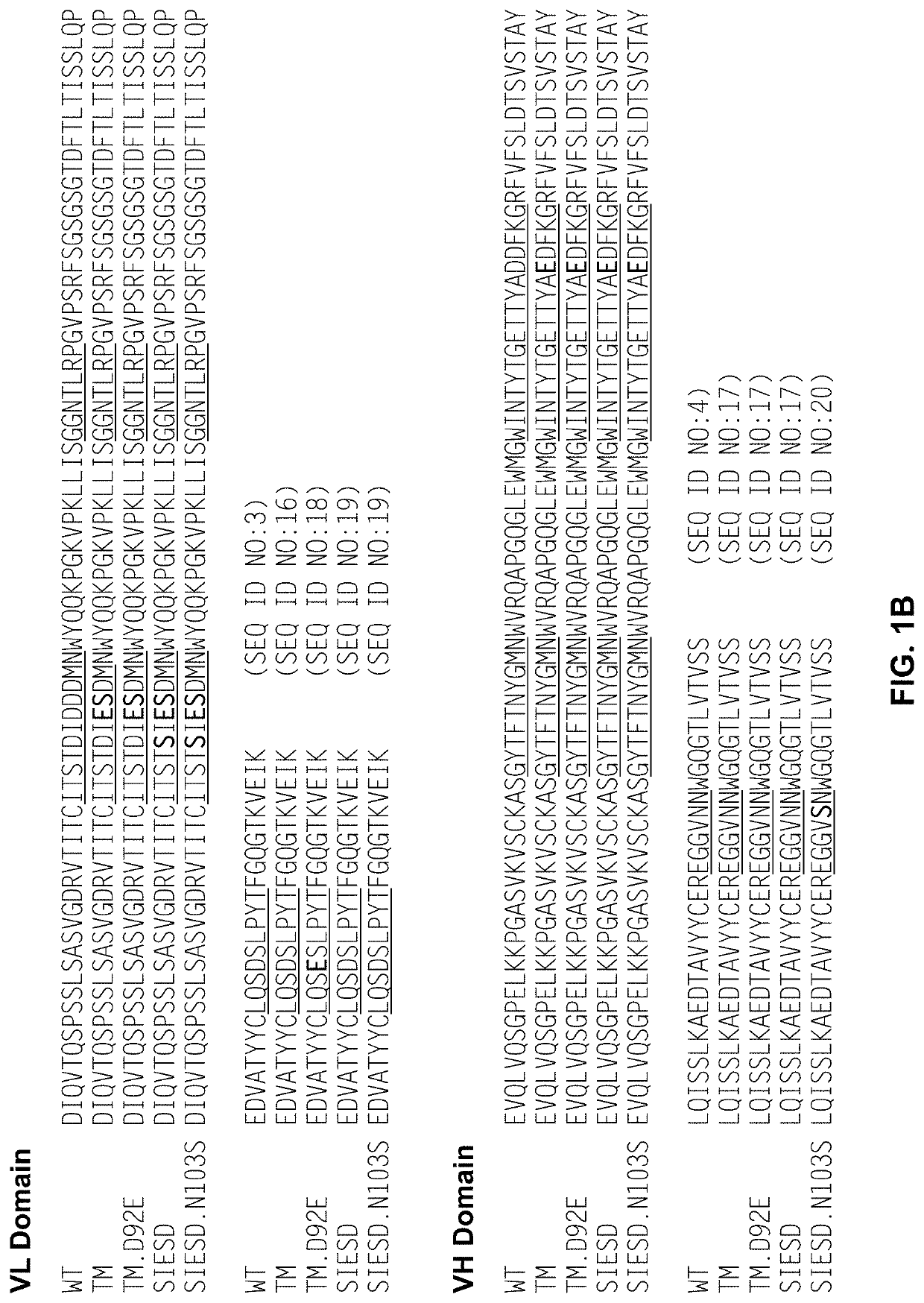
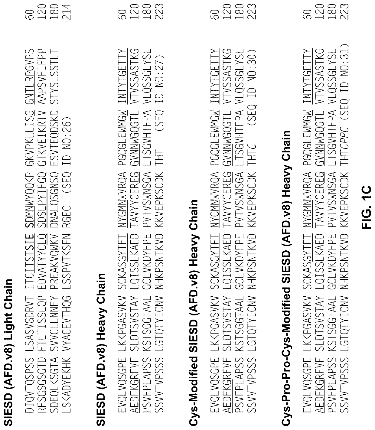
[0021]The anti-factor D antibody variants used in the conjugates of the present disclosure have improved stability. The anti-factor D antibody variants comprise substitution of at least one target aspartic acid (D or Asp) residue within a hypervariable region (HVR) of a reference anti-Factor D antibody, wherein the target Asp residue is identified as prone to isomerization and the substitution is Asp to Glutamic acid (E or Glu), and wherein the anti-Factor D antibody variant exhibits improved stability without significant loss of Factor D binding affinity when compared to the reference anti-Factor D antibody. In some aspects, the target Asp residue subject to substitution is within an Asp-Xaa motif, wherein Xaa is Asp, Gly, His, Ser or Thr. In some aspects, the target Asp residue is the first Asp of an Asp-Asp (DD) motif. In some aspects, the anti-factor D antibody variants comprise one or more substitutions at additional Asp sites within a HVR of a reference anti-Factor D antibody, wherein the substitution is Asp to Serine (S or Ser) in order to reduce the overall charges of the antibody, thereby improving the solubility of the antibody. In some aspects, the anti-factor D antibody variants comprise one or more substitutions at asparagine (N or Asn) sites identified as prone to deamidation, wherein the substitution is Asn to Ser in order to reduce or eliminate the antibody's deamidation.
[0022]In some embodiments, the anti-Factor D antibody variant is a Fab fragment, wherein the C-terminus of the heavy chain of the Fab fragment ends in the amino acids “CDKTHT,”“CDKTHL,”“CDKTH,”“CDKT,”“CDK,” or “CD.” In some embodiments, the C-terminus of the heavy chain of the Fab fragment ends in the sequence “CDKTHX,” wherein X is any amino acid except T. Truncations and / or mutations at the C terminus may be able to reduce or eliminate AHA-reactivity against the Fab, without compromising thermostability or expression. In some embodiments, the C-terminus of the heavy chain of a Fab fragment ends in the amino acids “CDKTHTC,”“CDKTHTCPPC,”“CDKTHTCPPS,”“CDKTHTSPPC”, “CDKTHTAPPC”, “CDKTHTSGGC” or “CYGPPC”. In some such embodiments, a free cysteine in the C-terminal amino acids may be amenable to conjugation, for example, to a polymer such as PEG. In some embodiments, a Fab fragment comprises a heavy chain constant domain amino acid sequence selected from the group consisting of SEQ ID NOs: 54 (ending in “CDKTHT”), 55-66 (ending in “CDKTHL,”“CDKTHTC,”“CPPC,”“CPPS,”“SPPC,”“APPC,”“SGGC,”“CYGPPC,”“CDKTH,”“CDKT,”“CDK,” or “CD”), and 116 (ending in “CDKTHX”). In some embodiments, a Fab is an IgG2 Fab fragment comprising a heavy chain constant domain amino acid sequence of SEQ ID NOs: 67 (ending in “VERK”) or an IgG2 Fab-C fragment comprising a heavy chain constant domain amino acid sequence of SEQ ID NO: 68 (ending in “VERKC”). In some embodiments, a Fab is an IgG4 Fab fragment comprising a heavy chain constant domain amino acid sequence selected from the group consisting of SEQ ID NOs: 69-73 (ending in “KYGPP”, “KYGP”, “KYG”, “KY”, or “K”) or an IgG4 Fab-C fragment comprising a heavy chain constant domain amino acid sequence of SEQ ID NO: 74 (ending in “KYGPPC”). As an alternative to truncating and / or mutation at the C terminus, to avoid pre-existing anti-hinge antibody (PE-AHA) responses, IgG2 or IgG4 Fab fragments may be used, since these do not show PE-AHA response.

Problems solved by technology

Successful development and viable use of antibody therapeutics pose many unique challenges compared to traditional medicines that are small organic and inorganic molecules.
Meanwhile, certain routes of administration are associated with limitations such as delivery time, volume and physical force that require the high dose protein to be in a high-concentration formulation (e.g., at least 100 mg / ml).
However, highly concentrated protein formulations pose particular challenges with respect to stability, solubility, viscosity and other protein properties.
Proteins can be unstable and become degraded via multiple physical and chemical degradation pathways.
Antibody instability is undesirable for drug development, as it can lead to decreased amount of active drug and lower in vivo efficacy, increased variability among batches of the therapeutics, and perhaps most importantly, immunogenicity in patients against aggregates and degradants.
Antibodies are large multidomain proteins, and factors contributing to their stability and propensity to aggregate are complex, including many extrinsic conditions such as temperature, pH, concentration, ionic strength and physical stress.
Late forms of the disease are the leading cause of vision loss in industrialized countries.
Advanced dry AMD can result in significant retinal damage, including geographic atrophy (GA), with irreversible vision loss.
Moreover, patients with dry AMD can progress to the wet form, in which abnormal blood vessels called choroidal neovascular membranes (CNVMs) develop under the retina, leak fluid and blood, and ultimately cause a blinding disciform scar in and under the retina.
Eyes have many unique biophysical and anatomic features that make the ocular drug delivery more challenging.
For example, blood-ocular barriers are defense mechanisms to protect the eye from infection, but at the same time make it hard for drug to penetrate, especially for diseases in the posterior segments of the eye.
Implants are produced using a hot-melt extrusion process such that the drug is briefly exposed to temperatures approaching 90° C. Although the drug remains in solid state for the duration of release, degradation of PLGA may expose the drug to a low pH environment.

Method used


Image

  • Anti-factor d antibody variant conjugates and uses thereof
  • Anti-factor d antibody variant conjugates and uses thereof
  • Anti-factor d antibody variant conjugates and uses thereof

Examples


example 1

n of Anti-Factor D Antibody Variants

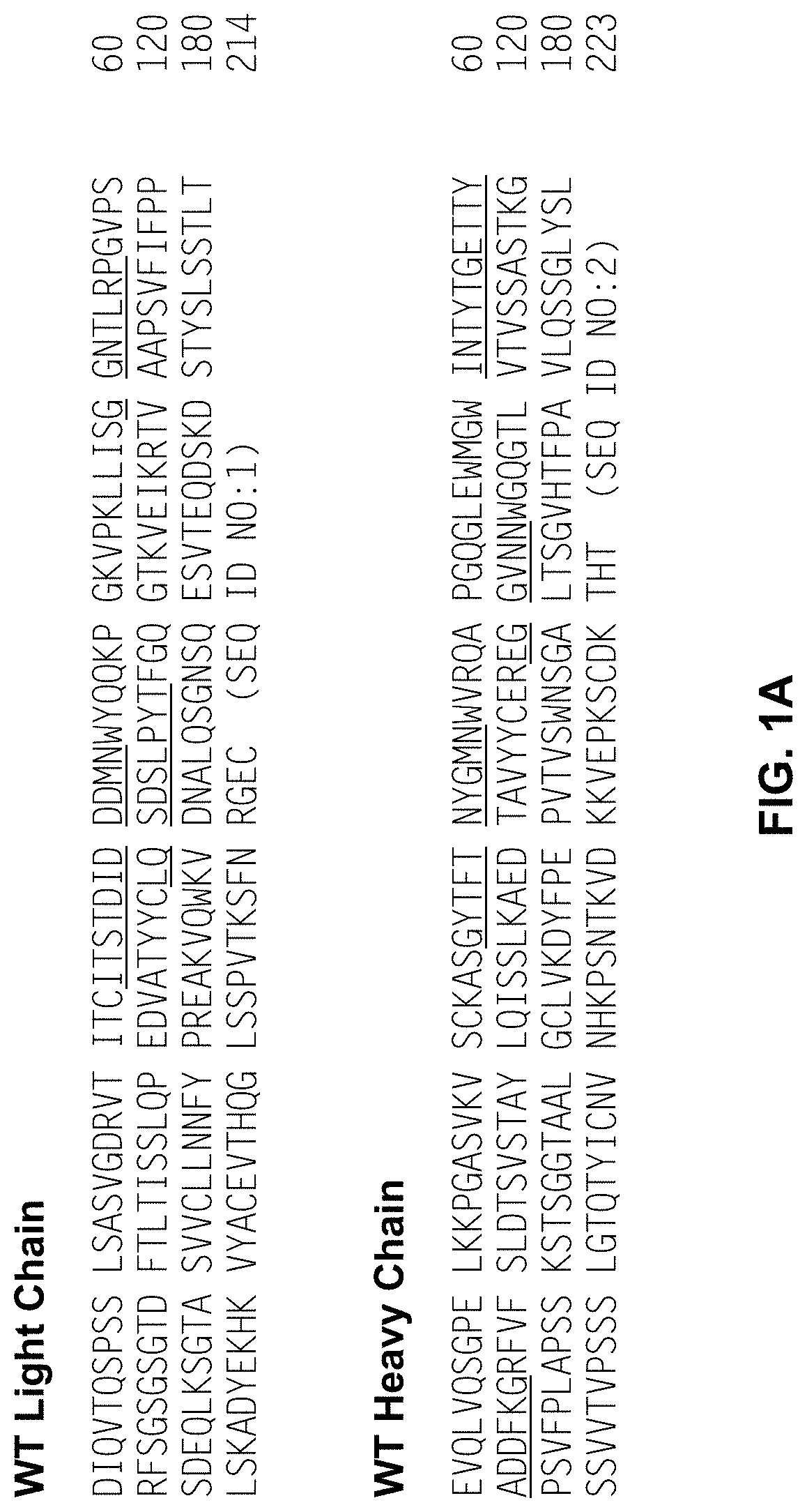
[0295]Lampalizumab, a humanized anti-Factor D Fab fragment that potently inhibits Factor D and the alternative complement pathway, through binding to an exosite on factor D is currently in clinical development for the treatment of geographic atrophy (GA), an advanced form of dry AMD. Lampalizumab (FCFD4515S; hereinafter “aFD”) is an antibody Fab fragment comprised of a 214 residue light chain (SEQ ID NO:1) and a 223 residue heavy chain (SEQ ID NO:2).

[0296]While results of a phase II human clinical trial in GA indicate that a treatment effect is obtained with monthly intravitreal injection of aFD, there exist incentives to use higher drug doses to achieve even better efficacy. Meanwhile, less frequent dosing would provide improved convenience to the patient, have potential benefits of decreased infection rate and increased clinical efficacy, and could facilitate treatment of patients with less advanced forms of dry AMD.

[0297]Efforts were made to fu...

example 2

ties of the Anti-Factor D Antibody Variants

[0301]Promising single and combination mutants were tested for factor D (fD) binding affinity and ability to inhibit factor D activities.

a. Factor D Binding Affinity by Surface Plasmon Resonance (SPR) Measurements

[0302]Kinetics and binding constant KD for factor D binding to immobilized aFD.WT and variants thereof was determined by surface plasmon resonance (SPR) measurements on a Biacore®T200 instrument. Antibody Fab fragments were immobilized on a Series S CMS sensor chip using the anti-huFab capture kit (GE healthcare Cat. #28-9583-25) following a protocol described by the manufacturer. Kinetics of binding were calculated from sensorgrams recorded for injection of 60 μL aliquots of solutions of human factor D varied in concentration from 0.39 nM to 25 nM in 2-fold increments. The flow rate was 30 μL / minute, the running buffer was HBS-P+, the temperature of analysis was 25° C., real-time reference cell subtraction was employed, and dissoc...

example 3

or D Antibody Variants with Improved Stability

[0309]Based on the affinity assays above, several single and combination anti-Factor D antibody variants were selected for further stability analysis.

a. Solubility

[0310]Samples were initially tested for solubility at low ionic strength and pH 6. Samples were first prepared in 20 mM His-HCl pH 5 buffer by concentration to ˜100 mg / mL using Amicon Centriprep YM-10 centrifugal filter units. These solutions at pH 5 and low ionic strength did not show turbidity upon visual inspection. Samples were centrifuged at 14,000×g for 10 minutes to pellet any insoluble material. No pellet was observed, and the protein concentration of the solution was determined by UV absorbance measurements. Samples (˜1 mL) were placed in Slide-A-Lyzer cassettes of 10 K MWCO (Pierce) and dialyzed overnight at 4° C. versus 1 L of 20 mM His buffer, pH 6, followed by visual inspection for turbidity. Photographs of the solutions were taken and are provided in FIG. 6. At pH...


PUM

PropertyMeasurementUnit
concentrationaaaaaaaaaa

Abstract

The disclosure relates to antibody-polymer conjugates comprising one or more anti-Factor D antibody variants, their production and their use in the preparation of compositions and medicaments for treatment of diseases and disorders associated with excessive or uncontrolled complement activation.

Description

CROSS-REFERENCE TO RELATED APPLICATIONS[0001]The instant application is a divisional of U.S. patent application Ser. No. 15 / 336,552, which was filed on Oct. 27, 2016, and which claims the benefit of priority to U.S. Provisional Application No. 62 / 249,020, which was filed on Oct. 30, 2015, and to U.S. Provisional Application No. 62 / 250,965, which was filed on Nov. 4, 2015, all of which are herein incorporated by reference in their entirety.SEQUENCE LISTING[0002]The instant application contains a Sequence Listing which has been submitted electronically in ASCII format and is hereby incorporated by reference in its entirety. Said ASCII copy, created on Oct. 27, 2016, is named P33044-US-02.txt and is 172,135 bytes in size.BACKGROUND OF THE DISCLOSURE[0003]The development of therapeutic antibodies represents a revolutionary era in the long history of human medicine. More than 30 antibodies have been approved for human therapy, and over 250 antibodies are in clinical development worldwide...

Claims


Application Information

Patent Timeline
Patent Type & Authority Applications(United States)
IPC IPC(8): C07K16/28C07K16/22C07K16/18A61K45/06A61K9/00A61K47/60
CPCA61K2039/505C07K16/22C07K16/18C07K2317/55C07K16/2863C07K2317/94A61K9/0048A61K47/60C07K2317/92A61K45/06A61P27/02A61P43/00A61P7/10A61P9/10
Inventor HASS, PHILIP E.KELLEY, ROBERT F.SCHEER, JUSTIN M.SHATZ, WHITNEYTESAR, DEVINVAN LOOKEREN CAMPAGNE, MENNO
Owner GENENTECH INC
Learn More