Method for rapidly and quantitatively detecting rabies virus antibody by using rabies virus G protein, coding gene of G protein and test paper
A rabies virus, quantitative detection technology, applied in the field of G protein coding gene and test paper, can solve the problems of modification, difference, and inability to carry out glycosylation, and achieve objective and accurate evaluation results
- Summary
- Abstract
- Description
- Claims
- Application Information
AI Technical Summary
Problems solved by technology
Method used
Image
Examples
Embodiment 1
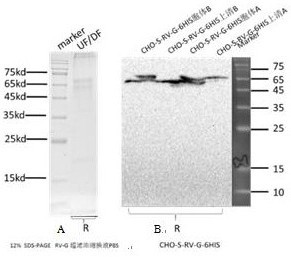
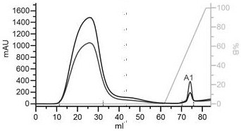
[0036] Embodiment 1: Preparation and identification of rabies virus G protein:
[0037] 1.1 Materials:
[0038] Donor plasmid pcDNA3.1, Escherichia coli Mach1T1 competent cells, DL2000 DNA Marker were purchased from Takara Company; T4 DNA ligase, restriction endonuclease Hind Ⅲ, EcoRI were purchased from NEB Company ; CHO-S cells were purchased from Invitrogen; plasmid mini-extraction kits, DNA gel recovery kits, and genome extraction kits were purchased from Axygen; G protein monoclonal antibodies were purchased from Beijing Sanlian Boyue Biotechnology Co., Ltd.
[0039] 1.2 Method:
[0040] 1.2.1 Synthesis of viral G gene, construction and identification of pcDNA3.1(+)-G recombinant expression vector:
[0041] Referring to NC_001542.1 (NCBI) and P03524-strain ERA (UniProt), the gene sequence was optimized in order to improve protein stability and increase expression in eukaryotes. The gene sequence is shown in SEQ No.1 (the amino acid sequence corresponding to the sequenc...
Embodiment 2
[0054] Embodiment 2: the preparation and use of colloidal gold detection test strip:
[0055] 2.1 Preparation and inspection of colloidal gold solution: 155mL of purified water was placed on a magnetic heating stirrer, heated and stirred until boiling. Add 5mL of 1% chloroauric acid solution, after boiling, add 6mL of newly prepared 1% trisodium citrate solution under constant stirring, continue heating and boiling for 5 minutes. Stop heating, cool to room temperature (15-25°C), and store in the dark at 2-8°C. It should be a red clear liquid without turbidity and surface suspended matter;
[0056] 2.2 Labeling and inspection of recombinant G protein: Take 1mL colloidal gold solution in a centrifuge tube, transfer it to a conical flask, and add 15µL 0.2mol / L potassium carbonate. Take 10 μg of recombinant protein G purified in Example 1, add it to the colloidal gold solution, mix quickly, and place at room temperature (15-25°C) for 30 minutes. Add 10µL 20% bovine serum albumi...
Embodiment 3
[0080] Embodiment 3: the evaluation of test strip quality:
[0081] 3.1 Specificity:
[0082] Detect the specific recognition of colloidal gold test strips to common viruses, such as Japanese encephalitis virus, influenza virus, canine distemper virus, canine parvovirus, feline parvovirus, and feline calicivirus.
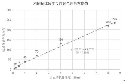
[0083] Materials and methods: 100 µL of serum containing positive antibodies to the following viruses were collected: Japanese encephalitis virus, H3N2, canine distemper virus, canine parvovirus, feline parvovirus, feline calicivirus, rabies virus (from dogs and cats, respectively) ), the above sera were all from the Beijing Animal Disease Prevention and Control Center, and SPF canine serum and healthy cat (healthy cats with common infection viruses detected by two methods of etiology and serology, all negative healthy cats) were used as negative controls; according to colloid Conditions of use of gold test strips, drop into the sample holes of the test strips resp...
PUM
| Property | Measurement | Unit |
|---|---|---|
| Concentration | aaaaa | aaaaa |
Abstract
Description
Claims
Application Information
 - R&D
- Intellectual Property
- Life Sciences
- Materials
- Tech Scout
- Unparalleled Data Quality
- Higher Quality Content
- 60% Fewer Hallucinations
Browse by: Latest US Patents, China's latest patents, Technical Efficacy Thesaurus, Application Domain, Technology Topic, Popular Technical Reports.
© 2025 PatSnap. All rights reserved.Legal|Privacy policy|Modern Slavery Act Transparency Statement|Sitemap|About US| Contact US: help@patsnap.com


