
Inactivation of West Nile virus and malaria using photosensitizers

Inactive Publication Date: 2006-05-23
TERUMO BCT BIOTECH
View PDF319 Cites 9 Cited by
  • Summary
  • Abstract
  • Description
  • Claims
  • Application Information

AI Technical Summary

Benefits of technology

[0044]Photoradiation may comprise light in the visible spectrum, the ultraviolet spectrum, or light in both the visible and ultraviolet spectra. Any suitable wavelength or wavelengths of light may be used in any proportion and energy that produces the desired level of inactivation of microorganisms. As used herein, “wavelength” does not necessarily mean one discrete wavelength. Wavelength may comprise a range of about ±100 nm centered around one wavelength. Preferably, if ultraviolet light is used, the amount of ultraviolet light is kept to a level that minimizes damage to desired fluid components. Generally, this is provided by using 50% or less ultraviolet light relative to the total light energy delivered. Also, the wavelengths of light may be centered around one peak wavelength (i.e., a range of ±10 nm centered around one wavelength).
[0108]Enhancers may also be added to the fluid to make the process more efficient and selective. Such enhancers include antioxidants or other agents to prevent damage to desired fluid components or to improve the rate of inactivation of microorganisms and are exemplified by adenine, histidine, cysteine, tyrosine, tryptophan, ascorbate, N-acetyl-L-cysteine, propyl gallate, glutathione, mercaptopropionylglycine, dithiothreotol, nicotinamide, BHT, BHA, lysine, serine, methionine, glucose, mannitol, trolox, glycerol, and mixtures thereof. These enhancers may be added in dried medium, including powder or pill form or in the form of liquids.
[0110]In decontamination systems of this invention, the photoradiation source may be connected to the photopermeable container for the fluid by means of a light guide such as a light channel or fiber optic tube which prevents scattering of the light between the source and the container for the fluid, and more importantly, prevents substantial heating of the fluid within the container. Direct exposure to the light source may raise temperatures as much as 10 to 15° C., especially when the amount of fluid exposed to the light is small, which can cause denaturization of blood components. Use of the light guide keeps any heating to less than about 2° C. The method may also include the use of temperature sensors and cooling mechanisms where necessary to keep the temperature below temperatures at which desired proteins in the fluid are damaged. Cooling mechanisms include flow of air or fluid, as well as other mechanisms known to the art. Preferably, the temperature is kept between about 0° C. and about 45° C., more preferably between about 22° C. and about 45° C., and preferably about 30° C., depending on the composition of the fluid. The heating of fluids from light exposure is known in the art and conditions to prevent damage to desired components of the fluids are known in the art without undue experimentation and described in the references cited herein. Usual operating temperature ranges for red blood cells are 32 to 36° C. and for platelets and plasma, below 30° C.

Problems solved by technology

Contamination of blood supplies with infectious microorganisms such as malaria, West Nile virus, HIV, hepatitis and other viruses and bacteria presents a serious health hazard for those who must receive transfusions of whole blood or administration of various blood components such as platelets, red cells, blood plasma, Factor VIII, plasminogen, fibronectin, anti-thrombin III, cryoprecipitate, human plasma protein fraction, albumin, immune serum globulin, prothrombin complex, plasma growth hormones, and other components isolated from blood.
Blood screening procedures may miss contaminants, and sterilization procedures which do not damage cellular blood components but effectively inactivate all infectious viruses and other microorganisms have not heretofore been available.
–514). Donated blood is not tested for infection with malaria, although there are no means to completely prevent the transmission of malaria by blood transfusion (Shulman I. Transmission of parasitic infections by blood trans
s:733–8). In the United States, the risk of transfusion-transmitted malaria is limited by excluding blood donors who have traveled to malaria-ende
; 31:798–804). In countries with a high prevalence of malaria infection, deferral of donors may no
However, most people who are infected with the West Nile Virus do not show symptoms and may not be excluded from the donation process, therefore, transmitting the virus through transfused blood.
Solvent detergent methods of blood component decontamination work by dissolving phospholipid membranes surrounding viruses such as HIV, and do not damage protein components of blood; however, if blood cells are present, such methods cannot be used because of damage to cell membranes.
Protoporphyrin, which occurs naturally within the body, can be metabolized to form a photosensitizer; however, its usefulness is limited in that it degrades desired biological activities of proteins.
Chlorpromazine is also exemplified as one such photosensitizer; however its usefulness is limited by the fact that it should be removed from any fluid administered to a patient after the decontamination procedure because it has a sedative effect.
A problem with the use of such photosensitizers is that they attack cell membranes of desirable components of fluids to be decontaminated, such as red blood cells, and the singlet oxygen also attacks desired protein components of fluids being treated.
These materials, although of fairly low toxicity, do have some toxicity e.g. to red blood cells.
This patent fails to disclose an apparatus for decontaminating blood on a flow-through basis.
However, no suggestion is made that such photosensitizers be used for decontamination of medical fluids.

Method used


Image

  • Inactivation of West Nile virus and malaria using photosensitizers
  • Inactivation of West Nile virus and malaria using photosensitizers
  • Inactivation of West Nile virus and malaria using photosensitizers

Examples


Embodiment Construction

[0128]The following applications are hereby incorporated by reference to the extent not inconsistent with the disclosure herewith: U.S. patent application Ser. No. 10 / 104,766, filed Mar. 21, 2002; U.S. patent application Ser. No. 10 / 247,262 filed Sep. 18, 2002; U.S. provisional application Ser. No. 60 / 368,778, filed Mar. 28, 2002; U.S. patent application Ser. No. 10 / 159,781, filed May 30, 2002; U.S. patent application Ser. No. 09 / 982,298, filed Oct. 16, 2001; U.S. patent application Ser. No. 10 / 328,717, filed Dec. 23, 2002; U.S. patent application Ser. No. 10 / 065,073, filed Sep. 13, 2002; U.S. patent application Ser. No. 09 / 962,029, filed Sep. 25, 2001; U.S. provisional application Ser. No. 60 / 353,223, filed Feb. 1, 2002; U.S. provisional application Ser. No. 60 / 355,393, filed Feb. 8, 2002; U.S. provisional application Ser. No. 60 / 377,697, filed May 3, 2002; U.S. patent application Ser. No. 10 / 325,402, filed Dec. 20, 2002; U.S. provisional application Ser. No. 60 / 353,319, filed Feb....


PUM

PropertyMeasurementUnit
Wavelengthaaaaaaaaaa
Depthaaaaaaaaaa
Wavelengthaaaaaaaaaa

Abstract

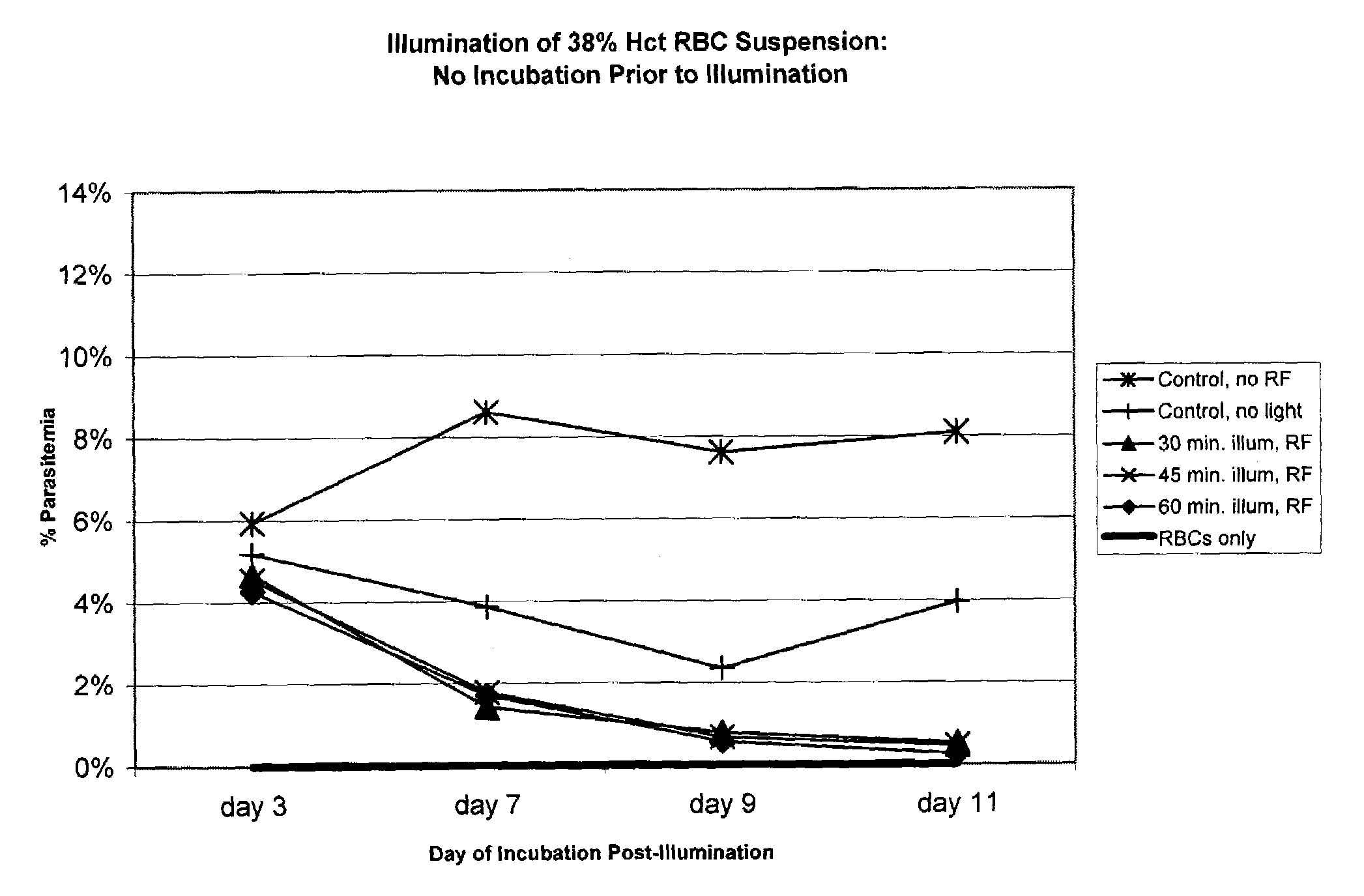
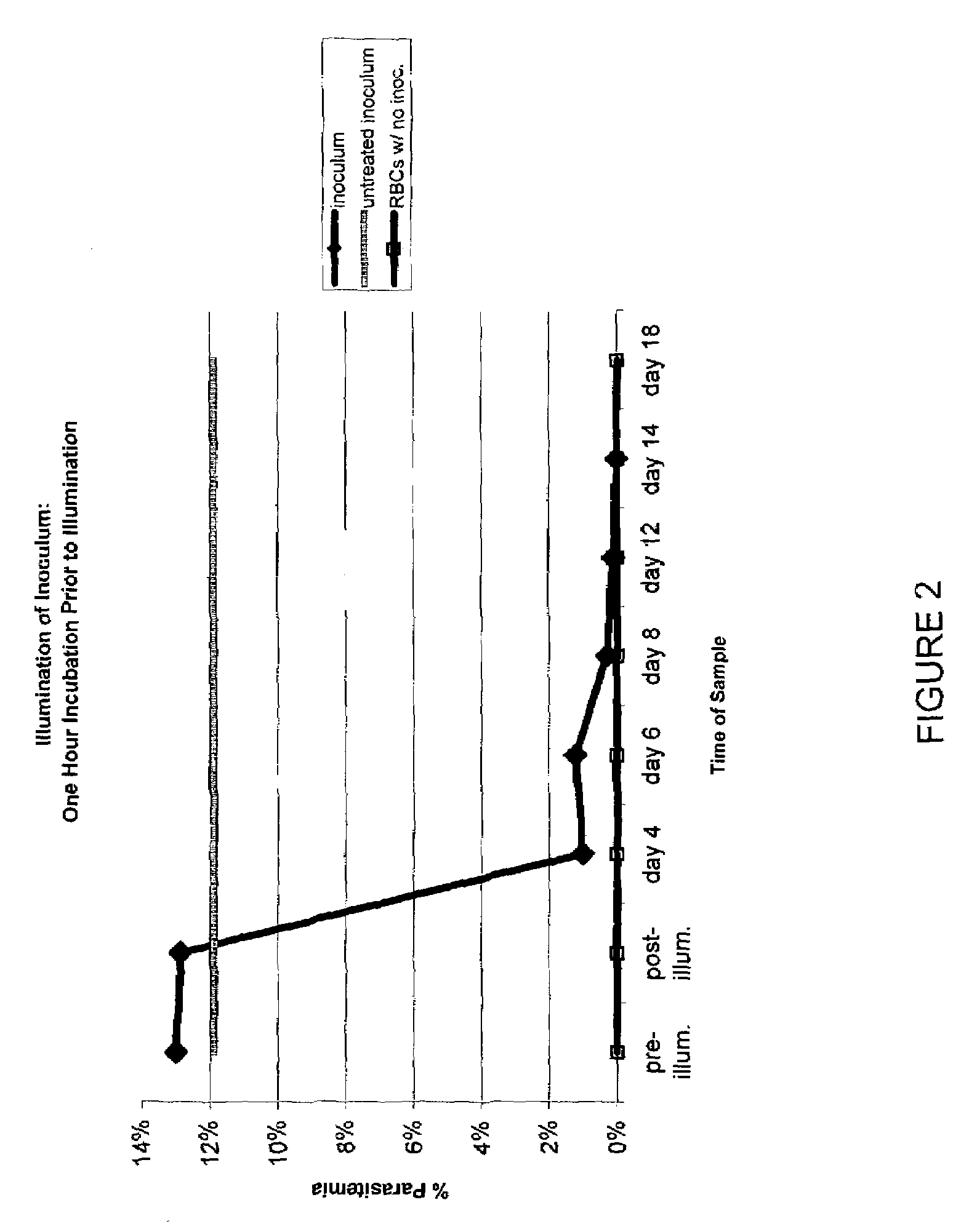
Methods and apparatuses are provided for inactivation of microorganisms in fluids or on surfaces. Preferably the fluids contain blood or blood products and comprise biologically active proteins. Preferred methods include the steps of adding an effective, non-toxic amount of a photosensitizer to a fluid and exposing the fluid to photoradiation sufficient to activate the photosensitizer whereby microorganisms are inactivated.

Description

CROSS-REFERENCE TO RELATED APPLICATIONS[0001]This application claims priority to U.S. provisional application 60 / 353,162, filed Feb. 1, 2002, and is a continuation-in-part of U.S. application Ser. No. 09 / 586,147, filed Jun. 2, 2000, now abandoned, which is a continuation-in-part of U.S. application Ser. No. 09 / 357,188 filed Jul. 20, 1999, now U.S. Pat. No. 6,277,337, which is a continuation-in-part of U.S. application Ser. No. 09 / 119,666 filed Jul. 21, 1998, now U.S. Pat. No. 6,258,577 which are hereby incorporated by reference to the extent not inconsistent with the disclosure herewith.BACKGROUND OF THE INVENTION[0002]Contamination of blood supplies with infectious microorganisms such as malaria, West Nile virus, HIV, hepatitis and other viruses and bacteria presents a serious health hazard for those who must receive transfusions of whole blood or administration of various blood components such as platelets, red cells, blood plasma, Factor VIII, plasminogen, fibronectin, anti-throm...

Claims


Application Information

Patent Timeline
IPC IPC(8): C12N13/00A61L2/08A61L2/10C12N7/04A61K41/00A61L2/00A61M1/02A61M1/36
CPCA61K41/0019A61K41/0057A61L2/0011A61L2/0082A61L2/0088A61M1/3683A61L2/10A61M1/3681A61M1/0213A61L2/08A61M1/3693A61M2205/123A61K41/17Y02A50/30
Inventor REDDY, HEATHERGOODRICH, RAYMOND P.
Owner TERUMO BCT BIOTECH
Learn More