Systems and methods for making non-invasive physiological assessments by detecting induced acoustic emissions

Inactive Publication Date: 2006-04-13
PHYSIOSONICS +1
View PDF68 Cites 148 Cited by
  • Summary
  • Abstract
  • Description
  • Claims
  • Application Information

AI Technical Summary

Benefits of technology

[0065] In many circumstances, a tissue site may not be terribly painful, but it may be enlarged or otherwise abnormal. Acoustic probing may be used to identify whether there are localized sites of pain within the enlarged or abnormal tissue site and thereby provide a positive diagnosis, or at least eliminate certain diagnoses. Enlarged tissue sites may result, for example, from tumors, other abnormal growths, inflamed tissue, or the like. Cancerous nodes are generally not painful, while enlarged nodes secondary to inflammatory conditions generally are painful. Thus, acoustic probing using the techniques described herein, provides a differential diagnosis of benign versus metastitic lymphadenopathy in patients with known head and neck primary tumors. This technique is also useful for providing a differential diagnosis in other anatomic locations, such as the mediastinum and the pelvis.
[0070] Brain tissue, and other CNS tissue, including, e.g., CSF, tissue adjacent to CSF or brain parenchyma, cranial nerves such as the optic nerve, and the like, are suitable target tissue sites for assessment of ICP. Elevated ICP causes brain and other CNS tissue to become relatively stiffer, or less compliant, when subjected to forces, such as intrinsic forces, exerted on the CNS tissue as a consequence of respiration, cyclic blood flow, compensating CSF and venous outflow, and autoregulatory-based changes in the cerebral vasculature, or when subjected to extrinsic (induced) forces exerted on the CNS tissue. The properties of blood vessels change—i.e. the vessel walls become stiffer or more pliable—as the tissue compresses or expands, or during vasoconstriction or vasodilation, respectively, producing, for example, local manifestations of the pulsatility of the cerebral vasculature.
[0076] Tissue target sites may be volumetrically large and provide data relating to large areas for gross assessment of CNS tissue properties. One of the advantages of the methods and systems of the present invention, however, is that target tissue sites may be volumetrically small, and spatially resolved, to provide data from localized tissue sites with a high degree of spatial resolution. In this way, localized differences in tissue properties may be identified and associated with a spatial location within the interrogated tissue. According to one embodiment, tissue sites of varying size and / or location are assessed simultaneously or sequentially. For most applications, the use of acoustic source(s) and. / or transducer(s) capable of interrogating and detecting target tissue sites having a volume of from 1 mm3 to 100 cm3 are suitable.
[0085] A variety of techniques may be used to analyze the acquired acoustic data relating to intrinsic and / or induced CNS tissue displacement or associated biological responses. For example, analytical techniques developed and employed in connection with ultrasound imaging, such as cross-correlation, auto-correlation, wavelet analysis, Fourier analysis, CW Doppler, sum absolute difference, and the like, may be employed to determine various properties of tissue deformation, and to relate tissue deformation to tissue properties. False peak correction techniques may be used to improve the accuracy of the assessment. Additionally, properties of the major and minor endogenous oscillations of brain tissue within a cardiac cycle, or relationships between major and minor endogenous oscillations within a cardiac cycle, or across several respiratory cycles, are empirically related to ICP and other tissue properties and conditions. These determinations may be made with, or without, additional information relating to ABP and / or respiration and / or exogenous tissue displacements.

Problems solved by technology

Characterization of internal tissues using non-invasive and non-traumatic techniques is challenging in many areas.
Non-invasive detection of various cancers remains problematic and unreliable.
Similarly, non-invasive assessment and monitoring of intracranial pressure is also a practical challenge, despite the efforts devoted to developing such techniques.
Under some conditions, elevated intracranial pressures may cause the brain to be mechanically compressed, and to herniate.
The most common cause of elevated intracranial pressure is head trauma.
Changes in intracranial pressure, particularly elevated intracranial pressure, are very serious and may be life threatening.
All of these methods and systems are invasive.
The subarachnoid bolt / screw technique requires minimal penetration of the brain, it has a relatively low risk of infection, and it provides a direct pressure measurement, but it does require penetration of an intact skull and it poorly drains CSF.
The ventriculostomy catheter technique provides CSF drainage and sampling and it provides a direct measurement of intracranial pressure, but the risks of infection, intracerebral bleeding and edema along the cannula track are significant, and it requires transducer repositioning with head movement.
Finally, the fiber optic catheter technique is versatile because the catheter may be placed in the ventricle or in the subarachnoid space, and it does not require adjustment of the transducer with head movement, but it requires a separate monitoring system, and the catheter is relatively fragile.
All of these conventional techniques require invasive procedures and none is well suited to long term monitoring of intracranial pressure on a regular basis.
Moreover, these procedures can only be performed in hospitals staffed by qualified neurosurgeons.
The hypothesis that ICP is altered in microgravity environments is difficult to test, however, as a result of the invasive nature of conventional ICP measurement techniques.
While this mode of measurement is simple and inexpensive to perform, it does not provide the most accurate measure of ABP, and it is susceptible to artifacts resulting from the condition of arterial wall, the size of the patient, the hemodynamic status of the patient, and autonomic tone of the vascular smooth muscle.
Additionally, repeated cuff measurements of ABP result in falsely elevated readings of ABP, due to vasoconstriction of the arterial wall.
While such catheters are very reliable and provide the most accurate measure of ABP, they require placement by trained medical personnel, usually physicians, and they require bulky, sophisticated, fragile, sterile instrumentation.
Additionally, there is a risk of permanent arterial injury causing ischemic events when these catheters are placed.
As a result, these invasive monitors are only used in hospital settings and for patients who are critically ill or are undergoing operative procedures.
Conventional clinical autoregulation determination techniques are inexact and burdensome.
Furthermore, measurement of CBF using transcranial Doppler techniques requires a skilled sonographer to find and maintain the focus of the equipment on large cerebral blood vessels while the patient, and the patient's CNS, may not be stationary.
Vasospasm is a condition in which the cerebral vasculature contracts to such an abnormal degree that blood flow through the affected vessel is significantly reduced, although measured blood flow velocity may actually increase, causing transient and often permanent neurologic deficits (e.g., strokes).
In practice, TCD techniques are generally limited to assessing vasospasm in the large blood vessels at the base of the skull, since TCD techniques are not sufficiently sensitive to assess vasospasm in smaller blood vessels throughout the brain.
This is an extensive and expensive procedure.
Pain is a frequent presenting symptom of numerous medical conditions, and although it plays an important role, often being the first alert that something is wrong, it can also be extremely nonspecific.
Unfortunately, it is difficult to identify the exact source of pain: several constituent pieces of a complex structure may be intimately adjoining, yet only one may be the source.
These tests are frequently uncomfortable for the patient and carry the risk of infection and contrast reaction.
Until recently, there were no good options for treatment.
But in patients with multiple fractures, identifying the painful one may be difficult.
The diagnosis of appendicitis is difficult and imprecise.
Moreover, manual probing or palpation of the abdomen, with its poor specificity, is still a standard test, with mixed results.
In the conditions described above, pain symptoms signal a problem but frequently do not pinpoint the location of that problem.

Method used


Image

  • Systems and methods for making non-invasive physiological assessments by detecting induced acoustic emissions
  • Systems and methods for making non-invasive physiological assessments by detecting induced acoustic emissions
  • Systems and methods for making non-invasive physiological assessments by detecting induced acoustic emissions

Examples


example 1

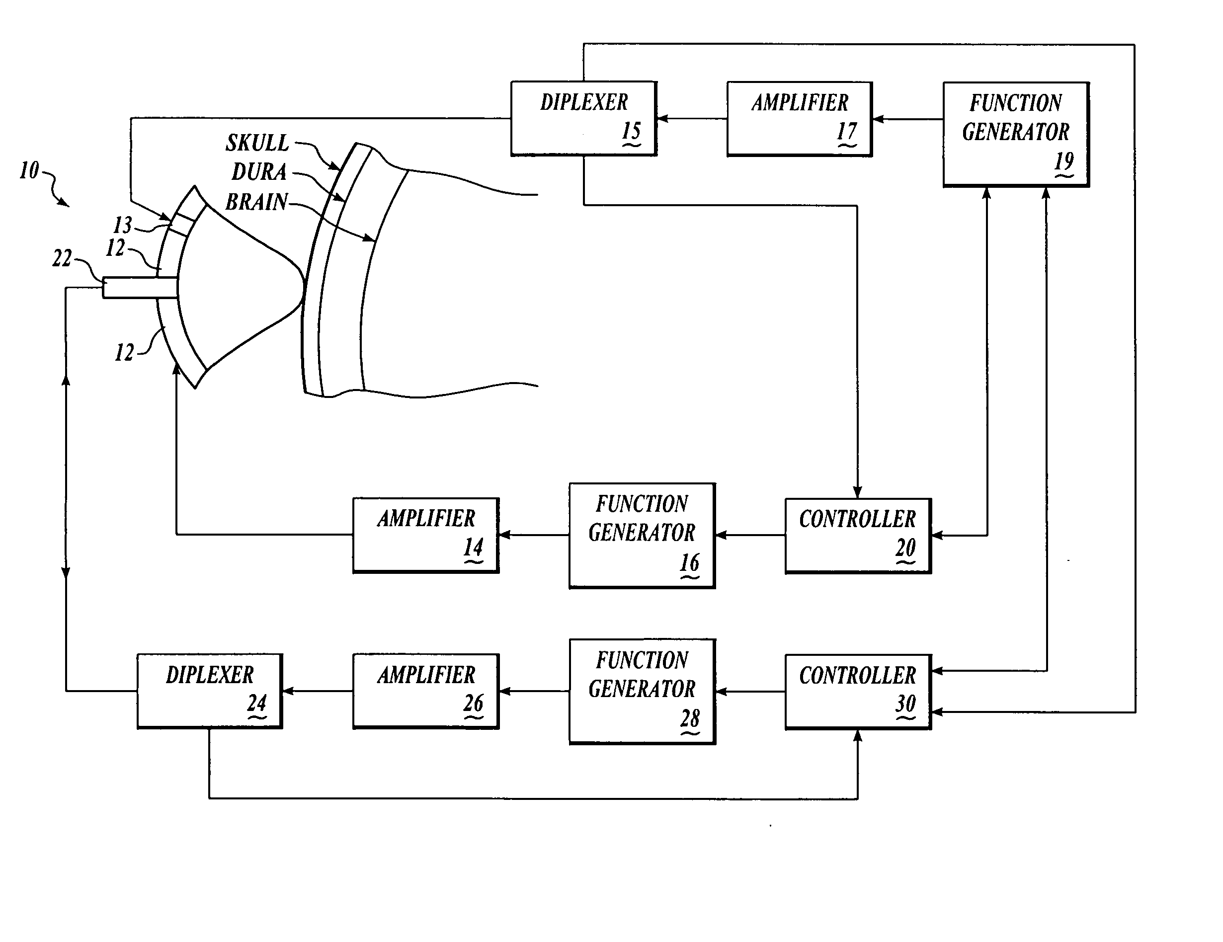
[0189] We have shown in vitro (FIG. 6A) and in vivo (FIG. 6B-D) and describe in detail below, that intrinsic displacements of brain tissue (e.g. compressions and distensions), and their various acoustic scatter properties, can be directly measured using a standard transcranial Doppler (TCD) transducer, off-the-shelf data acquisition systems, and novel analysis of the acoustic backscatter signal from brain.

[0190] An in vitro model for examining changes in ICP using acoustic techniques was constructed using fresh bovine brain immersed in fluid in a water-tight, visually and acoustically transparent bottle attached to a hand-pump for changing the pressure on the brain. An acoustic transducer (ATL / Philips Medical Systems, Bothell, Wash.), and the bottle, were placed in water so that the focus of the interrogation transducer was near the edge of the brain, but within the brain. Using a transducer whose amplifier was driven at 200 mV and a LeCroy Waverunner oscilliscope, we collected aco...

example 2

[0198] We have shown, in vitro, using a beef brain model similar to that described above, that a palpation pulse of ultrasound across a range of acoustic intensities can cause increasing displacements of brain without causing gross tissue damage.

[0199] Fresh bovine brain was immersed in fluid in a water-tight, visually and acoustically transparent bottle attached to a hand-pump for changing the pressure on the brain. ATL acoustic transducers (ATL-Philips Medical Systems, Bothell, Wash.), and the bottle, were placed in water so that the focus of the acoustic palpation and interrogation transducers were near the edge of the brain, but within the brain. Using LeCroy Waverunner oscilloscope, we collected acoustic interrogation waveforms backscattered from brain. For palpating and interrogating beef brain, in vitro, the interrogation pulses were administered as described with respect to FIG. 6A, while the palpation pulses had a pulse repetition frequency of 1 Hz, contained 30,000-50,000...

example 3

[0201] Existing transcranial Doppler (TCD) devices and controllers may be modified to process raw data relating to tissue displacement according to methods and systems of the present invention. As data, such as Doppler information, is acquired by an ultrasound transducer / receiver, it is conventionally passed through a set of filters designed to eliminate portions of the signal attributable to the motion of the vessel wall, tissue displacement, CSF perturbation etc., leaving only the portion of the signal attributable to blood flow for subsequent transcranial Doppler analysis. For the present application, the unfiltered signal acquired by a TCD device, including portions of the signal attributable to blood vessel wall motion, brain tissue displacement and CSF pertubation, as well as blood flow, may be used according to methods of the present invention to assess CSF tissue properties, such as ICP.

[0202] Unfiltered data acquired by an ultrasound transducer / receiver in a TCD or a simil...


PUM


Abstract

Systems and methods for assessing a physiological parameter of a target tissue wherein a pulse of focused ultrasound is applied to a target tissue site thereby inducing oscillation of the target tissue. By these systems and methods, a property of an acoustic signal emitted from the oscillating target tissue is measured and related to a physiological property of the tissue. Specific applications for systems and methods of the present invention include the assessment and monitoring of intracranial pressure (ICP), arterial blood pressure (ABP), CNS autoregulation status, vasospasm, stroke, local edema, infection and vasculitus, as well as diagnosis and monitoring of diseases and conditions that are characterized by physical changes in tissue properties.

Description

CROSS-REFERENCE TO RELATED APPLICATION(S) [0001] This application is a continuation application of U.S. patent application Ser. No. 09 / 995,897, filed Nov. 28, 2001, issued as U.S. Pat. No. ______ on ______, 2005, which claims priority under 35 U.S.C. 119(e) to U.S. Patent Application No. 60 / 253,959, filed Nov. 28, 2000.STATEMENT REGARDING FEDERALLY SPONSORED RESEARCH [0002] Subject matter disclosed in this application was supported by federally sponsored research and d evelopment funding. The U.S. Government may have certain rights in the invention as provided for by the terms of U.S. Navy Contract N00014-96-1-0630 issued by the Office of Naval Research.TECHNICAL FIELD OF THE INVENTION [0003] An objective of this invention is to assess medically relevant physiological properties of target tissues by detecting exogenous (induced) and / or endogenous (intrinsic) displacement and / or compression of tissue. Another objective is to spatially localize tissues having certain physiological pro...

Claims


Application Information

Patent Timeline
IPC IPC(8): A61B8/00A61B5/03A61B8/04A61B8/08
CPCA61B5/0048A61B5/031A61B5/415A61B5/418A61B8/04A61B8/08A61B8/485A61B8/488A61B8/0808A61B5/4058A61B5/4064A61B5/0051A61B8/00
Inventor MOURAD, PIERRE D.KLIOT, MICHELFREDERICKSON, ROBERT C.A.
Owner PHYSIOSONICS
Why Eureka
Try Eureka