Eureka AIR delivers breakthrough ideas for toughest innovation challenges, trusted by R&D personnel around the world.

Nanocell drug delivery system

a technology of nanocell and drug delivery system, which is applied in the direction of aerosol delivery, powder delivery, granular delivery, etc., can solve the problems of patient suffering from the side effects of the anti-neoplastic agent without receiving any of its benefits, and collapse of the vasculature feeding the tumor,

Inactive Publication Date: 2005-12-01
MASSACHUSETTS INST OF TECH
View PDF32 Cites 234 Cited by
  • Summary
  • Abstract
  • Description
  • Claims
  • Application Information

AI Technical Summary

Benefits of technology

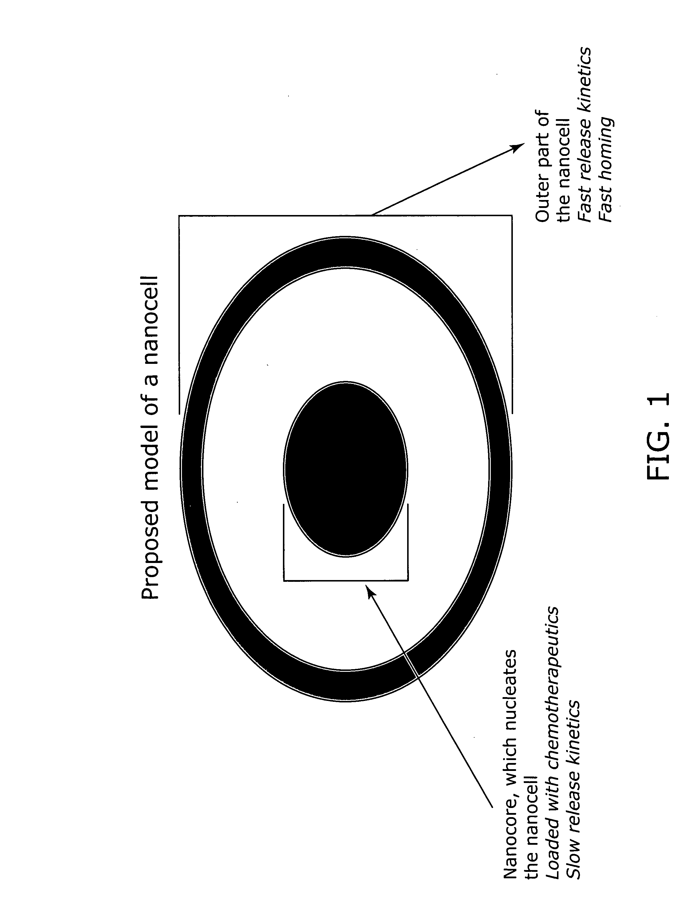
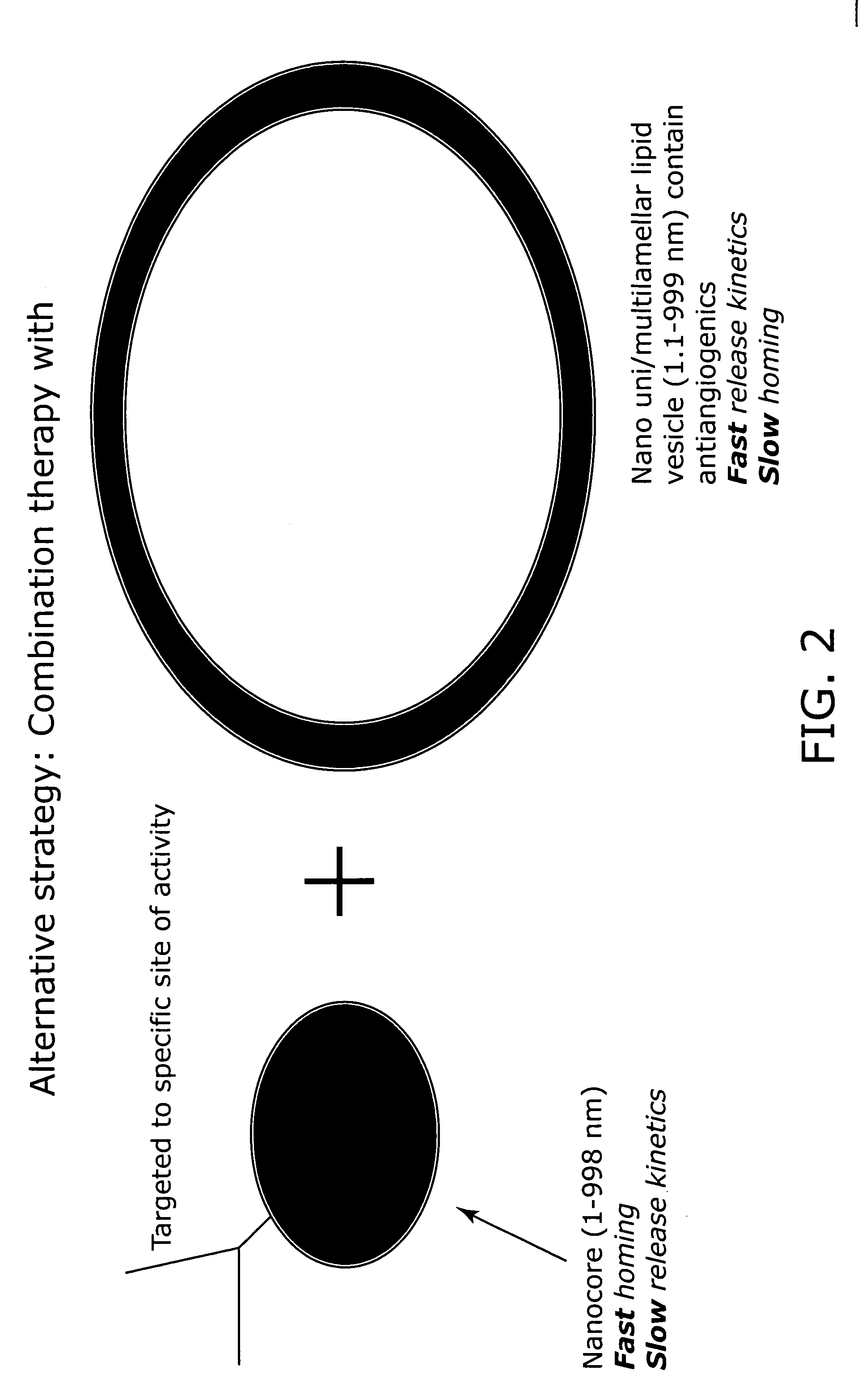
[0010] For example, in treating cancer, an antiangiogenic agent is loaded inside the lipid vesicle and is released before the anti-neoplastic / chemotherapeutic agent inside the inner nanoparticle. This results in the collapse of the vasculature feeding the tumor, and also leads to the entrapment of the anti-neoplastic agent-loaded nanocores inside the tumor with no escape route. The anti-neoplastic agent is released slowly resulting in the killing of the nutrient-starved tumor cells. In other words, this double balloon drug delivery system allows one to load up the tumor with an anti-neoplastic agent and then cut off the blood supply to the tumor. This sequential process results in the entrapment of the toxic chemotherapeutic / antineoplastic agent within the tumor, leading to increased and selective toxicity against the tumor cells, and less drug is present in the systemic circulation, since it cannot leak out from the functionally avascular tumor site, resulting in less side effects. This technique also overcomes the hypoxia caveat, as the tumor-entrapped cytotoxic chemotherapeutic cell kills off the tumor cells that would have otherwise survived in the hypoxic growth factor-rich environment resulting from the vascular shutdown.
[0011] The inner nanoparticle (also known as the nanocore) is approximately 10-20000 nm in its greatest dimension and contains a first therapeutic agent encapsulated in a polymeric matrix. These nanocores are prepared using any of the materials such as lipids, proteins, carbohydrates, simple conjugates, and polymers (e.g. PLGA, polyesters, polyamides, polycarbonates, poly(beta-amino esters), polyureas, polycarbamates, proteins, etc.) and methods (e.g., double emulsion, spray drying, phase inversion, etc.) known in the art. Pharmaceutical or diagnostic agents can be loaded in the nanocore, or covalently linked, or bound through electrostatic charges, or electrovalently conjugated, or conjugated through a linker. The result is a slow, sustained, and / or delayed release of the agent(s) from the nanocore. Preferably, if the agent is covalently linked to the nanocore, the linker or bond is biodegradable or hydrolysable under physiological conditions, e.g., susceptible to enzymatic breakdown. The nanocore can be a substantially spherical nanoparticle, nanoliposome, a nanowire, a quantum dot, or a nanotube.
[0012] To form a nanocell, the nanocores are coated with a lipid with a second therapeutic agent partitioned in the lipid phase. Nanocells may also be formed by coating the nanocores with a distinct polymer composition with a second therapeutic agent. Preferably, the nanoshell or the surrounding matrix of the nanocell should comprise a composition that allows a fast release of the agent / s that it entraps. Therefore, in certain embodiments, the effect of this agent begins before the active agent loaded in the nanocore reaches therapeutic level. Therefore, the second therapeutic agent is outside the nancore but inside the lipid membrane of the nanocell, which is approximately 50-20000 nm in its greatest diameter. The nanocell may be further coated to stabilize the particle or to add targeting agents onto the outside of the particle.
[0016] It was a further object of the current invention to provide an assay system that allows the screening of anti-angiogenic agents and chemotherapeutic agents together or separately in a situation similar to an in vivo environment. This includes cells growing on extra-cellular matrix, and accurately simulates in vivo condition. In this assay, the endothelial cells are seeded and allowed to grow on the extracellular matrix before the tumor cells are seeded on the tissue culture plate. To detect the tumor cells, they are transfected to express a fluorescent gene product such as green fluorescent protein (GFP). The endothelial cells are stained with a fluorescent dye. Kits with the necessary agents need to practice the inventive assay method are also provided by the present invention. Definitions

Problems solved by technology

Therefore, if a drug in a combination therapy cannot reach its target or does not reach its target at the appropriate time, much, if not all, of the efficacy of the drug is lost.
If the anti-neoplatic agent does not reach the tumor before the functional vasculature is shut down by the anti-angiogenic agent, the patient will suffer from the side effects of the anti-neoplastic agent without receiving any of its benefits.
This results in the collapse of the vasculature feeding the tumor, and also leads to the entrapment of the anti-neoplastic agent-loaded nanocores inside the tumor with no escape route.
The result is a slow, sustained, and / or delayed release of the agent(s) from the nanocore.

Method used


Image

  • Nanocell drug delivery system
  • Nanocell drug delivery system
  • Nanocell drug delivery system

Examples


example 1

Synthesis and Analysis of Nanocells

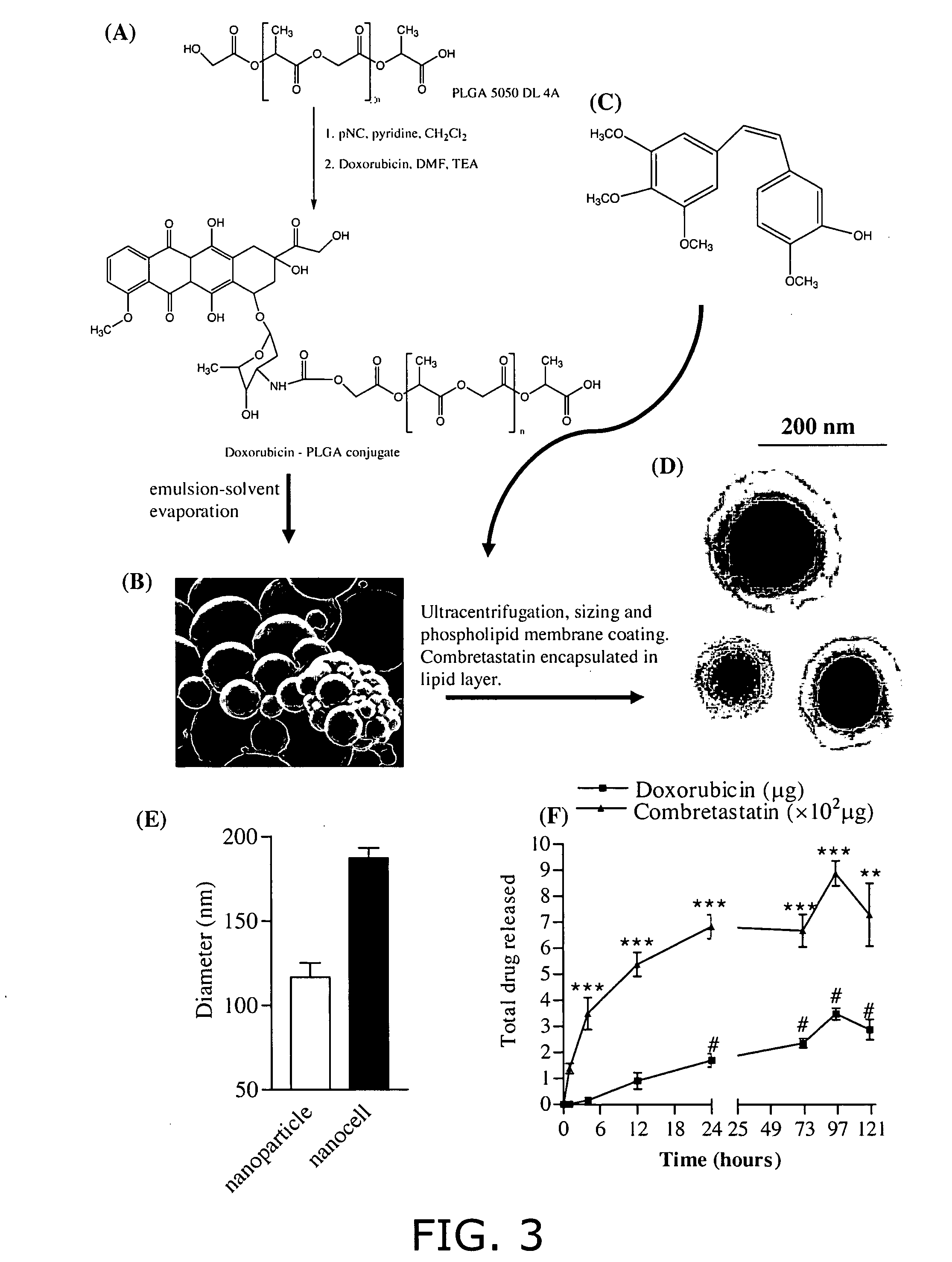
[0088] (A) Conjugation of Doxorubicin to PLGA (FIG. 3). Polylactic glycolic acid (PLGA) (Medisorb® 5050 DL 4A), having a lactide / glycolide molar ratio of 50 / 50, was obtained from Alkermes (Wilmington, Ohio). The average molecular weight of this polymer is reported to be 61 kDa, and it has free hydroxyl and carboxylic groups at its terminal ends. Doxorubicin hydrochloride, p-nitrophenyl chloroformate, and triethylamine were obtained from Sigma-Aldrich (St. Louis, Mo.). Briefly, 1.5 g of PLGA 5050 DL 4A was dissolved in 15 ml of methylene chloride and activated by the addition of 14 mg of p-nitrophenyl chloroformate and 9.4 mg (˜9.6 μL) of pyridine to the solution, kept in an ice bath at 0° C. (stoichiometric molar ratio of PLGA: p-nitrophenyl chloroformate: pyridine=1:2.8:4.7). The reaction was carried out for 3 hours at room temperature under nitrogen atmosphere. The resulting solution was diluted with methylene chloride and washed with 0.1% HCl a...

example 2

Developing the Novel in Vitro Assay System

[0096] Protocol: For setting up the system, human umbilical vein endothelial cells, pooled from three donors, were purchased from Cambrex, and used between passages 3-6. The cells were grown in endothelial basal medium supplemented with 20% fetal bovine serum (FBS) and bulletkit-2 (Sengupta et al. Cancer Res. 63(23):8351-59, Dec. 1, 2003). For the tumor component, we used B16 / F10 melanoma cells as the model cell line, which were stably transfected to express green fluorescent protein. Plasmid expressing enhanced green fluorescent protein (pEGFP-C2, Clontech) was linearized and lipofected (Lipofectamine 2000, Invitrogen) into B16-F10 cells. The stably integrated clones of B16-F10 cells were selected by 800 μg / ml G418. The green fluorescence of the G418 resistant clones was further confirmed by Flow Cytometry and epifluorescence microscopy. The GFP-B16 / F10 cells were regularly cultured in DMEM supplemented with 5% FBS. Sterile glass coverslip...

example 3

In Vitro Efficacy of Drug Loaded Nanocells (FIG. 9)

[0101] Sterile glass coverslips (Corning) were coated with matrigel (extracellular matrix extracted from murine Englebreth-Holms sarcoma, diluted 1:3 in phosphate buffer saline; Becton Dickinson) or collagen (type I from rat's tail, Becton Dickinson). Synchronized human umbilical vein endothelial cells were trypsinised and plated on the coverslips at a density of 2×10⁴ cells per well. The cells were allowed to adhere for 24 hours in endothelial basal media supplemented with 20% fetal bovine serum. At this time point, the media was replaced with EBM supplemented with 1% serum, and green fluorescent protein-expressing B 16 / F 10 cells were added to the system at a density of 5×10³ cells per well. The co-culture was allowed to incubate overnight, following which different treatments were added to the media. At 24 hours post-treatment, the cells were fixed in paraformaldehyde (4% on ice, for 20 min), and stained with propidium iodide. T...


PUM

PropertyMeasurementUnit
Diameteraaaaaaaaaa
Diameteraaaaaaaaaa
Diameteraaaaaaaaaa

Abstract

Nanocells allow the sequential delivery of two different therapeutic agents with different modes of action or different pharmacokinetics. A nanocell is formed by encapsulating a nanocore with a first agent inside a lipid vesicle containing a second agent. The agent in the outer lipid compartment is released first and may exert its effect before the agent in the nanocore is released. The nanocell delivery system may be formulated in pharmaceutical composition for delivery to patients suffering from diseases such as cancer, inflammatory diseases such as asthma, autoimmune diseases such as rheumatoid arthritis, infectious diseases, and neurological diseases such as epilepsy. In treating cancer, a traditional antineoplastic agent is contained in the outer lipid vesicle of the nanocell, and an antiangiogenic agent is loaded into the nanocore. This arrangement allows the antineoplastic agent to be released first and delivered to the tumor before the tumor's blood supply is cut off by the antianiogenic agent.

Description

RELATED APPLICATIONS [0001] The present application claims priority under 35 U.S.C. § 119(e) to U.S. provisional application, U.S. Ser. No. 60 / 549,280, filed Mar. 2, 2004, entitled “Nanocell Drug Delivery System,” which is incorporated herein by reference.BACKGROUND OF THE INVENTION [0002] The prerequisites for rational drug therapy are an accurate diagnosis, knowledge of the pathophysiology of the disease, the knowledge of basic pharmacotherapeutics in normal and diseased people, and the reasonable expectations of these relationships so that the drug's effects can be anticipated (DiPiro et al. Eds. Pharmacotherapy—A pathophysiologic approach, 2nd Ed). Advances made in biomedical sciences, in terms of the genome, proteome, or the glycome, have unraveled the molecular mechanisms underlying many diseases, and have implicated a complex network of signaling cascades, the transcriptome, and the glycome that are distinctly altered. In most pathophysiological conditions, this may manifest ...

Claims


Application Information

Patent Timeline
IPC IPC(8): A61K9/00A61K9/127A61K9/16A61K9/19A61K9/50A61K9/51A61K31/7012A61K31/737A61K38/18A61K38/19A61K38/21A61K38/46A61K45/00A61K45/06A61K49/00
CPCA61K9/0073G01N2500/10A61K9/167A61K31/7012A61K31/737A61K45/06A61K47/48192A61K47/488A61K47/48815B82Y5/00B82Y10/00A61K31/787A61K38/00A61K9/5153A61K47/482A61K31/704A61K31/09G01N33/5011A61K47/48869A61K9/1271A61K47/59A61K47/593A61K47/6907A61K47/6911A61K47/6925A61P11/00A61P11/08A61P17/06A61P19/02A61P25/00A61P25/08A61P29/00A61P35/00A61P43/00A61P9/00A61P9/10
Inventor SENGUPTA, SHILADITYAZHAO, GANLINCAPILA, ISHANEAVARONE, DAVIDSASISEKHARAN, RAM
Owner MASSACHUSETTS INST OF TECH
Why Eureka
Eureka Blog
Learn More