Eureka AIR delivers breakthrough ideas for toughest innovation challenges, trusted by R&D personnel around the world.

Viruses comprising mutant ion channel protein

a technology of mutant ion channel protein and virus, which is applied in the direction of viruses, inactivation/attenuation, peptides, etc., can solve the problems of inability to directly demonstrate the requirement for this activity in the replication of influenza a viruses, obviate difficulties, and often misdefinition of mutations of attenuated viruses, so as to promote viral replication in vivo, the effect of high attenuation and attenuation

Inactive Publication Date: 2011-11-15
WISCONSIN ALUMNI RES FOUND
View PDF13 Cites 18 Cited by
  • Summary
  • Abstract
  • Description
  • Claims
  • Application Information

AI Technical Summary

Benefits of technology

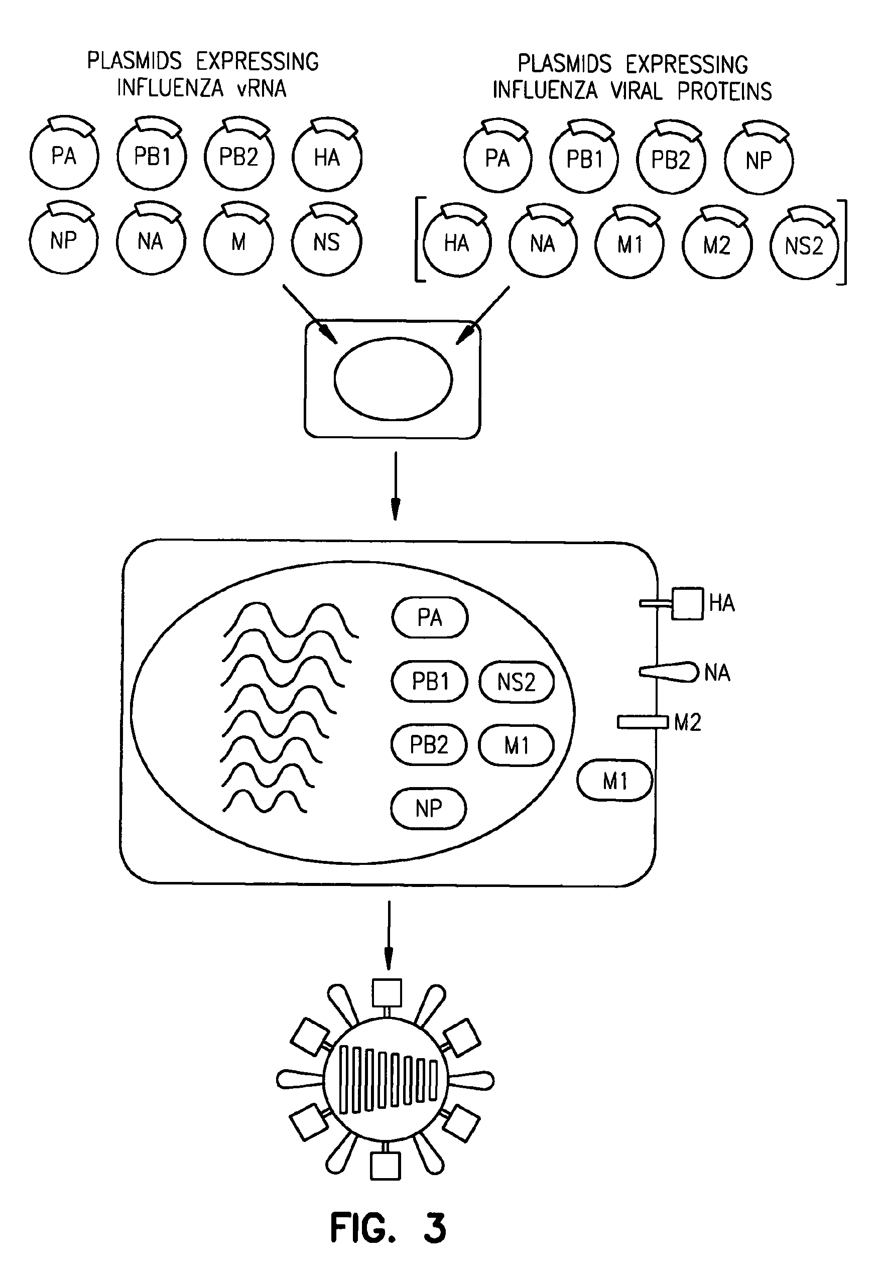
[0009]As described hereinbelow, recombinant influenza A viruses with defective M2 ion channel activity were prepared using a reverse-genetics system (see Example 1 and Neumann et al., 1999). Unexpectedly, all of the M2 ion channel mutants replicated as efficiently as the wild-type virus in vitro, although their growth was attenuated in mice. Recombinant viruses were also prepared which comprise a mutant M2 ion channel protein which is a chimeric protein, e.g., a chimeric protein in which the TM domain of M2 was replaced with the TM domain from HA or NA. These recombinant viruses replicated well in tissue culture, but were highly attenuated in mice. Thus, M2 ion channel activity is not essential for the life cycle of influenza A viruses. Rather, it may serve an auxiliary function that could, for example, promote viral replication in vivo. Given that the administration of E. coli-derived particles, spontaneously formed from a fusion protein containing the M2 ectodomain and a portion of the hepatitis B core protein, to mice resulted in 90-100% protection against lethal virus challenge (Neirynck et al., 1999), and that cold-adapted live vaccines are efficacious in humans, the attenuated growth of mutant M2 ion channel viruses in vivo, but not in vitro, indicates that these mutant viruses may be useful in the development of live influenza vaccines. Thus, the invention further provides a vaccine or immunogenic composition comprising the recombinant virus of the invention, and a method of using the vaccine or immunogenic composition to immunize a vertebrate or induce an immune response in a vertebrate, respectively.

Problems solved by technology

However, a requirement for this activity in the replication of influenza A viruses has not been directly demonstrated.
Live virus vaccines activate all phases of the immune system and stimulate an immune response to each of the protective antigens, which obviates difficulties in the selective destruction of protective antigens that may occur during preparation of inactivated vaccines.
However, the mutations in attenuated virus are often ill-defined.

Method used


Image

  • Viruses comprising mutant ion channel protein
  • Viruses comprising mutant ion channel protein
  • Viruses comprising mutant ion channel protein

Examples


example 1

Materials and Methods

[0037]Cells and viruses. 293T human embryonic kidney cells and Madin-Darby canine kidney cells (MDCK) were maintained in Dulbecco's modified Eagle medium (DMEM) supplemented with 10% fetal calf serum and in modified Eagle's medium (MEM) containing 5% newborn calf serum, respectively. All cells were maintained at 37° C. in 5% CO2. Influenza viruses A / WSN / 33 (H1N1) and A / PR / 8 / 34 (H1N1) were propagated in 10-day-old eggs.

[0038]Construction of plasmids. To generate RNA polymerase I constructs, cloned cDNAs derived from A / WSN / 33 or A / PR / 8 / 34 viral RNA were introduced between the promoter and terminator sequences of RNA polymerase I. Briefly, the cloned cDNAs were amplified by PCR with primers containing BsmBI sites, digested with BsmBI, and cloned into the BsmBI sites of the pHH21 vector which contains the human RNA polymerase I promoter and the mouse RNA polymerase I terminator, separated by BsmBI sites (FIG. 2). The PB2, PB1, PA, HA, NP, NA, M, and NS genes of the ...

example 2

Materials and Methods

[0055]Cells and viruses. 293T human embryonic kidney cells and Madin-Darby canine kidney cells (MDCK) were maintained in DMEM supplemented with 10% FCS and in MEM containing 5% newborn calf serum, respectively. The 293T cell line is a derivative of the 293 line, into which the gene for the simian virus 40 T antigen was inserted (DuBridge et al., 1987). All cells were maintained at 37° C. in 5% CO2. Influenza virus A / Udorn / 307 / 72 (H3N2) (Udorn) was propagated in 10-day-old eggs.

[0056]Construction of plasmids. The cDNA of Undorn virus was synthesized by reverse transcription of viral RNA with an oligonucleotide complementary to the conserved 3′ end of viral RNA, as described by Katz et al. (1990). The cDNA was amplified by PCR with M gene-specific oligonucleotide primers containing BsmBI sites, and PCR products were cloned into the pT7Blueblunt vector (Novagen, Madison, Wis.). The resulting construct was designated pTPo1UdM. After digestion with BsmBI, the fragmen...

example 3

Materials and Methods

[0081]Cells and viruses. 293T human embryonic kidney cells and Madin-Darby canine kidney (MDCK) cells were maintained in DMEM supplemented with 10% FCS and in MEM containing 5% newborn calf serum, respectively. The 293T cell line is a derivative of the 293 line, into which the gene for the simian virus 40 T antigen was inserted (Dubridge et al., 1987). All cells were maintained at 37° C. in 5% CO2. M2del29-31 and WSN-UdM (wild-type) viruses were propagated in MDCK cells. A / WSN / 33 (H1N1) virus was propagated in 10-day-old embryonated chicken eggs.

[0082]Immunization and protection tests. BALB / c mice (4-week-old female) were intranasally immunized with 50 μl of 1.1×10⁵ PFU per ml of M2del29-31 or wild-type WSN-UdM viruses. On the second week, four mice were sacrificed to obtain sera, trachea-lung washes, and nasal washes. Two weeks and one or three months after the vaccination, immunized mice were challenged intranasally, under anesthesia, with 100 LD50 doses of th...


PUM


Abstract

A method to prepare viruses lacking ion channel activity is provided.

Description

CROSS-REFERENCE TO RELATED APPLICATION[0001]This application is a continuation of U.S. patent application Ser. No. 09 / 834,095, filed Apr. 12, 2001, now U.S. Pat. No. 6,872,395, issued Mar. 29, 2005, which application claims priority under 35 U.S.C. 119(e) from U.S. Provisional Application Ser. No. 60 / 197,209 filed Apr. 14, 2000, which applications are incorporated herein by reference.STATEMENT OF GOVERNMENT RIGHTS[0002]This invention was made with a grant from the Government of the United States of America (grants AI-29599, AI-42774 and AI-44386 from the National Institute of Allergy and Infectious Diseases). The Government may have certain rights in the invention.BACKGROUND OF THE INVENTION[0003]Cell membranes consist of a double layer of lipid molecules in which various proteins are embedded. Because of its hydrophobic interior, the lipid bilayer of cell membranes serves as a barrier to the passage of most polar molecules and therefore is crucial to cell viability. To facilitate t...

Claims


Application Information

Patent Timeline
Patent Type & Authority Patents(United States)
IPC IPC(8): C12N7/02A61K39/285A61K35/76C12N15/09A61K39/00A61K39/145A61K48/00A61P31/16C07K14/005C07K14/11C12N5/10C12N7/00C12N7/01C12N7/04C12R1/93
CPCC07K14/005C12N7/00A61K2039/5256A61K2039/53C07K2319/00C12N2760/16122C12N2760/16134C12N2760/16161A61P31/16
Inventor KAWAOKA, YOSHIHIRO
Owner WISCONSIN ALUMNI RES FOUND
Why Eureka
Eureka Blog
Learn More