
Nanoparticle probes with raman spectroscopic fingerprints for analyte detection

a technology of raman spectroscopic fingerprints and nanoparticle probes, which is applied in the field of nanoparticle probes with raman spectroscopic fingerprints for analyte detection, can solve the problems of conspicuous technological absence and difficult to distinguish spectra

Active Publication Date: 2011-07-26
NORTHWESTERN UNIV
View PDF136 Cites 8 Cited by
  • Summary
  • Abstract
  • Description
  • Claims
  • Application Information

AI Technical Summary

Benefits of technology

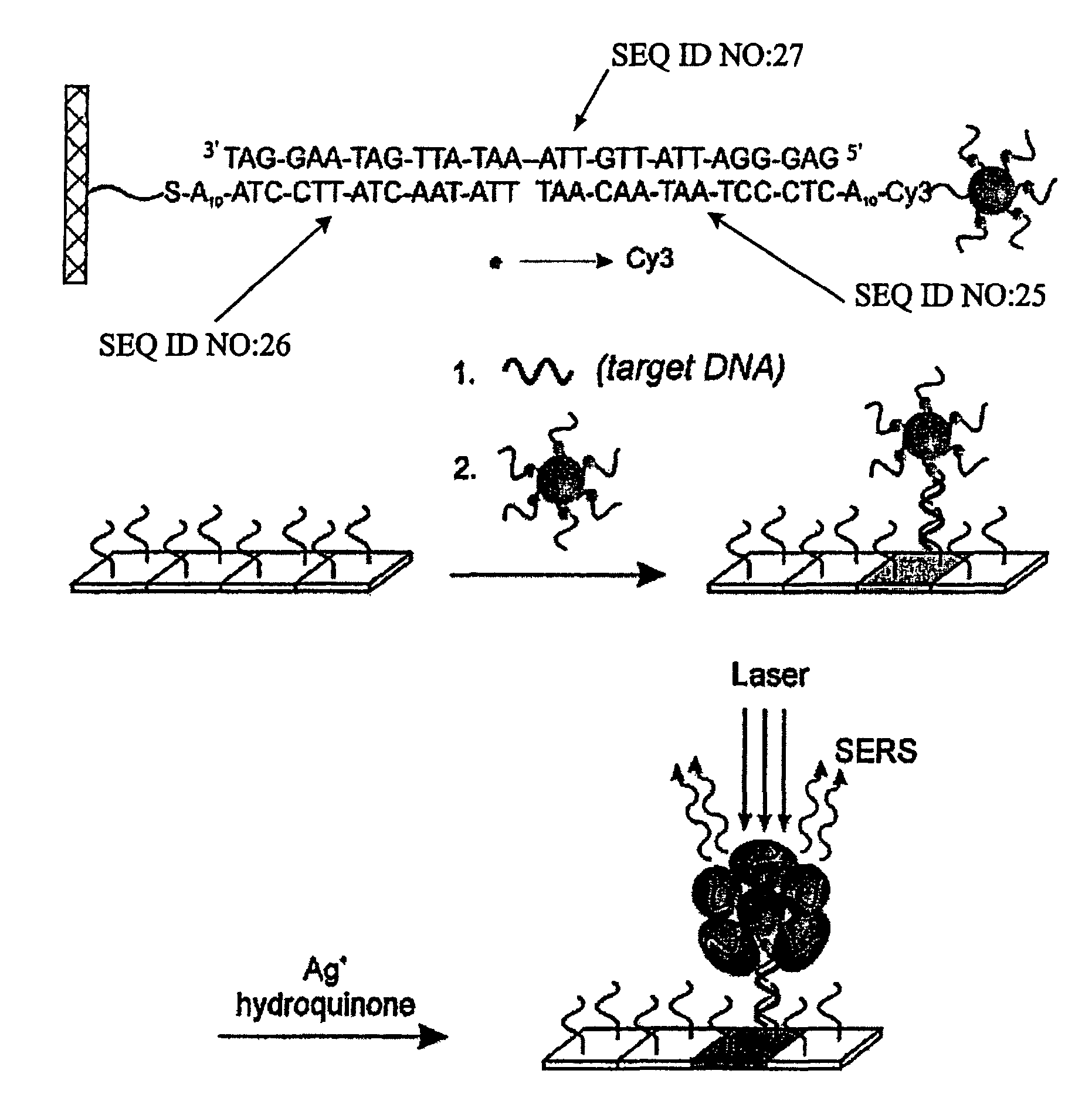
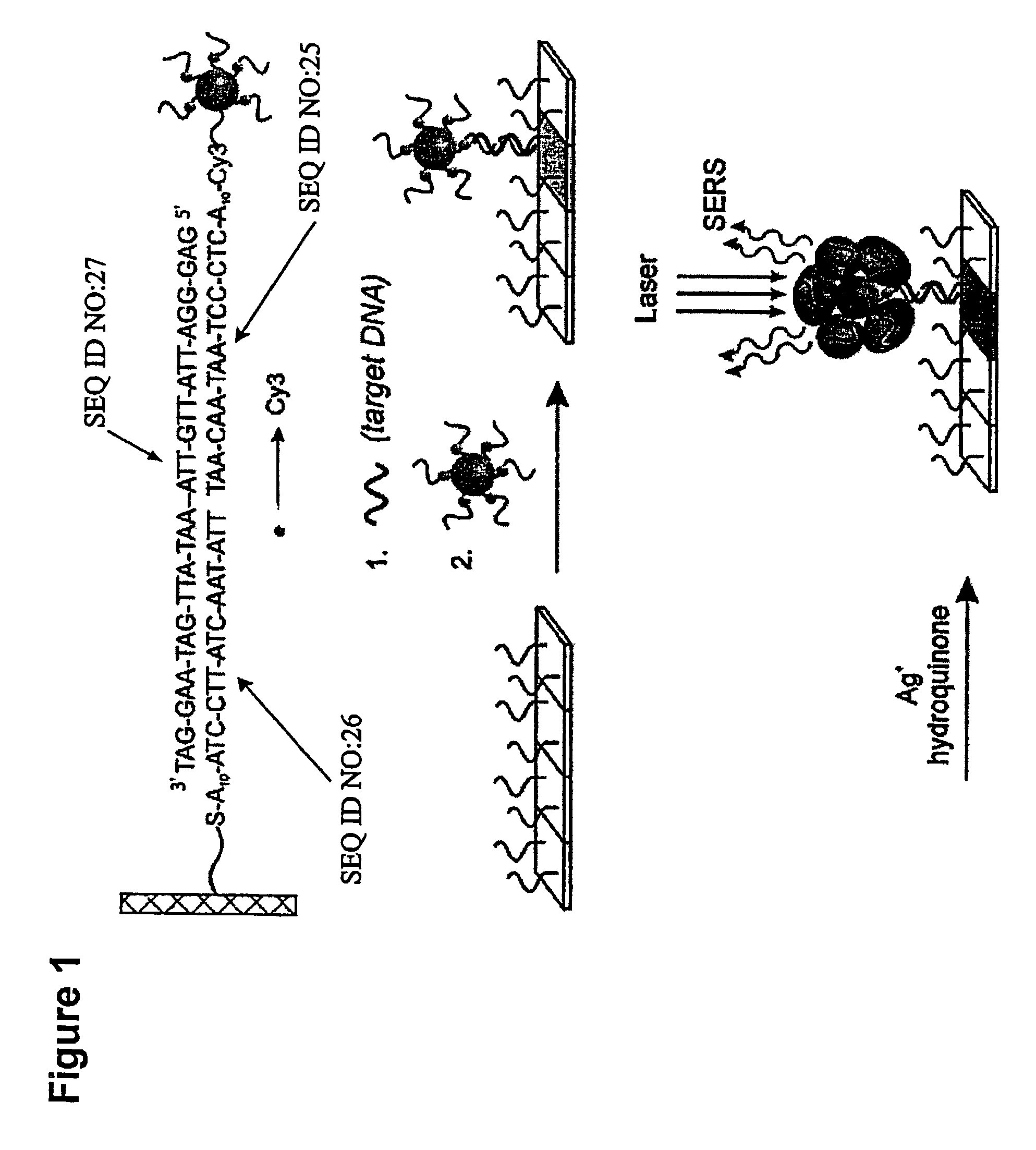
[0037]Accordingly, in one embodiment of the invention, detection reagents or probes are provided. In one aspect of this embodiment, the detection reagent comprises particles having specific binding members and Raman labels bound to the particle. The Raman label may be bound directly or indirectly to the particle in any suitable manner including the use of functional groups such as thiols and linkers having functional groups. Alternatively, the Raman labels can bound to the specific binding member and the resulting entity can be attached to the particle. When utilized in a SERS-detection sandwich-based assay, the detection reagent can be treated with an enhancing stain such as silver, gold or copper to provide a SERS effect when irradiated.

Problems solved by technology

Although oligonucleotides can be directly detected by SERS on aggregated particles,26 the structural similarities of oligonucleotides with different sequences results in spectra that are difficult to distinguish.
In part, this conspicuous technological absence is due to the difficulty in reproducibly generating and functionalizing stable SERS-active substrates23 as well as a lack of an appropriate probe design strategy to enable multiplexed detection.

Method used


Image

  • Nanoparticle probes with raman spectroscopic fingerprints for analyte detection
  • Nanoparticle probes with raman spectroscopic fingerprints for analyte detection
  • Nanoparticle probes with raman spectroscopic fingerprints for analyte detection

Examples


example 1

Microarray Fabrication

[0149]Oligonucleotide capture strands were immobilized onto SMPB (succinimidyl-4-(maleimidophenyl)-butyrate) functionalized glass slides by spotting 5′-hexyl-thiol-capped oligonucleotides (1 mM in a 0.15 M NaCl, pH 6.5 phosphate buffer solution (PBS, 10 mM phosphate)) with a commercial arrayer (GMS 417 arrayer, Genotic MicroSystems, Inc). After spotting the chip with the capture oligonucleotides (˜200 μm spots), the chip was kept in a humidity chamber for 12 hours to effect the coupling reaction between SMPB and the hexylthiol-capped oligonucleotides. Then the chip was washed copiously with Nanopure water. Passivation of the areas of the chip surrounding the oligonucleotide spots was carried out by immersing the chip in a solution of hexylthiol-capped poly-adenine (A15) (0.1 mM) for 4 h and then in a solution of 3-mercapto-propane sulfonic acid, sodium salt (0.2 M) for 30 minutes to cap off the remaining SMPB sites. Finally, the chip was washed with Nanopure wa...

example 2

Synthesis and Purification of Cy3-Labeled-(propylthiol)-Capped Oligonucleotides

[0150]The Cy3-modified, (propylthiol)-capped oligonucleotides were synthesized on a 1 mmol scale using standard phosphoramidite chemistry5 with a Thiol-Modifier C3 S—S CPG (controlled-pore glass) solid support on a commercial synthesizer (Expedite). The Cy3-CE phosphoramidite (Indodicarbocyanine 3, 1′-O-(4-monomethoxytrityl)-1-O-(2-cyanoethyl)-(N,N-diisopropyl)-phosphoramidite (Glen Research) was used to incorporate the Cy3 unit in the oligonucleotides. To aid purification, the final dimethoxytrityl (DMT) protecting group was not removed. After synthesis, the CPG-supported oligonucleotides were placed in 1 mL of concentrated ammonium hydroxide for 8 h at 55° C. to cleave the oligonucleotide from the solid support and remove the protecting groups from the bases. In each case, cleavage from the solid support via the succinyl ester linkage produced a mixed disulfide composed of the (mercaptopropyl) oligonucl...

example 3

Synthesis and Purification of TMR-, Cy3.5- and Cy5-labeled-(propylthiol)-capped Oligonucleotides

[0151]This Example describes the syntheses of three oligonucleotides having Raman labels bound thereto: 3′ HS—TMR-A10-AAC CGA AAG TCA ATA [SEQ ID NO. 2 in FIG. 19a]; 3′ HS—Cy3.5-A10-CCT CAT TTA CAA CCT [SEQ ID NO. 3 in FIG. 19a]; and 3′HS—Cy5-A10-CTC CCT AAT AAC AAT [SEQ ID NO. 4 in 19b]. Because the dyes are sensitive to the standard cleavage reagent (ammonia), ultramild base monomers (from Glen Research) were used here to allow the deprotection reaction under ultramild conditions: phenoxyacetyl (Pac) protected dA, 4-isopropyl-phenoxyacetyl (iPr-Pac) protected dG, and acetyl (Ac) protected dC. TAMRA-dT (TMR-dT, 5′-Dimethoxytrityloxy-5-[N-((tetramethylrhodaminyl)-aminohexyl)-3-acrylimido]-2′-deoxyUridine-3′-[2-cyanoethyl)-(N,N-diisopropyl)]-phosphoramidite), Cy3.5-CE phosphoramidite (Indodicarbocyanine 3.5, 1′-O-(4-monomethoxytrityl)-1-O-(2-cyanoethyl)-(N,N-diisopropyl)-phosphoramidite), ...


PUM

PropertyMeasurementUnit
Diameteraaaaaaaaaa
Spectrumaaaaaaaaaa

Abstract

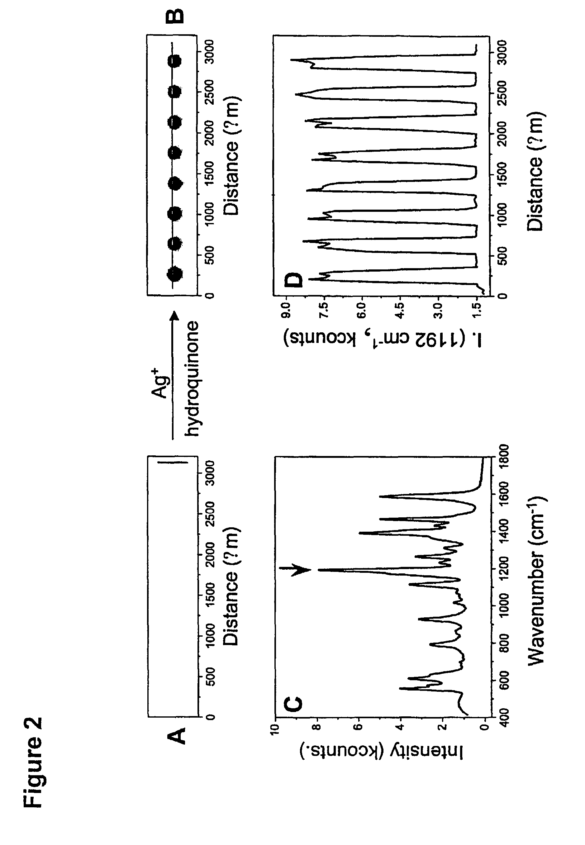
The invention encompasses reagents comprising particles with at least one Raman dye and a specific binding members bound thereto and methods of using such reagents. The invention also encompasses reagents of a specific binding member and two or more different Raman dyes and methods for using such reagents. New types of particle probes having a specific binding member bound thereto are described. These reagents are used in a novel detection strategy that utilizes the catalytic properties of the Au nanoparticles to generate a silver coating that can behave as a surface-enhanced Raman scattering (SERS) promoter for the dye-labeled particles that have been captured by target and an underlying chip in microarray format. The strategy provides the high sensitivity and high selectivity attributes of grey-scale scanometric detection but provides a route to multiplexing and ratioing capabilities since a very large number of probes can be designed based upon the concept of using a Raman tag as a spectroscopic fingerprint in detection. These spectra are used as fingerprints to differentiate oligonucleotide or other targets in one solution. This method has been used to distinguish six dissimilar DNA targets with six Raman labeled nanoparticle probes, and also two RNA targets with single nucleotide polymorphisms (SNPs).

Description

CROSS-REFERENCE[0001]This application is a continuation-in-part of U.S. patent application Ser. No. 10 / 172,428, filed Jun. 14, 2002, abandoned, which claims the benefit of priority based on U.S. provisional applications Nos. 60 / 378,538, filed May 7, 2002; and 60 / 383,630, filed May 28, 2002, all which are hereby incorporated by reference in their entirety.[0002]The work described in this application has been supported in part from grants from the Air Force Office of Scientific Research, DARPA, and the NSF. Accordingly, the United States government may have some rights to the invention.BACKGROUND OF THE INVENTION[0003]The development of high-sensitivity, high-selectivity detection formats for chemical and biological molecules is of paramount importance for realizing the full potential of genomics and proteomics advances made over the past decade.1-4 High density gene chips have made it possible to monitor the levels of expression of thousands of genes simultaneously. Lower density chi...

Claims


Application Information

Patent Timeline
IPC IPC(8): C12Q1/68G01N33/53C12M1/00C12N15/09G01N21/65C12Q1/02G01N33/543G01N33/553G01N33/566G01N33/58
CPCC12Q1/6816G01N33/54373G01N33/583G01N33/587C12Q2565/632C12Q2565/519C12Q2537/125Y02A50/30
Inventor MIRKIN, CHAD A.CAO, YUNWEIJIN, RONGCHAO
Owner NORTHWESTERN UNIV
Learn More