
Self-assembling nanoparticle drug delivery system

a drug delivery system and nanoparticle technology, applied in the field of self-assembling drug delivery systems, can solve problems such as system non-replicability, and achieve the effect of facilitating membrane transduction

Inactive Publication Date: 2011-06-21
CHIMEROS
View PDF93 Cites 22 Cited by
  • Summary
  • Abstract
  • Description
  • Claims
  • Application Information

AI Technical Summary

Benefits of technology

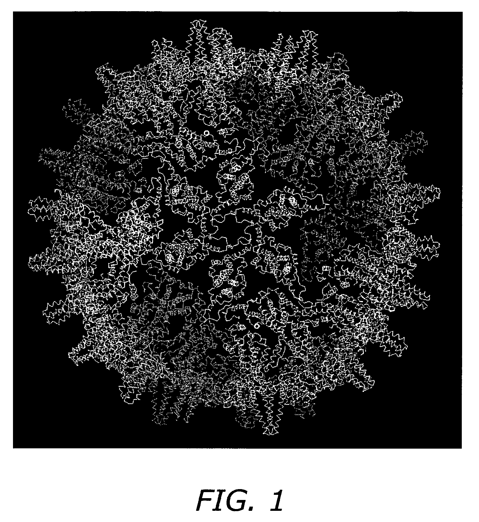
[0010]The present invention provides for a novel nanoparticle drug delivery system that can be administered across mucosal barriers and is able to transport a wide range of molecules including therapeutic proteins into the circulatory system. The nanoparticles of the present invention comprise building blocks re-engineered from natural proteins self-assemble to form nanocages. During the assembly process, drugs of choice will be captured by the specific chemistries of the inward facing surfaces of the cage-forming blocks by simple diffusion / concentration mechanics. The assembled cage has special functionalities to guide the assembly of a surrounding envelope, which is an encapsulating self-assembling double layer of neutral, anionic or cationic lipids. Peptides that facilitate membrane transduction will be integrated into the lipid bi-layer envelope to endow the system with the ability to pass through cell walls. Polyethylene glycol (PEG) of varying chain lengths will next be anchored into the membrane for the purpose of eluding the immune system and to fend off attacking degradative enzymes. This multilayered delivery system orchestrates a complex arrangement of biomolecules and is entirely self-assembling. The nanoparticle drug delivery system can by administered by any route including, but not limited to, subcutaneous, intravenous and intramuscular routes and passage through a mucosal layer such as oral, transdermal, intranasal and buccal routes.
[0011]The present invention represents a synthetically enveloped non-viral capsule composed of re-engineered biological molecules and enhanced with synthetic chemical components. Although this design is inspired by the natural behavior of viruses, this system is non-replicating. In addition, all of the proteins used to make the building blocks of the system were all re-engineered to exhibit desired characteristics by altering stabilities and removing or adding disulfide linkages. The building blocks are designed so that once the cage starts to disintegrate, they are degraded quickly so as to limit any potential immune response. A characteristic of this drug delivery system is its ability to create the building blocks of the cage with therapeutic proteins attached to every unit. Yet another important feature of this system is the use of the beneficial characteristics of a virus to deliver molecules that no virus could deliver, such as synthetic drugs, without pathogenic potential. The nanoparticle drug delivery system does not incorporate an attenuated virus, but just a shell of proteins that form regular geometric shapes.

Problems solved by technology

Although this design is inspired by the natural behavior of viruses, this system is non-replicating.

Method used


Image

  • Self-assembling nanoparticle drug delivery system
  • Self-assembling nanoparticle drug delivery system
  • Self-assembling nanoparticle drug delivery system

Examples


example 1

Core Protein Expression and Purification

[0089]A pET-11a vector containing the full-length HBV C-protein gene, is transformed into E. coli DE3 cells and grown at 37° C. in LB media, fortified with 2-4% glucose, trace elements and 200 ug / mL carbenicillin. Protein expression is induced by the addition of 2 mM IPTG (isopropyl-beta-D-thiogalactopyranoside). Cells are harvested by pelleting after three hours of induction. SDS-PAGE is used to assess expression of C-protein.

[0090]Core protein is purified from E. coli by resuspending in a solution of 50 mM Tris-HCl, pH 7.4, 1 mM EDTA, 5 mM DTT, 1 mM AEBSF, 0.1 mg / mL DNase1 and 0.1 mg / mL RNase. Cells are then lysed by passage through a French pressure cell. The suspension is centrifuged at 26000×G for one hour. The pellet is discarded and solid sucrose added to the supernatant to a final concentration of 0.15 M and centrifuged at 10000×G for one hour. The pellet is discarded and solid (NH4)2SO4 is then added to a final concentration of 40% sa...

example 2

Protocol for Phospholipid Conjugation via SMPB Intermediate (FIG. 2)

[0091]1. Dissolve 100 micromoles of phosphatidyl ethanolamine (PE) in 5 mL of argon-purged, anhydrous methanol containing 100 micromoles of triethylamine (TEA). Maintain the solution under an argon or nitrogen atmosphere. The reaction may also be done in dry chloroform.

[0092]2. Add 50 mg of SMPB (succinimidyl-4-(p-maleimidophenyl)butyrate, Pierce) to the PE solution. Mix well to dissolve.

[0093]3. React for 2 hours at room temperature, while maintaining the solution under an argon or nitrogen atmosphere.

[0094]4. Remove the methanol from the reaction solution by rotary evaporation and redissolve the solids in chloroform (5 mL).

[0095]5. Extract the water-soluble reaction by-products from the chloroform with an equal volume of 1% NaCl. Extract twice.

[0096]6. Purify the MPB-PE derivative by chromatography on a column of silicic acid (Martin F J et al., Immunospecific targeting of liposomes to cells: A novel and efficient...

example 3

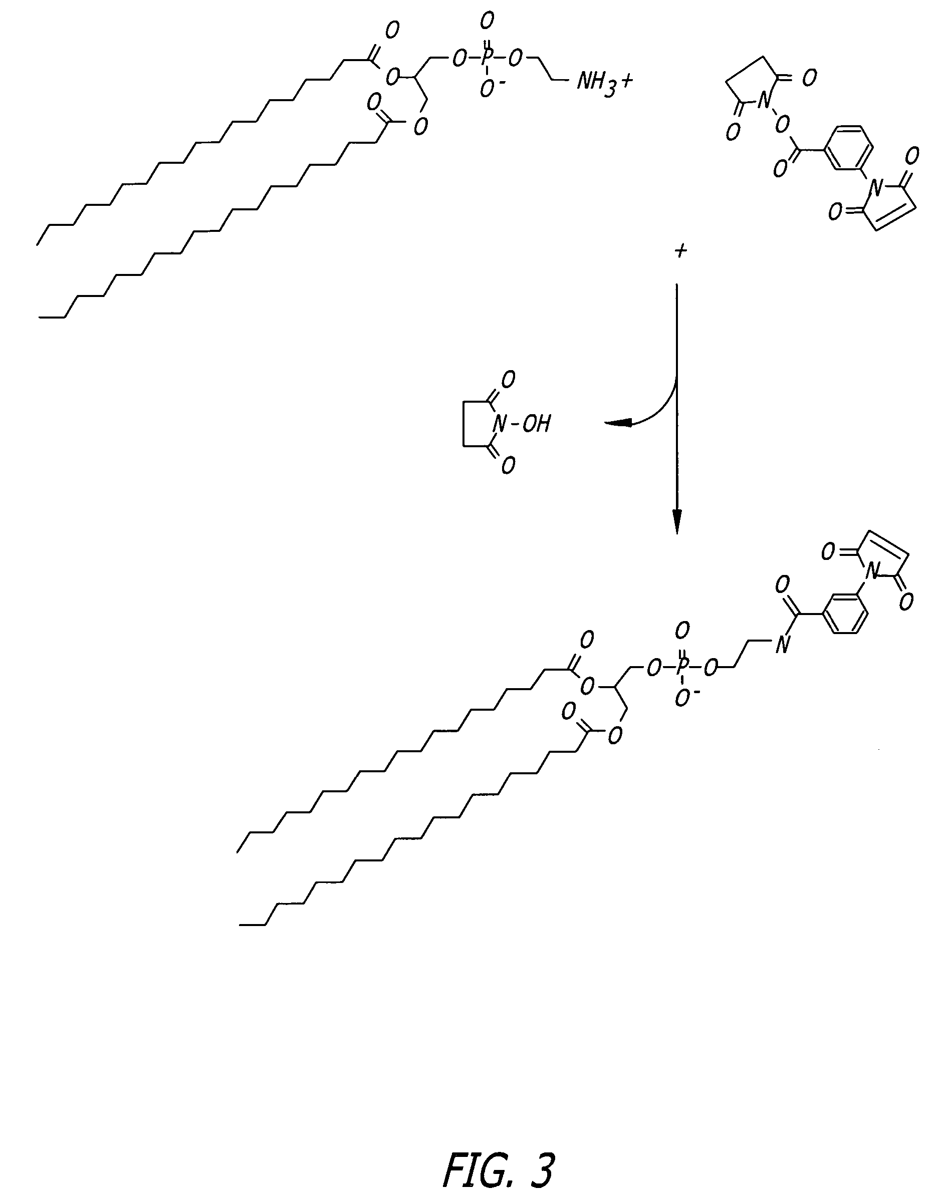
Protocol for Phospholipid Conjugation via MBS Intermediate (FIG. 3)

[0098]1. Dissolve 40 mg of PE in a mixture of 16 mL dry chloroform and 2 mL dry methanol containing 20 mg triethylamine, maintain under nitrogen.

[0099]2. Add 20 mg of m-maleimidobenzoyl-N-hydroxysuccinimide ester (MBS) to the lipid solution and mix to dissolve.

[0100]3. React for 24 hours at room temperature under nitrogen.

[0101]4. Wash the organic phase three times with PBS, pH 7.3, to extract excess cross-linker and reaction by-products.

[0102]5. Remove the organic solvents by rotary evaporation under vacuum.


PUM

PropertyMeasurementUnit
sizeaaaaaaaaaa
diameteraaaaaaaaaa
ionic strengthaaaaaaaaaa

Abstract

A self-assembling nanoparticle drug delivery system for the delivery of drugs including peptides, proteins, nucleic acids or synthetic chemical drugs is provided. The self-assembling nanoparticle drug delivery system described herein includes viral capsid proteins, such as Hepatitis B Virus core protein, encapsulating the drug, a lipid bi-layer envelope and targeting or facilitating molecules anchored in the lipid bilayer. A method for construction of the self-assembling nanocparticle drug delivery system is also provided.

Description

RELATED APPLICATIONS[0001]This application claims the benefit of priority under 35 U.S.C. §119(e) of U.S. Provisional Patent Application No. 60 / 574,409 filed May 25, 2004.FIELD OF THE INVENTION[0002]The present invention relates to methods for drug delivery. Specifically, the present invention relates to a self-assembling drug delivery system comprised of pharmacologic drugs captured within viral capsid proteins and encapsulated in a lipid envelope.BACKGROUND OF THE INVENTION[0003]Nanotechnology, the term derived from the Greek word nano, meaning dwarf, applies the principals of both physical and biological sciences at a molecular or submicron level. The materials at nanoscale can be a device or system, or supramolecular structures, complexes or composites. Nanotechnology is making significant advances in biomedical applications, including drug delivery techniques.[0004]The development of drug delivery systems for small molecules, proteins and DNA have been greatly influenced by nan...

Claims


Application Information

Patent Timeline
Patent Type & Authority Patents(United States)
IPC IPC(8): A61K39/29A61K39/385C07K14/02
CPCA61K9/5184A61K47/48776B82Y5/00C12N7/00C07K14/005C12N2730/10123C07K2319/00C12N2730/10122Y10S977/795Y10S977/801Y10S977/802Y10S977/797Y10S977/798Y10S977/80A61K47/6901A61K47/6913A61P1/04A61P1/08A61P11/00A61P11/06A61P11/08A61P17/04A61P19/02A61P19/06A61P25/00A61P25/04A61P25/06A61P25/08A61P25/16A61P25/18A61P25/20A61P25/22A61P25/24A61P29/00A61P3/02A61P31/00A61P31/04A61P31/10A61P31/12A61P35/00A61P3/06A61P3/08A61P37/02A61P37/06A61P7/00A61P7/02A61P7/04A61P9/00A61P9/06A61P9/10A61P9/12A61P3/10A61K9/1277A61K9/5192
Inventor DE LOS RIOS, MIGUELOH, KENNETH J.
Owner CHIMEROS
Learn More