Fluorometric detection using visible light

a fluorometric and visible light technology, applied in the field of can solve the problems of skin tissue damage, dna sample damage, and the use of laser light is not applicable to the simple and direct viewing of dna gel by the human eye, so as to reduce the occurrence of photo-bleaching, reduce degradation and modification of fluorescent materials, and avoid damage to fluorescent and phosphorescent materials.

Inactive Publication Date: 2005-07-05
CLARE CHEM RES
View PDF35 Cites 29 Cited by
  • Summary
  • Abstract
  • Description
  • Claims
  • Application Information

AI Technical Summary

Benefits of technology

[0041]The optical characteristics of the excitation sources of the present invention provide substantial versatility in design and function, which make the present methods and devices compatible to a diverse range of operating conditions and commercial applications. First, use of excitation radiation substantially free of ultraviolet light, preferably having a wavelength greater than or equal to 380 nm and more preferably greater than or equal to 400 nm, avoids damage to fluorescent and phosphorescent materials induced by the absorption of high-energy ultraviolet radiation. For example, the excitation radiation of the present invention substantially reduces the occurrence of photo-bleaching, which is commonly observed upon excitation of fluorescent and phosphorescent materials with ultraviolet light. The present invention, therefore, provides efficient methods of exciting photolytically-labile and photosensitive compounds without substantially degrading or chemically modifying these materials. Reducing degradation and modification of fluorescent materials is beneficial because it increases the observed lifetime of fluorophors present in an illuminated sample and thereby provides fluorescent devices capable of repeatedly exciting a sample containing fluorescent materials for long periods of operation. Therefore, the present invention provides methods and devices for exciting fluorescence providing improved fluorophor longevity.
[0042]Second, use of fluorescence excitation radiation substantially free of ultraviolet light minimizes generation of harmful ultraviolet radiation. Ultraviolet radiation is a known hazard to most organisms and has been associated with the occurrence of erythema, degenerative and neoplastic changes in the skin, retinal damage and cataracts, and modification of the immunologic system of the skin. Ultraviolet light also is damaging to most materials including but not limited to plastics, glasses, and ceramics. As scattering occurs to some extent in almost every interaction of light waves and matter, scattered excitation radiation is unavoidable upon illumination of a sample containing fluorophors. The devices and methods of exciting fluorophors of the present invention, however, minimize scattered ultraviolet radiation by employing excitation radiation with very low intensities in the ultraviolet region. In a preferred embodiment, the intensity of light having wavelengths less than about 380 nm is less than about 1% of the total luminous intensity of the excitation source of the present invention. The methods and devices of exciting fluorescence of the present invention have the benefit, therefore, of reducing the exposure to ultraviolet light of the surroundings. Reference to “surroundings” in this context refers to all areas other than the intended illumination region. This benefit allows for embodiments of the present invention wherein scattered excitation radiation is in optical contact with the surroundings. Particularly, the present invention includes embodiments wherein scattered excitation radiation is in optical contact with the user, viewer or the user interface. This benefit also eliminates the need for costly and cumbersome light scattering sheaths and other light shielding devices employed to reduce the exposure of the surroundings to scattered excitation light.
[0043]Third, excitation with visible radiation also permits use of a wide class of materials for coupling an excitation beam to a sample containing fluorophors and for filtering light from and excitation source. Most materials efficiently absorb and scatter ultraviolet light. In contrast, a wide class of materials exhibits high transmittance throughout the visible spectrum, particularly from about 380 nm to about 800 nm. Accordingly, the devices and methods of exciting fluorophors of the present invention may employ optical components, such as filters, substrate holders and diffusion plates composed of a wide range of materials. Preferred materials for transmitting or filtering excitation radiation include ordinary glass, PYREX materials, gels, liquids and liquid crystals, and transparent plastics, such as polystyrene, polyethylene, acrylic, polymethylmethacrylate, polyimide, polycarbonate and polychlorotrifluoroethylene. Although these materials efficiently absorb and scatter ultraviolet radiation, they efficiently transmit light in the visible region of the electromagnetic spectrum. Further, these materials may be easily modified by the incorporation of pigments or other visible light-absorbing compounds to provide transmission of only selected wavelengths of light in the visible spectrum. Therefore, the methods, devices, and device components of the present invention are highly compatible with a wide range of optical materials. Particularly useful for the present invention are optical components constructed from transparent plastics, which may assume a great variety of shapes. The ability to use transparent plastics in the fabrication of devices of the present

Problems solved by technology

The use of UV light for viewing molecules in gels has two major disadvantages: (1) It is dangerous.
More prolonged exposure to UV light results in damage to the skin tissues (sunburn) and care must be taken to minimize skin exposure by wearing gloves, long-sleeved jackets and a full-face mask.
(2) DNA samples are damaged by exposure to UV light.
However, the use of laser light is not applicable to the simple and direct viewing of a DNA gel by the human eye.
However, no enabling disclosure for making such devices is provided.
None of these references provides devices or systems for viewing fluorescence patterns using visible light.
Ultraviolet radiation is a known hazard to most orga

Method used


Image

  • Fluorometric detection using visible light
  • Fluorometric detection using visible light
  • Fluorometric detection using visible light

Examples


example 1

Sensitivity

[0195]The sensitivity of an optimized device of this invention was measured by detection of known quantities of DNA on gels stained with SYBR® Green I and ethidium bromide, both by eye and by photography. What is seen by the eye and what is recorded on photographic film are not necessarily one and the same, especially when using black-and-white photography. For example, photographic film is able to accumulate an image over many seconds and, after processing, the image can be quantitated. On the other hand, the interpretative skill of the human eye when directly viewing an image is unparalleled. Though scientists use photographs of DNA gels for their laboratory records and for detailed analysis such as calculation of the sizes of DNA fragments, much of the analysis of a DNA band pattern on a gel is achieved using the naked eye. Furthermore, the excision of gel slices containing DNA is always done by eye. Therefore, it is important that the sensitivity of any apparatus for ...

example 2

Blue Compact Fluorescent Lamps and SYBR® Gold

[0225]Various dilutions of a DNA cut with HindIII (Boehringer Mannheim) in 10 mmol / L Tris-Cl, 1 mmol / L EDTA were incubated at 60° C. for three minutes. The samples were placed on ice and sample loading buffer (0.25% bromophenol blue, 0.25% xylene cyanol, 15% Ficoll type 400 in 10 mM Tris-Cl, 1 mmol / L EDTA, pH 7.5) was added to each mix. Various amounts of the DNA samples (from 428 ng to 0.85 ng) were loaded onto a 1% agarose gel 7.6×12.7 cm (3″×5″ in.) in 89 nmol / L Tris borate, pH 7.82, and 2 mmol / L EDTA (TBE). The gel was run at 80 V for two hours and then placed in 100 mL of 1:10000 dilution (in TAE) of SYBR® Gold for 30 minutes. The gel was photographed using a Polaroid® camera (Polaroid Corporation, Cambridge, Mass. with Polaroid 667 film on either a Fisher UV 312 nm variable intensity transilluminator (Fischer Scientific, Pittsburgh, Pa., Model No. FBTTV-816), set to maximum intensity in all cases, a Wratten #12 on the camera, f-stop...

example 3

Ethidium Bromide Gel

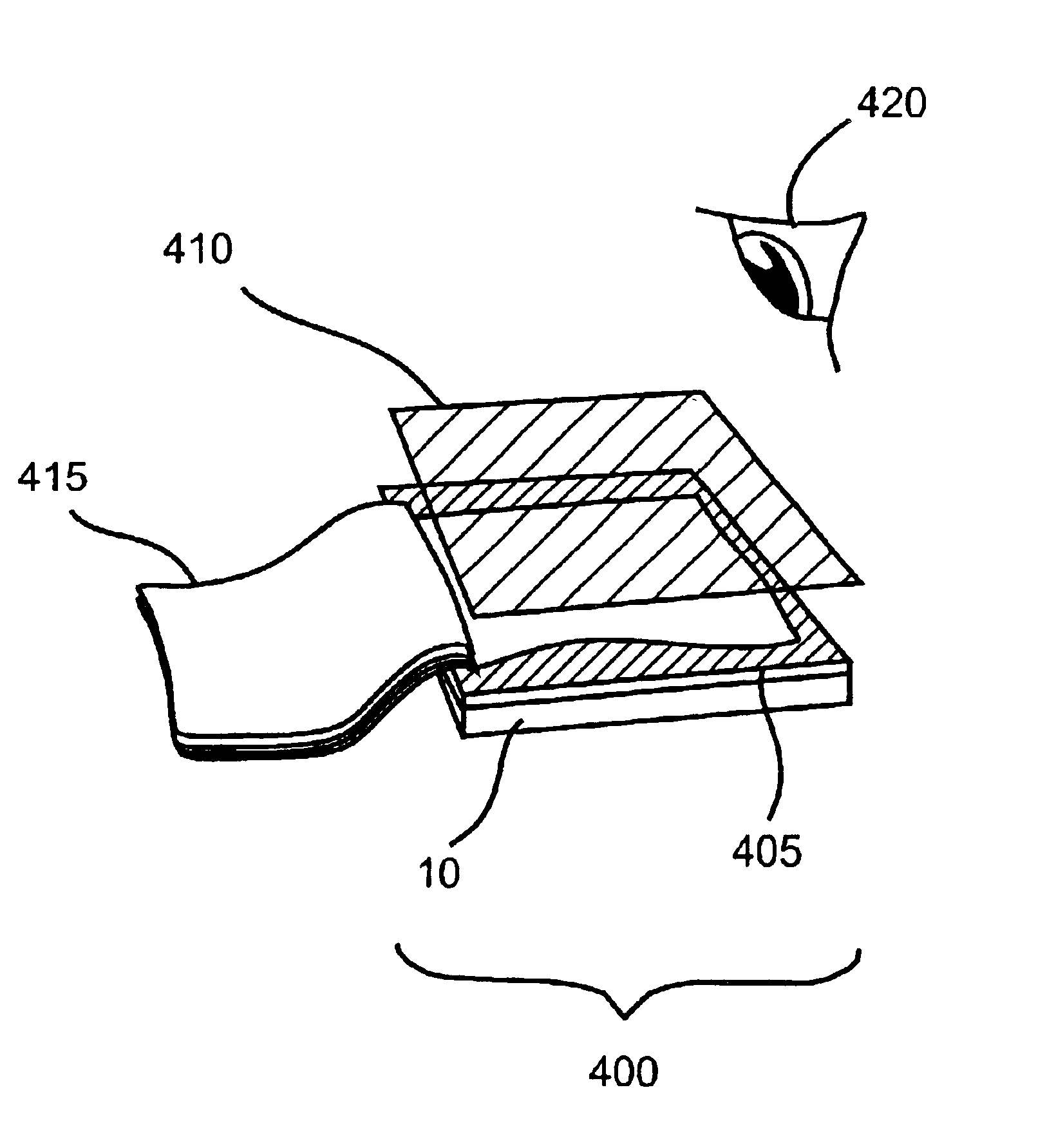
[0230]DNA cut with HindIII (Boehringer Mannheim) in 10 mmol / L Tris-Cl, 1 mM EDTA was mixed with sample loading buffer and various amounts of DNA (from 125 ng to 15.6 ng) were loaded onto a 0.7% agarose gel (3″×5″) in TAE. Ethidium bromide was added to both the gel and running buffer to a final concentration of 0.25 μg / mL. The gel was run at 110 V for two hours and then examined by eye and photographed using a Polaroid camera with Polaroid 667 film on either a UV 312 nm transilluminator (Fisher Scientific) set to maximum lamp intensity using a red Tiffen 40.5 mm 23A filter (Tiffen Manufacturing Corp., Hauppauge, N.Y.) on the camera (f-stop=5.6, exposure time=2 seconds) or an embodiment of the present invention as depicted in FIG. 9 equipped with a CF9DS / blue lamp, a #668-0GP first filter, and a Perspex® #300 second filter. A Wratten #21 (Kodak) was used as an additional second filter for photography (f-stop=5.6, exposure time=30 seconds). The gel was also observed...


PUM


Abstract

Systems, devices and methods of the present invention are for viewing a pattern of fluorophors capable of fluorescing when exposed to visible light, e.g., fluorescently stained DNA, protein or other biological material. The system includes a light source emitting light in the visible spectrum, such as a fluorescent lamp used in domestic lighting, a first optical filter capable of transmitting light from the source at wavelengths capable of exciting the fluorophors and of absorbing light of other wavelengths, and a second optical filter capable of blocking substantially all the light from the source not blocked by the first filter, so that the only light reaching the viewer is light produced by fluorescence of the fluorophors.

Description

CROSS-REFERENCE TO RELATED APPLICATIONS[0001]This application takes priority from U.S. Provisional Application No. 60 / 040,124, filed Mar. 7, 1997, and is a continuation-in-part of U.S. Ser. No. 09 / 753,783, filed Jan. 2, 2001, which is now U.S. Pat. No. 6,512,236, which is a continuation of U.S. Ser. No. 09 / 036,034, filed on Mar. 6, 1998, which is now U.S. Pat. No. 6,198,107, all of which are hereby incorporated by reference in their entireties to the extent not inconsistent herewith.BACKGROUND OF THE INVENTION[0002]The separation of DNA fragments by polyacrylamide or agarose gel electrophoresis is a well-established and widely used tool in molecular biology (Sharp, P. A. et al., “Detection of two restriction endonucleases activities in Haemophilus parainfluenzae using analytical agarose-ethidium bromide electrophoresis,” (1973) Biochemistry 12:3055). The standard technique for viewing the positions of the separated fragments in a gel involves the use of an ultra-violet (UV) transill...

Claims


Application Information

Patent Timeline
IPC IPC(8): G01N21/64
CPCG01N21/6428G01N21/6447
Inventor SEVILLE, MARK
Owner CLARE CHEM RES
Why Eureka
Try Eureka