
Receptor-mediated uptake of an extracellular BCL-xL fusion protein inhibits apoptosis

a fusion protein and fusion protein technology, applied in the direction of peptide/protein ingredients, antibody medical ingredients, peptide sources, etc., can solve the problems of unsatisfactory clinical work, treatment with standard apoptosis inhibitory molecules, and unelucidated molecular mechanisms of apoptosis, so as to prevent cell death. the effect of the consequences

Inactive Publication Date: 2004-05-18
US DEPT OF HEALTH & HUMAN SERVICES
View PDF31 Cites 35 Cited by
  • Summary
  • Abstract
  • Description
  • Claims
  • Application Information

AI Technical Summary

Benefits of technology

Disclosed herein are apoptosis-modifying fusion proteins constructed by fusing a protein, or an apoptosis-modifying fragment or variant thereof, from the Bcl-2 protein family with a cell-binding, targeting domain such as one derived from a bacterial toxin. Using this approach, apoptosis-modifying fusion proteins can be delivered effectively throughout the body and targeted to select tissues and cells. In certain embodiments, fusing various cell-binding domains to Bcl-2 family proteins (such as Bcl-x.sub.L or Bad) allows targeting to specific subsets of cells in vivo, permitting treatment and / or prevention of the cell-death related consequences of various diseases and injuries. The delivery of other Bcl-2 homologues to the cell permits regulation of cell viability either positively (using anti-death Bcl-2 family members), or negatively (using pro-death members of the Bcl-2 family).
The apoptosis-modifying fusion proteins disclosed herein have specifiable cell-targeting and apoptosis-modifying activities. Thus, they may be used clinically to treat various disease and injury conditions, through inhibition or enhancement of an apoptotic cellular response. For instance, apoptosis-inhibiting fusion proteins are beneficial to minimize or prevent apoptotic damage that can be caused by neurodegenerative disorders (e.g., Alzheimer's disease, Huntington's disease, spinal-muscular atrophy), stroke episodes, and transient ischemic neuronal injury (e.g., spinal cord injury). The apoptosis-enhancing fusion proteins n can be used to inhibit cell growth, for instance uncontrolled cellular proliferation.
The apoptosis modifying fusion proteins may also include a third domain from one of the two original proteins, or from a third protein. This third domain may improve the fusion protein's ability to be integrated into or otherwise cross a cellular membrane of the target cell. An example of such a third domain is the translocation region (domain or sub-domain) of diphtheria toxin.
Methods also are provided for producing functional recombinant apoptosis-modifying fusion proteins capable of binding to a target cell, integrating into or otherwise translocating across the cell membrane, and modifying an apoptotic response of the target cell. Such a protein can be produced in a prokaryotic or eukaryotic cell, for instance by transforming or transfecting such a cell with a recombinant nucleic acid molecule comprising a sequence which encodes a disclosed bispecific fusion protein. Appropriate cukaryotic cells include yeast, algae, plant or animal cells. Such transformed cells can then be cultured under conditions that cause production of the fusion protein, which is then recovered through protein purification means. The protein can include a molecular tag, such as a six histidine (hexa-his) tag, to facilitate its recovery.
FIGS. 4A-B shows that Bcl-x.sub.L -DTR inhibits of apoptosis induced by .gamma.-radiation, but not that induced by .alpha.-Fas antibody. FIG. 4A is a graph showing that the addition of Bcl-x.sub.L -DTR prior to irradiation of Jurkat cells reduces apoptotic death in response toy-radiation. Control cells were not irradiated and not treated with Bcl-x.sub.L -DTR. FIG. 4B is a graph that shows that, in Jurkat cells, Bcl-x.sub.L -DTR had little inhibitory effect on apoptosis induced by anti-Fas antibody. Control cells were treated with PBS and no anti-Fas antibody.

Problems solved by technology

In spite of much study, the molecular mechanisms of apoptosis are not fully elucidated.
Treatment with standard apoptosis inhibitory molecules, for instance peptide-type caspase inhibitors (e.g., DEVD-type), though useful for laboratory experiments where microinjection can be employed, has proven unsatisfactory for clinical work due to low membrane permeability of these inhibitors.
First, transfection is usually not cell-specific, and thus may disrupt apoptotic processes non-specifically in all cells.
Second, transfection tends to provide long term alterations in the apoptotic process, in that once a transgene is integrated and functional in the genome of target cells, it may be difficult to turn off.
Promising though they might have seemed, these and similar hybrid immunotoxins have proven to be substantially less effective at targeted cell death than the toxins from which they were generated.
In addition, in vivo results have been particularly poor using such hybrid constructs (Fulton et al., Fed. Proc. 461:1507, 1987).

Method used


Image

  • Receptor-mediated uptake of an extracellular BCL-xL fusion protein inhibits apoptosis
  • Receptor-mediated uptake of an extracellular BCL-xL fusion protein inhibits apoptosis
  • Receptor-mediated uptake of an extracellular BCL-xL fusion protein inhibits apoptosis

Examples


example 2

Expression and Purification of Functional Apoptosis-modifying Fusion Proteins

A. Prokaryotic Expression

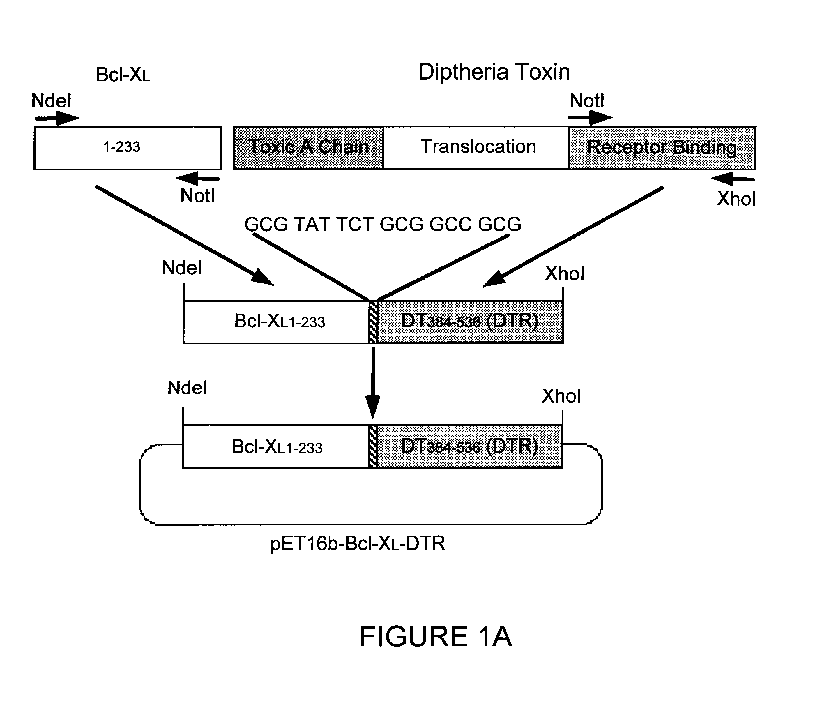
To produce proteins for extracellular addition to cells, the Bcl-xi gene, the DTR domain gene and the Bcl-x.sub.L -DTR fusion gene were cloned into pET16b. E. coli BL2 1 (DE3) strain was used to express Bcl-x.sub.L -DTR, Bad-DTTR, Bcl-x.sub.L and DTR, with addition of 1 mM IPTG when the OD260 reached 0.5-0.7. After two hours incubation and lysis by French press the inclusion bodies were collected and dissolved in 6M guanidine-HCl.

B. Eukaryotic Expression

Transfection of HeLa cells with the fusion constructs was performed as reported previously (Wolter et al., J Cell Biol 139:1281-1292, 1997). HeLa cells were harvested and lysed in 1 ml buffer containing 100 .mu. / ml leupeptin 20 hours after transfection, centrifuged to remove cell debris, and 15 .mu.l aliquots of the supernatant loaded onto 10-20% SDS-PAGE. The plasmid encoded proteins were visualized by immunoblotting with anti-Bcl-x...

example 3

Assays for Measuring Fusion Protein Binding to, and Translocation Into, Target Cells

A. Competitive Binding Assay

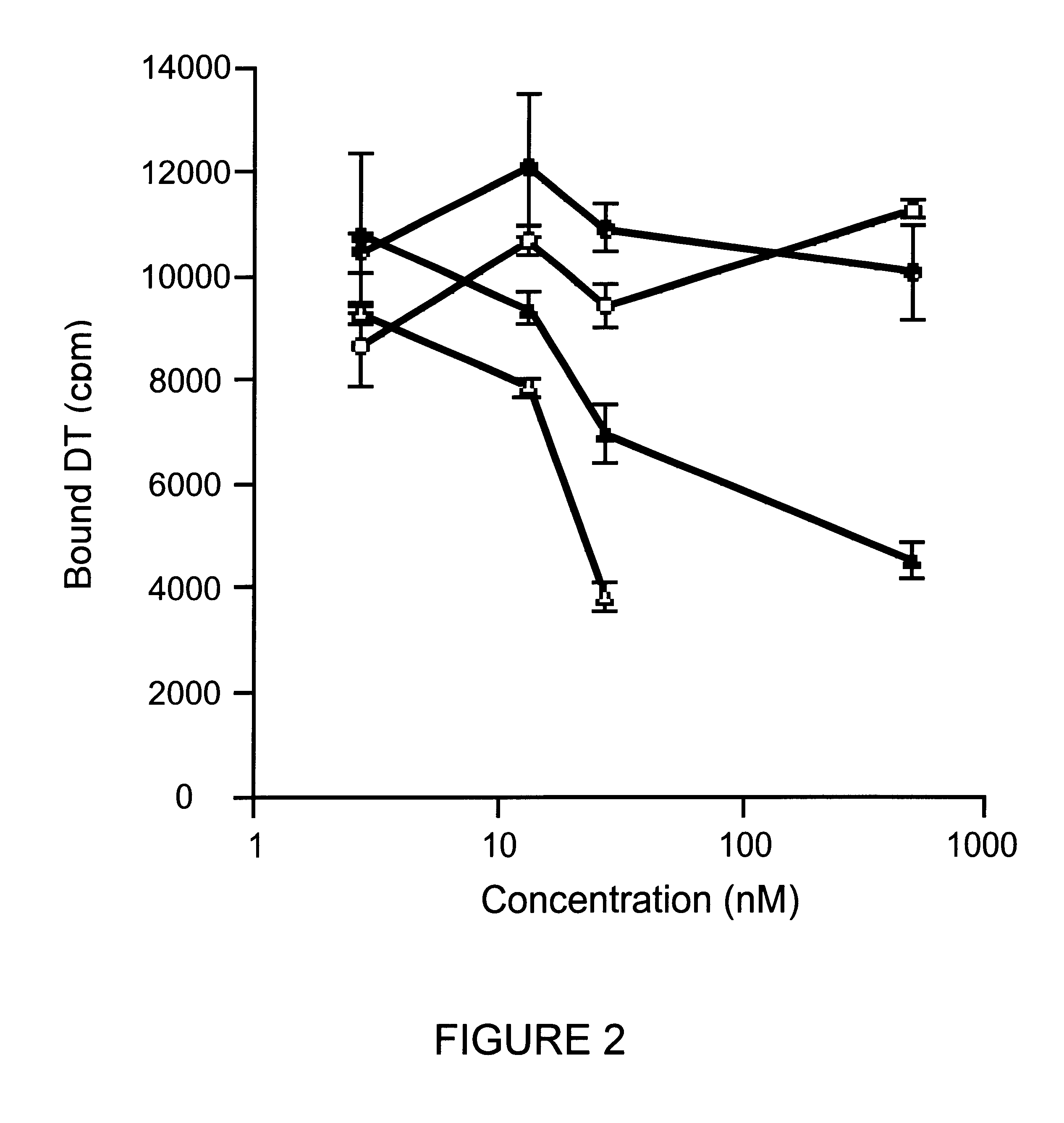
Protein binding to the diphtheria toxin receptor was performed as previously reported (Greenfield et al, Science 238:536-539, 1987) with the following modifications. DT was radiolabeled with I.sup.125 using iodobeads (Pierce Chem. Co., Rockford, Ill.) as described by the manufacturer. Cos-7 cells, grown to confluency in 12 well costar plates, were analyzed for receptor binding and competition by incubation for three hours on ice. Results are reported in FIG. 2. Cold competitor proteins, native DT (.DELTA.), Bcl-x.sub.L -DTR (.tangle-solidup.), Bcl-x.sub.L (.smallcircle.), and DTR (.circle-solid.), were used to displace I.sup.125 labeled DT tracer.

Native DT and Bcl-x.sub.L -DTR compete for DT receptor binding in the nanomolar concentration range. DT and the Bcl-x.sub.L -DTR fusion protein competed for I.sup.125 -DT binding to its receptor to a similar extent although the af...

example 4

Measurement of Bcl-x.sub.L -DTR Apoptosis-inhibiting Activity

A. Apoptosis Inhibition After Transient Cell Transfection

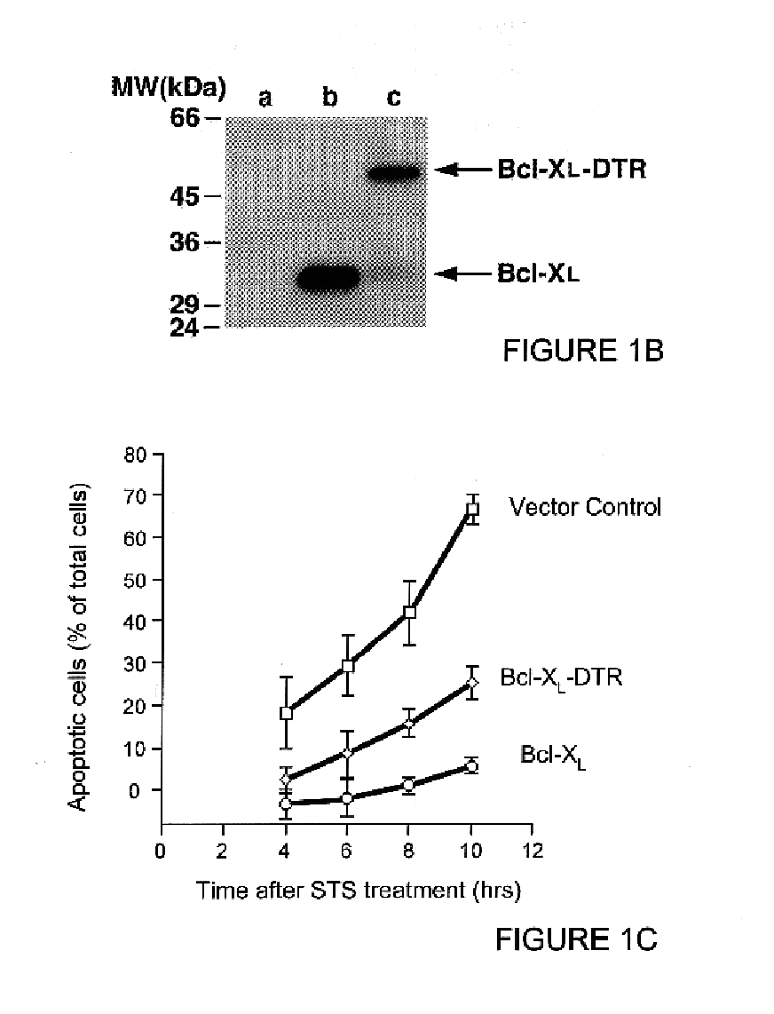
To demonstrate that Bcl-x.sub.L -DTR is effective at inhibiting apoptosis when expressed from within the target cell, this construct and the control construct containing Bcl-x.sub.L were transiently transfected into HeLa cells. Assay of apoptosis inhibition after transient transfection was performed as reported previously (Wolter et al., J. Cell Biol. 139:1281-1292, 1997). The Bcl-x.sub.L -DTR fusion gene blocked apoptosis after transient transfection into HeLa cells (FIG. 1C) to an extent similar to that of the Bcl-x.sub.L gene after C-terminal tail truncation (Wolter et al., J Cell Biol 139:1281-1292, 1997).

B. Inhibition of STS-induced Apoptosis by Extracellular Treatment With Bcl-x.sub.L -DTR

Hoechst dye no. 33342 staining: The effectiveness of extracellular delivery of Bcl-x.sub.L or the Bcl-x.sub.L -DTR fusion protein for inhibiting the rate of cell death by apop...


PUM

PropertyMeasurementUnit
Molar densityaaaaaaaaaa
Fractionaaaaaaaaaa
Fractionaaaaaaaaaa

Abstract

Apoptosis-modifying fusion polypeptides, and the corresponding nucleic acid molecules, are disclosed. Pharmaceutical compositions comprising these polypeptides, and the use of these polypeptides to modify apoptosis are also provided.

Description

FIELDThis invention relates to modification of the apoptotic response of target cells, for instance target cells in a subject. More specifically, it relates to apoptosis-modifying fusion proteins with at least two domains, one of which targets the fusion protein to a target cell, and another of which modifies an apoptotic response of the target cell.Tissue and cell homeostasis in multicellular organisms is largely influenced by apoptosis, the phenomenon of programmed cell death by which an intra- or extra-cellular trigger causes a cell to activate a biochemical "suicide" pathway. Morphological indicia of apoptosis include membrane blebbing, chromatin condensation and fragmentation, and formation of apoptotic bodies, all of which take place relatively early in the process of programmed cell death. Degradation of genomic DNA during apoptosis results in formation of characteristic, nucleosome sized DNA fragments; this degradation produces a diagnostic .about.180 bp laddering pattern wh...

Claims


Application Information

Patent Timeline
IPC IPC(8): C07K14/34C07K14/195C07K14/435C07K14/47A61K38/00A61K39/00
CPCC07K14/34C07K14/4747A61K38/00A61K39/00C07K2319/00C07K2319/01A61P43/00Y02A50/30
Inventor YOULE, RICHARD J.XIUHUAI, LIUCOLLIER, R. JOHN
Owner US DEPT OF HEALTH & HUMAN SERVICES
Learn More