
Allogenic tumor cell vaccine

Active Publication Date: 2018-07-05
ALLOPLEX BIOTHERAPEUTICS
View PDF0 Cites 9 Cited by
  • Summary
  • Abstract
  • Description
  • Claims
  • Application Information

AI Technical Summary

Benefits of technology

The patent describes a method of treating cancer in patients by using an allogeneic tumor cell line that has been modified to express multiple immunomodulator peptides. These peptides include IgG1, CD40L, TNF-alpha, GM-CSF, and Flt-3L. The tumor cell line is generated by transfecting or transducing the cells with recombinant DNA sequences coding for these peptides. The resulting tumor cell line variants are selected based on their ability to stimulate the immune system, including T cells, B cells, and dendritic cells. When these modified tumor cells are administered to cancer patients as a vaccine, they can improve the clinical outcome and overall survival of patients compared to placebo controls. The vaccine can be effective in melanoma, prostate cancer, and breast cancer. The tumor cell line variant vaccine elicits an immune response that reduces the risk of cancer progression and improves survival. This method of treating cancer offers a promising approach to immunotherapy and cancer vaccine development.

Problems solved by technology

However, immune responses against infectious agents do not always lead to elimination of the pathogen.
Fourth, unlike the situation with infections, cancer cells do not provide any immune adjuvant.
Without activation of the dendritic cell, contact between the dendritic cell and T cells (immune synapse) fails to result in optimal activation of the T cell.
One difficulty of developing this immunotherapy is that target antigens are often tissue specific molecules found on both cancer cells and normal cells, and either do not elicit immunity or show non-specificity regarding cell killing (Kaufman and Wolchok eds., General Principles of Tumor Immunotherapy, Chpt 5, 67-121 (2007)).
Furthermore, tumor cells have features that make immune recognition difficult, such as loss of expression of antigens that elicit immune response, lack of major histocompatibility (MHC) class II, and downregulation of MHC class I expression.
This method of therapy, however, can only be successful if a pre-existing antitumor immune response is present within a patient (Pardoll, D., The blockade of immune checkpoints in cancer immunotherapy, Nature Reviews: Cancer, Vol. 12, April 2012, 253).
CAR T therapy, however, has been shown to cause several types of toxicities, including cytokine release syndrome, neurological toxicity, non-tumor recognition, and anaphylaxis (Bonifant C L, et al., Toxicity and management in CAR T-cell therapy, Molecular Therapy—Oncolytics (2016) 3, 16011).
However, GVAX™ yielded only limited clinical responses.
The DC-tumor cell fusions provide a greater variety of tumor antigens, but have been met with limited success in human trials, likely due to the autologous components required, the heterogeneity of the product caused by maturation of DC cells, and variations in antigen loading (Browning, M., Antigen presenting cell / tumor cell fusion vaccines for cancer, Human Vaccines & Immunotherapeutics 9:7, 1545-1548; July 2013; Butterfield, L., Dendritic Cells in Cancer Immunotherapy Clinical Trials: Are We Making Progress?, Frontiers of Immunology, 2013 4: 454).
Murine models have been highly useful in discovering immunomodulatory pathways, but clinical utility of these pathways does not always translate from an inbred mouse strain to an outbred human population, since an outbred human population may have individuals that rely to varying extents on individual immunomodulatory pathways.
The cytokines can be directly toxic to target cells and can mobilize potent inflammatory mechanisms.
Furthermore, transfer of these Tregs prevents development of autoimmune disease.
This step in lymphocyte education is detrimental to preventing autoimmunity.
Inappropriate reactivity toward a normal self-antigen that was not eliminated in the thymus can occur, since the T cells that leave the thymus are relatively, but not completely, safe.
This results in recruitment of phagocytic cells to the site of an infection, phagocytosis of pathogens by immune cells, and / or formation of a membrane attack complex (MAC) that disrupts pathogen cell membrane and causes cell lysis.
Despite these promising results, human clinical trials of B7-containing vaccines have demonstrated increased immune response, but with only limited clinical benefit.
Human clinical trials of anti-CTLA-4 antibodies have demonstrated potential as an immune therapy, but because CTLA-4 plays a role in controlling T-cell responses, blocking its activity has the potential to lead to autoimmunity.
Ligation of 4-1BB is ineffective, however, when CD28 is present and an immune response is already present.
Second, their efficacy tends to be limited by the fact that while tumors express neoantigens (i.e. new, non-self elements) that are foreign and new to the individual, they are also undoubtedly human tumor cells and thus not always recognized as foreign (i.e. non-self) by the individual.

Method used


Image

  • Allogenic tumor cell vaccine
  • Allogenic tumor cell vaccine
  • Allogenic tumor cell vaccine

Examples


example 1

[0367]Examples 2-5 make use of, but are not limited to, the methods described hereinbelow.

Western Blotting

[0368]Briefly, cells are lysed with cold lysis buffer and centrifuged to pellet cellular debris. Protein concentration of the supernatant is determined by a protein quantification assay (e.g., Bradford Protein Assay, Bio-Rad Laboratories). The lysate supernatant is then combined with an equal volume of 2×SDS sample buffer and boiled at 100° C. for 5 minutes. Equal amounts of protein in sample buffer are loaded into the wells of an SDS-PAGE gel along with molecular weight marker and electrophoresed for 1-2 hours at 100 V. Proteins are then transferred to a nitrocellulose or PVDF membrane. The membrane is then blocked for 1 hour at room temperature using 5% non-fat dry milk in TBST blocking buffer. The membrane is then incubated with a 1:500 dilution of primary antibody in 5% non-fat dry milk in TBST blocking buffer, followed by three washes in 20 Mn Tris, Ph 7.5; 150 mM NaCl, 0.1...

example 2

[0389]The described invention provides an approach for restoring immunologic balance in, for example, treating cancer, by targeting multiple immunomodulators with a single cellular platform. This approach enables the simultaneous modulation of multiple signals, and affords a spatially and temporally restricted method of modulating the immune response, an important feature that differentiates this methodology from traditional approaches using systemic administration of biologic agents to act on a single immunomodulatory pathway at a time.

[0390]According to one aspect of the disclosed invention, a tumor-type specific cell line variant expressing five or more recombinant peptides may be generated for use as a tumor cell vaccine to treat that cancer type. For example, a tumor cell line may be selected for modification and lentiviral transfection of recombinant immunomodulator sequences may be used to stably integrate immune modulators into the cell genome. Example 3 below describes 7 le...

example 3

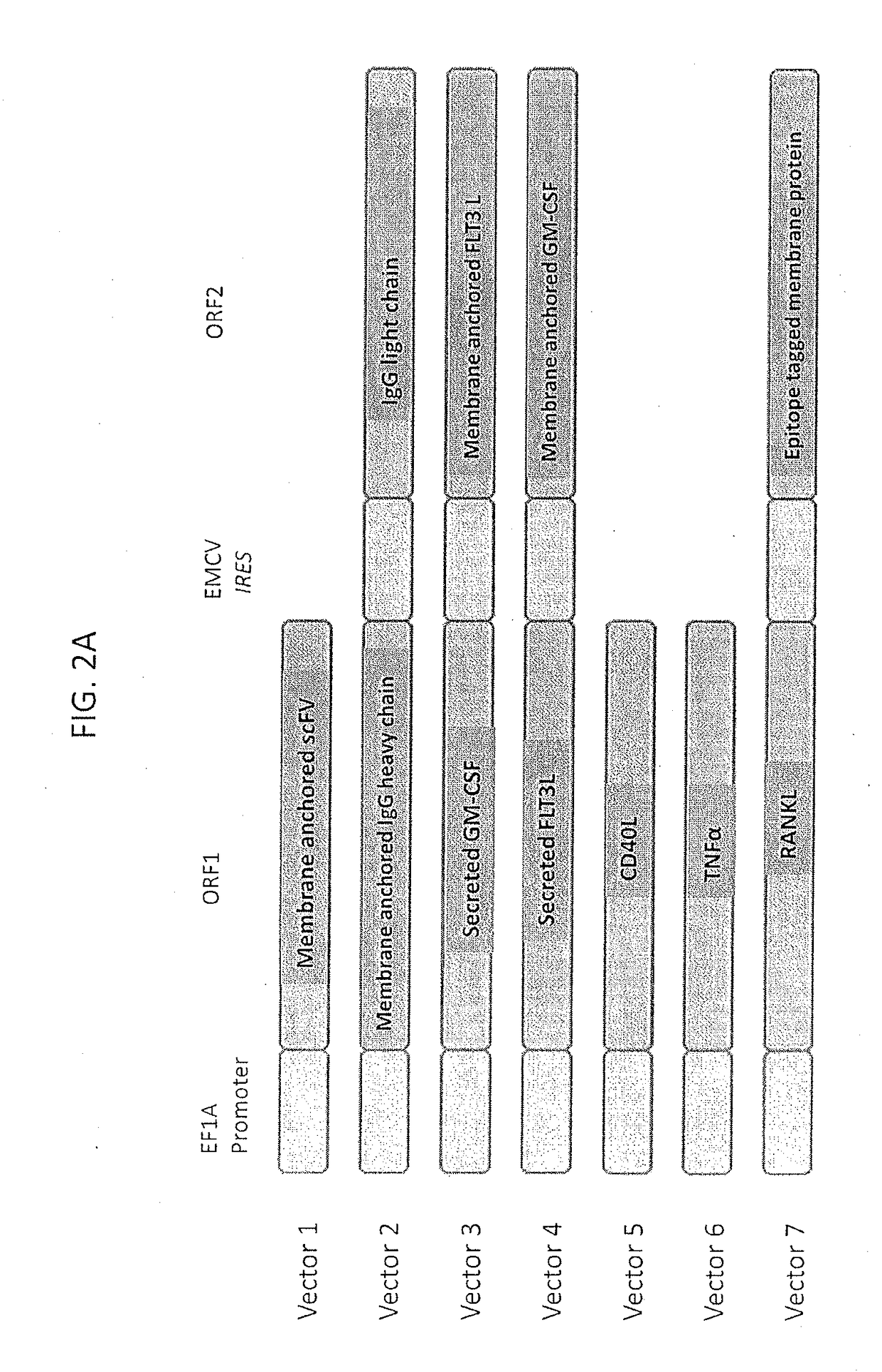
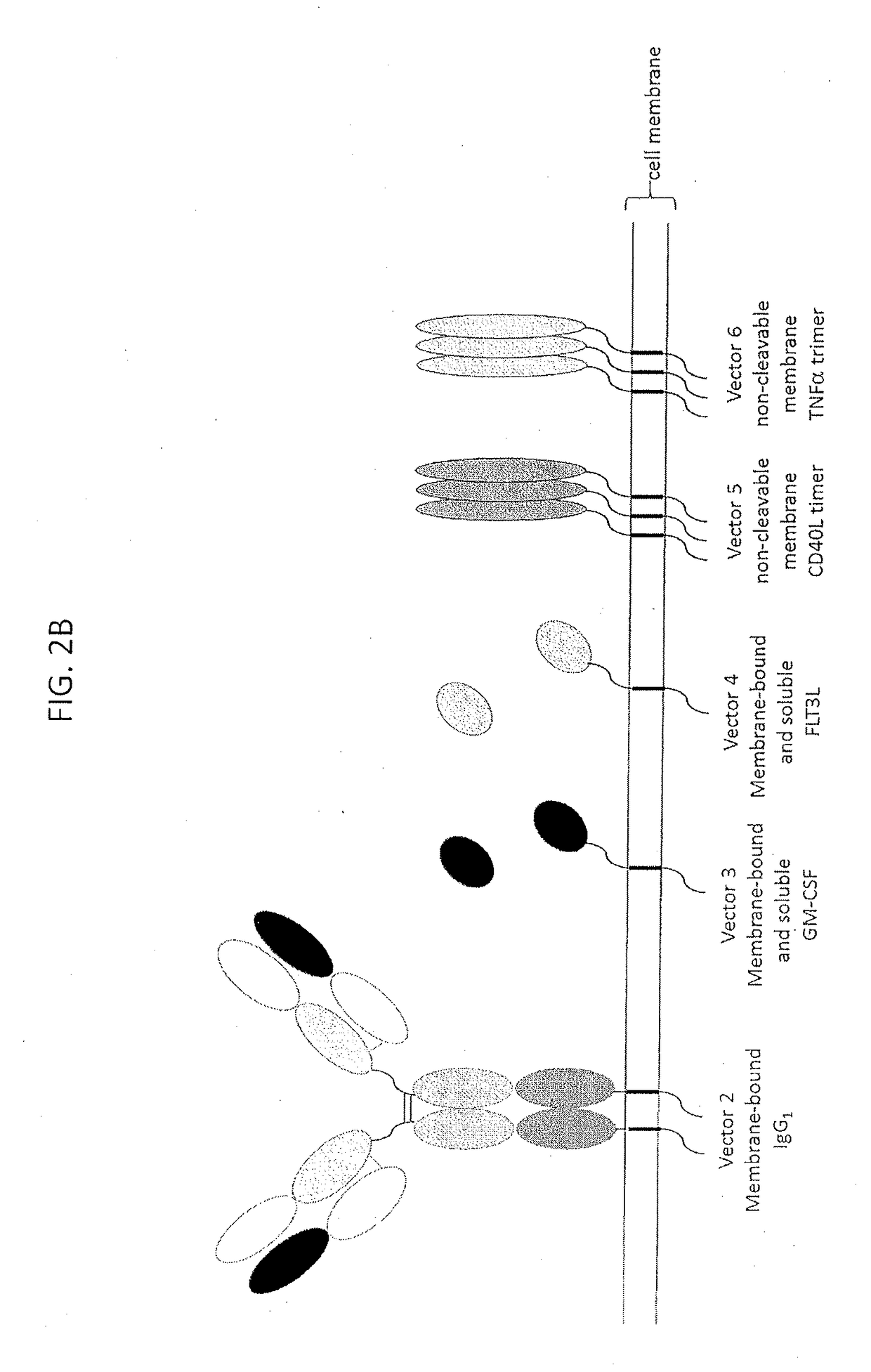
[0420]A schematic of the core lentiviral vectors employed in the experiments described herein is shown in FIG. 2A and a schematic of the encoded proteins is shown in FIG. 2B. The promoter is human elongation factor 1 alpha (EF1a) promoter and the internal ribosomal entry sequence (IRES) is derived from encephalomyocarditis virus (EMCV). The core vectors are described in detail hereinbelow as follows:

Vector 1. Immunomodulator: scFv-Anti-Biotin-G3hinge-mIgG1 (to Generate Surface IgG)

[0421]A schematic of the organization of vector 1, used for the immunomodulator scFv-anti-biotin-G3hinge-mIgG1 is shown in FIG. 3A. The nucleotide sequence of vector 1 (SEQ ID NO. 47) is shown in FIG. 3B. Table 2, below, shows the vector component name, the corresponding nucleotide position in SEQ ID NO. 47, the full name of the component and a description.

TABLE 2NucleotideComponent NamePositionFull NameDescriptionRSV promoter 1-229Rous sarcoma virus (RSV)Allows Tat-independent productionenhancer / promotero...


PUM

PropertyMeasurementUnit
Fractionaaaaaaaaaa
Volumeaaaaaaaaaa
Lengthaaaaaaaaaa

Abstract

The described invention provides a tumor cell vaccine comprising genetically modified tumor cell line of a particular tumor type that stably expresses high levels of two or more immunomodulators. According to some embodiments, an immunogenic amount of the tumor cell line variants may be selected for concomitant expression of two or more of recombinant membrane expressed IgG1, CD40L, TNF-alpha, as well as membrane and soluble forms of GM-CSF, and Flt-3L peptides that are effective to elicit an anti-tumor immune response compared to the parent unmodified tumor cell line as measured in vitro by a one-way mixed lymphocyte tumor reaction assay using human peripheral blood mononuclear cells and the genetically modified allogeneic cell vaccine candidate. According to some embodiments, the tumor cell vaccine candidate will induce an immune response in the recipient cancer patient that cross reacts with the patient's own (autologous) tumor cells, the effects of which will be sufficient to result in enhanced anti-tumor immunity contributing to the increased survival of a vaccinated patient cohort compared to a matched unvaccinated patient cohort.

Description

RELATED APPLICATIONS[0001]This Application claims priority to U.S. Provisional Application No. 62 / 425,424, filed on Nov. 22, 2016, the entire contents of which are incorporated by reference in their entirety herein.FIELD OF THE INVENTION[0002]The described invention relates generally to immunological approaches to the treatment of cancer, and more particularly to cancer vaccines comprising modified tumor cells.BACKGROUND OF THE INVENTIONImmune Response[0003]Generally speaking, immune responses are initiated by an encounter between an individual and a foreign antigenic substance, e.g., an infectious microorganism. The infected individual rapidly responds with both a humoral immune response with the production of antibody molecules specific for the antigenic determinants / epitopes of the immunogen and a cell mediated immune response with the expansion and differentiation of antigen-specific regulatory and effector T-lymphocytes, including both cells that produce cytokines and killer T ...

Claims


Application Information

Patent Timeline
IPC IPC(8): A61K39/00A61P35/00
CPCA61K39/0011A61P35/00A61K2039/5152C07K16/44C07K2317/524C07K2317/53C07K2317/56C07K2317/622C07K2317/72C12N5/0693C12N2501/25C12N2501/52A61K2039/5156A61K2039/55516A61K2039/55522A61K39/001138A61K39/001139A61K39/001129C07K14/525C07K14/535A61K2039/585A61K2039/876A61K2039/884A61K2039/812C12N2510/00
Inventor BORRIELLO, FRANK
Owner ALLOPLEX BIOTHERAPEUTICS
Learn More