
Preparation of extracellular matrix-modified tissue engineered nerve grafts for peripheral nerve injury repair

Active Publication Date: 2015-12-10
NANTONG UNIVERSITY
View PDF3 Cites 5 Cited by
  • Summary
  • Abstract
  • Description
  • Claims
  • Application Information

AI Technical Summary

Benefits of technology

The patent describes a method for creating a better type of nerve graft for repairing peripheral nerve injuries. The graft is made by culturing support cells in a 3D microgravity environment and then removing the cells to leave behind a natural extracellular matrix (ECM). This natural ECM is better for promoting nerve cell adhesion and regeneration. The graft also contains a large amount of collagen, which is important for nerve growth. The graft is biodegradable and can be easily accepted by patients. The method also involves implanting a scaffold made of silk fibroin filaments in the nerve conduit to guide the growth of nerve cells. Overall, this method may offer a better solution for repairing peripheral nerve injuries and restoring function.

Problems solved by technology

With the progress of social modernization and the pace acceleration of people's daily life, an increasing number of accidents, resulting from traffic, industry, and sports, local wars, violent events, and natural disasters (e.g. earthquakes), will lead to peripheral nerve injury.
Despite being a golden standard for peripheral nerve repair, autologous nerve grafting can not be widely used in the clinic due to its several limitations, such as deficient supply of donor nerves, mismatch between the injured nerve and the donor nerve in the structure and size, as well as donor site morbidity and secondary deformities.
Although there are a large number of studies on the preparation technology of ANGs achieving the faster progress, many preparation methods have complicated procedures, and the fine structure and mechanical properties of biomaterials will be impacted during processing.
At present, support cells used in the tissue engineered nerves include Schwann cells and various stem cells, which are allogeneic cells, and may cause immunogenicity, which is not suitable for clinic applications.
On the other hand, the in vivo fate and biological effects of support cells after they are implanted into the body are not fully clear, and they may be inactivated in the environment of the body, thus failing to achieve expected biological effects.
All the above issues limit the development of tissue engineered nerve grafts.
(1) The ECM is generally composed of collagen (Col), laminin (LN), fibronectin (FN), hyaluronic acid, and proteoglycan (such as chondroitin sulfate, heparan sulfate proteoglycan). ECM provides mechanical support and physical strength for the integrity of tissues, organs, and the whole organism. Evidence shows that LN, FN and Col can provide suitable “adhesiveness” for nerve growth, make axons grow along a matrix bridge, and guide the directional growth of nerve fibers. One or more ECM components, such as LN and FN, have been mostly adopted in the current field of tissue engineering. The commercially available, synthesized ECM, however, are expensive and their components are relatively single. The natural, cell-derived ECM, obtained in embodiments of the present invention after decellularization of cultured cells, can keep various important components and framework of ECM, and is more beneficial to the adhesion of nerve cells, and has a certain guiding effect on oriented axonal regeneration, thus promoting nerve regeneration. Moreover, it has the cheaper price than an synthesized ECM, and is easier to be accepted by patients.
(2) Support cells used in tissue engineered nerve grafts include Schwann cells and various stem cells. Most of these cells are allogeneic cells, which may cause immunogenicity during implantation. The ECM contained in tissue engineered nerve grafts developed in embodiments of the present invention is obtained after decellularizing cultured cells, thus leading to less or no immunogenicity. The tissue engineered nerve grafts are suitable for a large population of users.
(3) Tissue engineered nerve grafts developed by embodiments of the present invention are prepared by a three-dimensional microgravity cell culture method, which enables support cells to be uniformly adhered to the inner and outer surfaces of luminal filaments, and the ECM obtained after decellularization is also uniformly distributed on the inner and outer surfaces of luminal filaments, thus facilitating nerve regeneration.
(4) Tissue engineered nerve grafts developed in embodiments of the present invention contain no exogenic toxic substances, which may be probably introduced during preparation process, and also have excellent biocompatibility and biodegradability, as well as good mechanical property. The wall of nerve conduits exhibits a 3-D microporous structure, which facilitates transportation of nutrients required by nerve growth. The silk fibroin filaments included in the conduit lumen are prepared by electrostatic spinning, having a large specific surface area to serve as a required space for the growth of nerve cells.

Method used

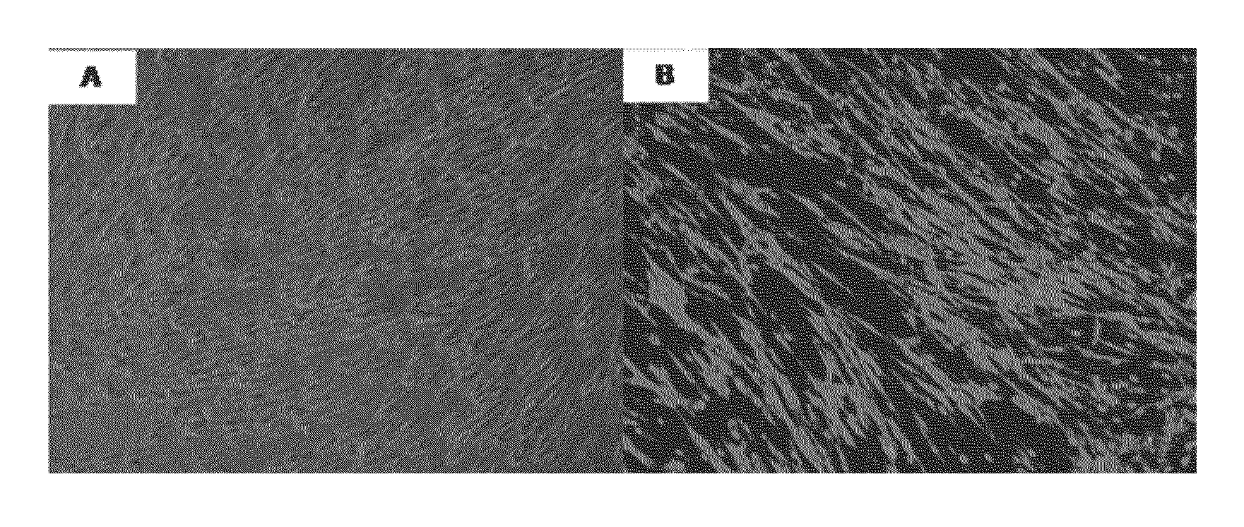

Image

  • Preparation of extracellular matrix-modified tissue engineered nerve grafts for peripheral nerve injury repair
  • Preparation of extracellular matrix-modified tissue engineered nerve grafts for peripheral nerve injury repair
  • Preparation of extracellular matrix-modified tissue engineered nerve grafts for peripheral nerve injury repair

Examples


working example 2

Culture of Skin-Derived Fibroblastes

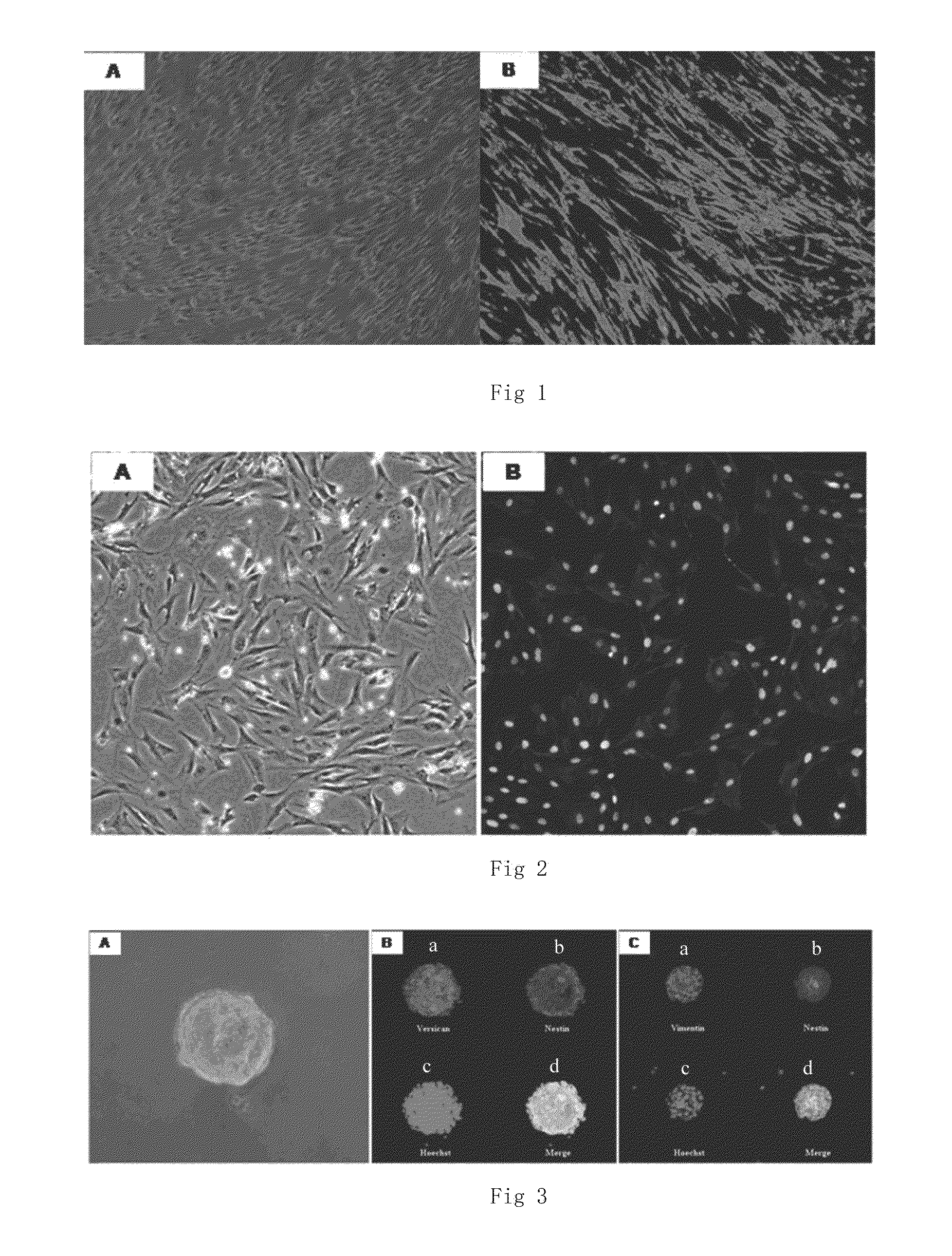
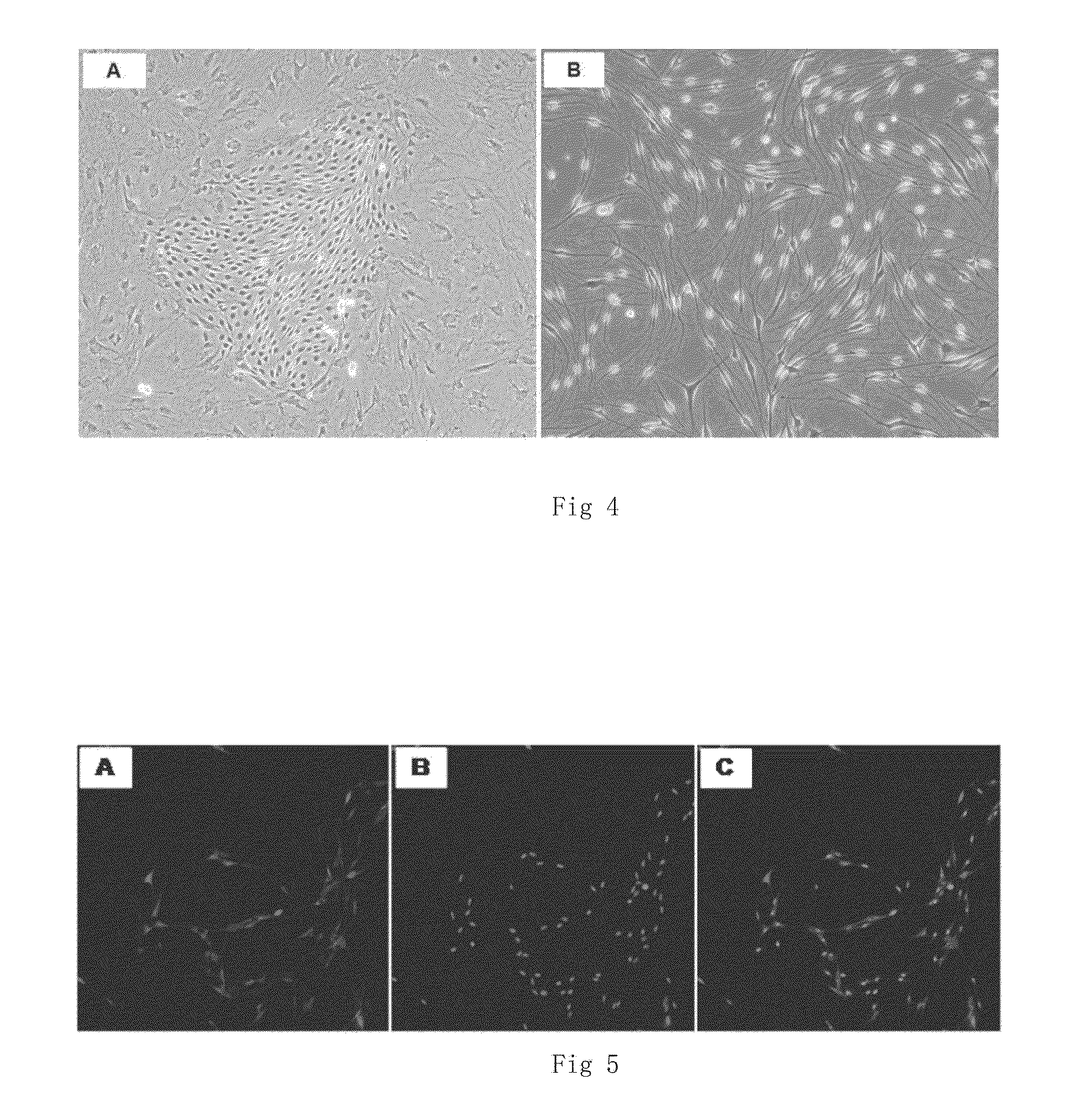
[0061]Newborn SD rats (1 day old) are killed and disinfected with alcohol Skins in the back are taken out and placed in a pre-cooled dissection solution to remove subcutaneous tissue (fat, hypodermic fascia layer, and blood vessels). After wash thrice with PBS, the skins are cut into pieces (<1 mm×1 mm) using a surgical blade knife, and completely digested using I type collagenase (1 mg / ml). The supernatant is abandoned. The cells are resuspended through a complete medium and seeded to a culture dish. After 90% cells are fused, the cells are subcultured. Epithelium contamination may occur during the process of primary culture, and most of parenchyma cells (epithelial and endothelial cells) will die gradually after several subculture. Therefore, the cells used in embodiments of the present invention are cells that have been subcultured for more than three generations (cell culture and identification are as shown in FIG. 2).

working example 3

Culture of Skin Stem Cells and Directional Differentiation Toward Schwann Cells

[0062]This is conducted as previously described (Jeffrey A Biernaskie, Ian A McKenzie, Jean G Toma & Freda D Miller, Skin-derived precursors (SKPs) and differentiation and enrichment of their Schwann cell progeny NATURE PROTOCOLS, 2006(1): 2803-2812). In brief, newborn SD rats (1 day old) are killed and disinfected with alcohol Skins in the back are taken out and placed in a pre-cooled dissection solution to remove subcutaneous tissue (fat, hypodermic fascia layer, and blood vessels). After wash thrice with PBS, the skins are cut into pieces (<1 mm×1 mm) using a surgical blade knife, and completely digested 0.1% pancreatin or XI type collagenase (1 mg / ml) for 45-60 min at 37° C., and then the digestion is terminated via a complete medium. The centrifugation is conducted and the supernatant is abandoned. Suspension culture is conducted in DMEM / F12 (3:1) containing 0.1% secondary antibody, 40 μg / ml fungizon...

working example 4

Culture of Bone Marrow Mesenchymal Stem Cells

[0064]Adult SD rats are killed by dislocation, soaked for 5 min with 75% alcohol. Femur and fetal bone are taken at an aseptic condition, and the marrow cavities are exposed and flushed by IMDM basal culture solution to collect the bone marrow. The bone marrow obtained is repeatedly pumped with an injector and prepared into a single-cell suspension, which is filtered through a 200-mesh sieve, placed in a horizontal centrifuge for centrifugation (1000 rpm×5 min). After the supernatant is abandoned, The cells are seeded at a density of 4×10⁵ / cm2 to an IMDM complete culture solution (containing 10% fetal calf serum) for culture. Full-dose solution change is conducted after 24 h, and the cells not adhered to the wall are removed. Afterwards, half-dose solution change is conducted every 3 d. The cell modality and growth situation are daily observed under an inverted microscope. When the cells overspread the bottom of the culture dish and are f...


PUM

PropertyMeasurementUnit
Temperatureaaaaaaaaaa
Timeaaaaaaaaaa

Abstract

A tissue engineered nerve graft for repairing peripheral nerve injury consists of a nerve conduit and a extracellular matrix (ECM) that is secreted by autologous or allogeneic support cells and obtained by decellularization. A preparation method of the ECM-modified tissue engineered nerve grafts containing support cells, nerve conduit and constructing a ECM-modified tissue engineered nerve graft.

Description

CROSS-REFERENCE TO RELATED APPLICATIONS[0001]This application claims priority to PCT Application No. PCT / CN2013 / 076239, having a filing date of May 27, 2013, based on CN 201310028903.6 having a filing date of Jan. 25, 2013, the entire contents of which are hereby incorporated by reference.FIELD OF TECHNOLOGY[0002]The following is in the technical field of medical biomaterials used for transplants into the human body, refers to the preparation of extracellular matrix (ECM)-modified tissue engineered nerve grafts for peripheral nerve injury repairBACKGROUND[0003]With the progress of social modernization and the pace acceleration of people's daily life, an increasing number of accidents, resulting from traffic, industry, and sports, local wars, violent events, and natural disasters (e.g. earthquakes), will lead to peripheral nerve injury. When the formed nerve defect of middle and long distance cannot be treated by end-to-end suture in the clinic, peripheral nerve grafting has to be ap...

Claims


Application Information

Patent Timeline
IPC IPC(8): A61L27/36A61L27/22C12N5/00A61L27/20A61L27/18A61L27/38A61L27/24
CPCA61L27/3675A61L27/3687A61L27/3878A61L27/3895A61L27/227A61L27/24C12N2527/00A61L27/18C12N5/0062A61L2430/32C12N2513/00C12N2525/00A61L27/20A61L27/3683A61L27/3633A61L27/3604
Inventor GU, XIAOSONGDING, FEIGU, YUNXUE, CHENGBINYANG, YUMINWANG, YONGJUNGONG, LEILEI
Owner NANTONG UNIVERSITY
Learn More