
Magnetic Resonance Imaging and Spectroscopy Means and Methods Thereof

Inactive Publication Date: 2008-11-20
BRAIN WATCH
View PDF3 Cites 61 Cited by
  • Summary
  • Abstract
  • Description
  • Claims
  • Application Information

AI Technical Summary

Benefits of technology

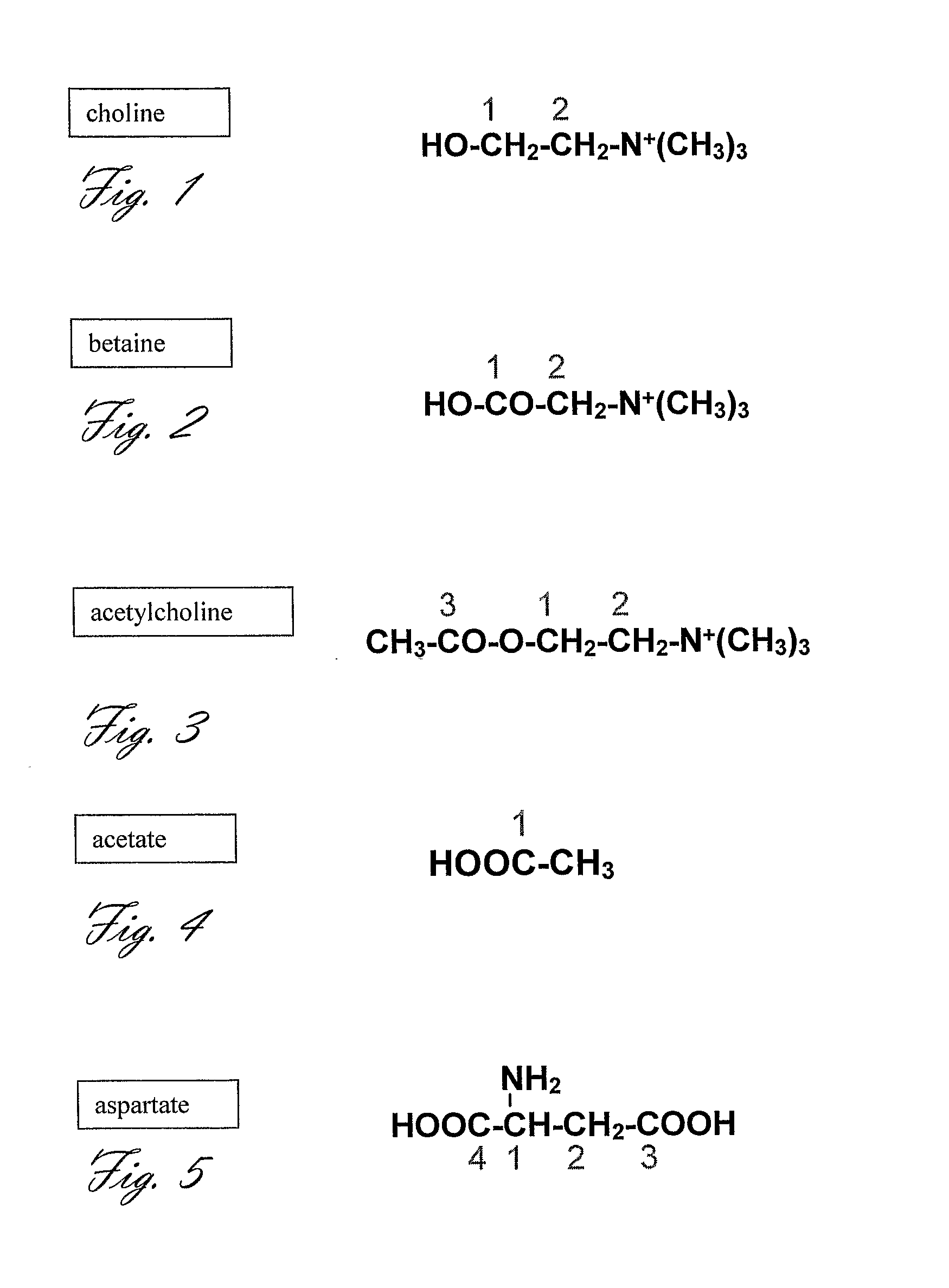
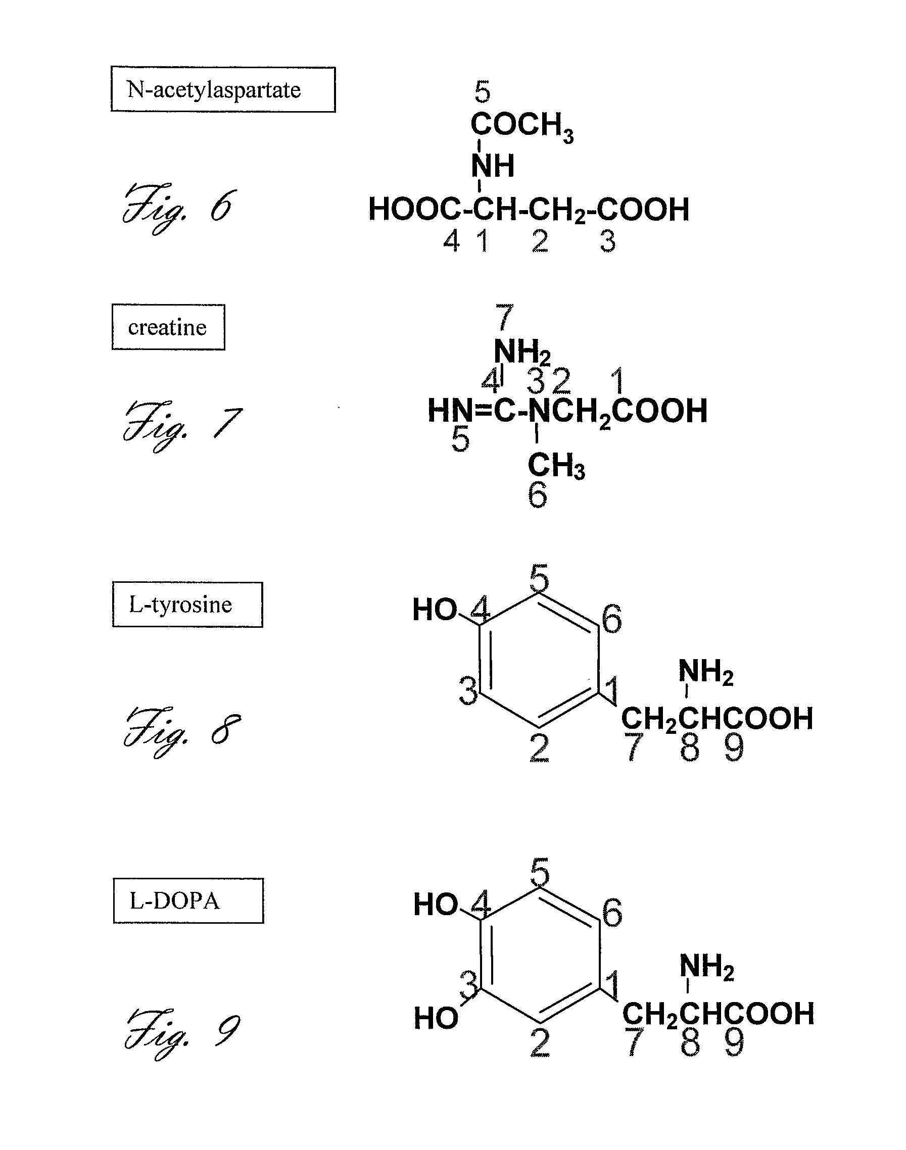
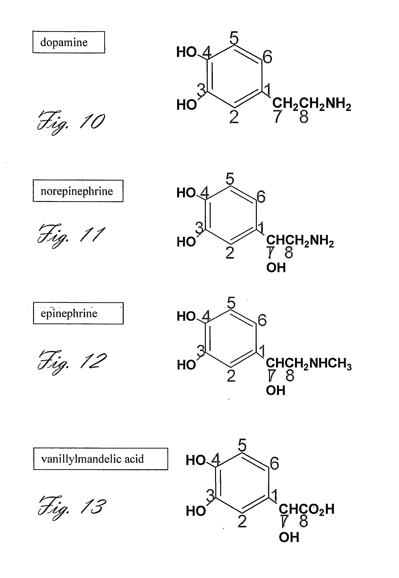
[0049]It has now been found that in vivo methods of magnetic resonance imaging and spectroscopy may be improved by using ex-vivo polarized MR agents comprising nuclei capable of emitting magnetic resonance signals in a uniform magnetic field (e.g. MR nuclei such as 13C, 15N, or 19F nuclei) and capable of exhibiting a long T1 relaxation time, preferably additionally a long T2 relaxation time, ability to cross the blood brain barrier, and optionally, an ability to be metabolized in the brain or body. Such agents will be referred to hereinafter as “high T1 neurochemical agents” or HTNC agents. Typically the HTNC agent molecules will contain MR imaging / spectroscopic nuclei in an amount greater than the natural abundance of said nuclei in said molecules (i.e. the agent will be enriched with said nuclei).
[0141]The resulting DNP-polarized HTNC agent in liquid form may be administered either alone or with additional components such as additional HTNC agents, or agents that will prevent its degradation in the peripheral circulation, increase its blood-brain-barrier permeability, prevent its uptake by peripheral organs, or modify its effect in the brain or body.

Problems solved by technology

However, as in the case of any drug there are side effects and cases where the patient's symptoms are not alleviated within a reasonable amount of time.
Despite the wide use of SSRIs, the exact biochemical effect of the drug on the individual's brain is not known and can not be quantified with existing technology.
However, despite an overwhelming need for better means to quantify the effects of psychiatric drugs on the brain, in situ, the technological means for doing so had not surfaced.
Despite their usefulness in diagnosis and in treatment monitoring, such tests do not provide a direct quantifiable biochemical measure of brain activity.
Serotonin syndrome is rare, but it is a serious, potentially life-threatening medical condition.
It may go unrecognized because it is often mistaken for a viral illness, anxiety, neurological disorder or worsening psychiatric condition.
The effects of this pharmacologic intervention are symptomatic and compensatory.
Tacrine, the first of the cholinesterase inhibitors to undergo extensive trials for this purpose, was associated with significant adverse effects including hepatotoxicity.
Ultimately, the benefits of such therapy decline as the neurodegenerative process progresses.
Placebo-controlled clinical trials exploring the efficacy and safety have shown that the effects of AChE inhibitors are dose-dependent.
Improvements in activities of daily living (ADL) are more difficult to assess.
As in the case of SSRIs, there is no available test to directly determine the effects of AChE inhibitors within the individual's brain, non-invasively.
Because of the lack of such a test, and because the efficiency of these drugs can be evaluated only after several weeks or months, it is not uncommon that patients are loosing valuable time in which the disease progresses irreversibly and is not stabilized because the patient is being given a treatment that is inefficient to them.
However, direct evidence of brain regions in which dopamine synthesis or metabolism are altered is not available.
However, direct evidence for NMDA dysfunction or altered glutamate synthesis and metabolism in schizophrenia is still lacking.
Similarly to the cases of SSRIs and AChE inhibitors, neuroleptics have side effects, not all patients respond to a specific treatment, and many times patients have to switch between drug regimes until the best drug for them is found by educated trial and error.
This phase of trial and error could last several weeks to several months because there is no test for determining the direct drug action and efficacy in the individual's brain.
However, most of these processes, including neuromodulators' metabolism, can not be directly detected in a non-invasive manner.
However, this reaction, as well as other aspects of NO metabolism) have not been directly observed in the living human brain or body in a non-invasive manner.
However, it is still not clear whether a decrease in NAA levels is a cause or effect of neurodegeneration and how well the total NAA level can be used in the diagnosis of a neurodegenerative state in the individual's brain.
However, the evaluation of the need for this treatment and the localization of such electrodes within the brain are lacking objective biomarker for the location of the dysfunctional neuromodulatory area within the brain.
However, the relationship between these changes and neuronal activity remains unclear, especially in the case of neuromodulation.
However, currently, the low (micro-molar range) concentration of neuromodulators prevents in vivo detection by MRS at high resolution.
However, the clinical use of MRI has to date been restricted to 1H, for reasons of sensitivity.
After polarization it may be dissolved in heated water or saline or melted and removed or separated from the OMRI contrast agent where the latter may be toxic and cannot be administered.

Method used


Image

  • Magnetic Resonance Imaging and Spectroscopy Means and Methods Thereof
  • Magnetic Resonance Imaging and Spectroscopy Means and Methods Thereof
  • Magnetic Resonance Imaging and Spectroscopy Means and Methods Thereof

Examples


example 1

Acetylcholine Synthesis in the Brain

[0199]The subject is pretreated with atropine prior to choline injection to prevent cholinergic intoxication.

[0200][2-13C, 15N]-choline (99% 13C-labeled, 99% 15N-labeled 10 mg) is dissolved in 40 mg of 50:50 glycerol:H2O. The trityl radical (Tris{8-carboxyl-2,2,6,6-tetra[2-(1-hydroxyethyl)]-benzo(1,2-d:4,5-d′)bis(1,3)dithiole-4-yl}methyl sodium salt) is added to reach concentrations of either 15 or 20 mM. The mixture is placed in an open top chamber.

[0201]The mixture is polarized by microwaves for at least one hour at a field of 2.5 T at a temperature of 4.2 K (or lower 1.2 K). The progress of the polarization process is followed by in situ NMR recording, according to previously published procedure (Ardenkjaer-Larsen, J. (2001) U.S. Pat. No. 6,278,893).

[0202]When a suitable level of polarization has been reached, the chamber is rapidly removed from the polarizer and, while handled in a magnetic field of no less than 50 mT, the contents are quickly...

experiment 8

[0228]Experiments 1, or 2, or 3, or 4 are performed in a patient that has been diagnosed with a brain tumor. The level and rate of [2-13C, 15N]-choline transport, [2-13C, 15N]-phosphocholine synthesis, and [2-13C, 15N]-betaine synthesis in the investigated tissue aid in the characterization of the tumor or the malignant potential at the tissue surrounding the tumor, as it is known in the art that choline metabolism is altered in malignant tissues. An extension of this experiment is the characterization of tumors in the body, such as tumors in the breast, prostate, and kidney.

example 2

Dopamine Synthesis in the Brain

[0229][13C6]-L-DOPA (99% 13C-labeled phenyl, 10 mg) is hyperpolarized and dissolved according to the procedure described in Example 1.

[0230]The subject is pretreated with a single dose or several doses of aromatic-L-amino-acid decarboxylase inhibitor such as carbidopa or benserazide, or difluoromethyldopa, or α-methyldopa (20 mg, 40 mg, 60 mg, or 80 mg) given orally.

[0231]1 hour after pretreatment with carbidopa, the hyperpolarized solution (cooled to 37° C.), is quickly injected to the subject (preferably in less than 10 sec, or as described in Example 1).


PUM


Abstract

The present invention discloses neurochemical agents and biochemical agents for human or mammalian neuro- and body-metabolic imaging, comprising chemicals involved in neuronal or glial function, neuromodulatory processes in the brain of said human or mammalian, vascular function, or organ specific metabolic processes; said neurochemical and biochemical agents are labeled with stable isotopes selected from a group including carbon-13, nitrogen-15, deuterium, fluorine-19 or a combination thereof in predetermined positions, so as to enhance the detectability of the agents and their metabolic successors.

Description

FIELD OF THE INVENTION[0001]This invention generally relates to MRI and spectroscopy means and methods thereof, and especially to magnetic resonance imaging and spectroscopy, and brain function as related to metabolism, psychobiology, psychiatry, neurology, and neurodegeneration.BACKGROUND OF THE INVENTION[0002]People suffering from psychiatric or neurodegenerative diseases are thought to have altered levels of some of the chemical messengers in the brain, called neurotransmitters and neuromodulators. In depression, the two principal chemical compounds involved are noradrenaline and serotonin. Nerve cells in the brain constantly produce, release and reabsorb serotonin. Lower levels of serotonin are thought to lead to the transmission of faulty messages and to be responsible for some of the symptoms of depression. Drugs such as selective serotonin reuptake inhibitors (SSRIs) increase the levels of noradrenaline and serotonin. This increased brain activity is intended to improve mood....

Claims


Application Information

Patent Timeline
IPC IPC(8): A61B5/055
CPCA61K49/10
Inventor KATZ-BRULL, RACHEL
Owner BRAIN WATCH
Learn More