
Method of immobilizing membrane-associated molecules

a membrane-associated molecule and membrane technology, applied in the field of membrane-associated molecule immobilization methods, can solve the problems of reducing the natural immobilization structure, limiting the development of new sensors and high-throughput screening technologies that utilize these cellular receptors, and achieving the effect of high-throughput screening

Inactive Publication Date: 2008-10-23
BRENNAN JOHN D +2
View PDF0 Cites 3 Cited by
  • Summary
  • Abstract
  • Description
  • Claims
  • Application Information

AI Technical Summary

Benefits of technology

[0014]The methods of entrapment and for detecting modulators of membrane-associated molecules of the present invention provide a general method for analyzing these molecules and their inhibitors, agonists and / or antagonists. The ability to immobilize membrane-associated molecules will allow development of bioaffinity chromatography or microarray technologies that will be useful for high throughput screening of potential inhibitors or effectors.

Problems solved by technology

The major problems limiting the development of new sensors and high-throughput screening technologies that utilize these cellular receptors arise due to the inherently low stability of such receptors and the difficulties associated with transducing receptor-ligand binding events into measurable signals.
Previous immobilization methods have been observed to reduce the natural dynamic motions of the bilayers, and lead to unstable immobilized structures.28,29,30,31,32,33,34,35 Problems can also arise due to the coupling of the lipid bilayer to the solid support, which can produce an unstable structure with a lifetime that is too short for functional purposes.
Furthermore, the structure of intrinsic membrane-proteins relies on hydrophobic interactions internal to the lipid bilayer, as well as hydrophilic interactions on either side of the lipid membrane.5 Very often with conventional supported BLMs, what would be considered the hydrophilic interior surface for the membrane protein is replaced by the solid substrate.5,36 This situation results in destabilization of the membrane protein with a concomitant loss in activity, or in the worst-case scenario complete loss of activity due to full denaturation of the protein.
These issues have been partially addressed by covalent attachment of a lipid monolayer to a solid substrate, which alleviates membrane dissociation; however, this method does not address the second issue mentioned, and furthermore decreases the natural dynamic behavior of the bilayer.4,5
However, the sol-gel method has found much less use for the immobilization of membrane-bound proteins.
These byproducts will readily dissolve or destabilize existing bilayer structures.43 Without a stable liposome, ion flux or membrane potential cannot be developed and therefore cannot be measured.

Method used


Image

  • Method of immobilizing membrane-associated molecules
  • Method of immobilizing membrane-associated molecules
  • Method of immobilizing membrane-associated molecules

Examples


example 1

Tryptophan Fluorescence of gA

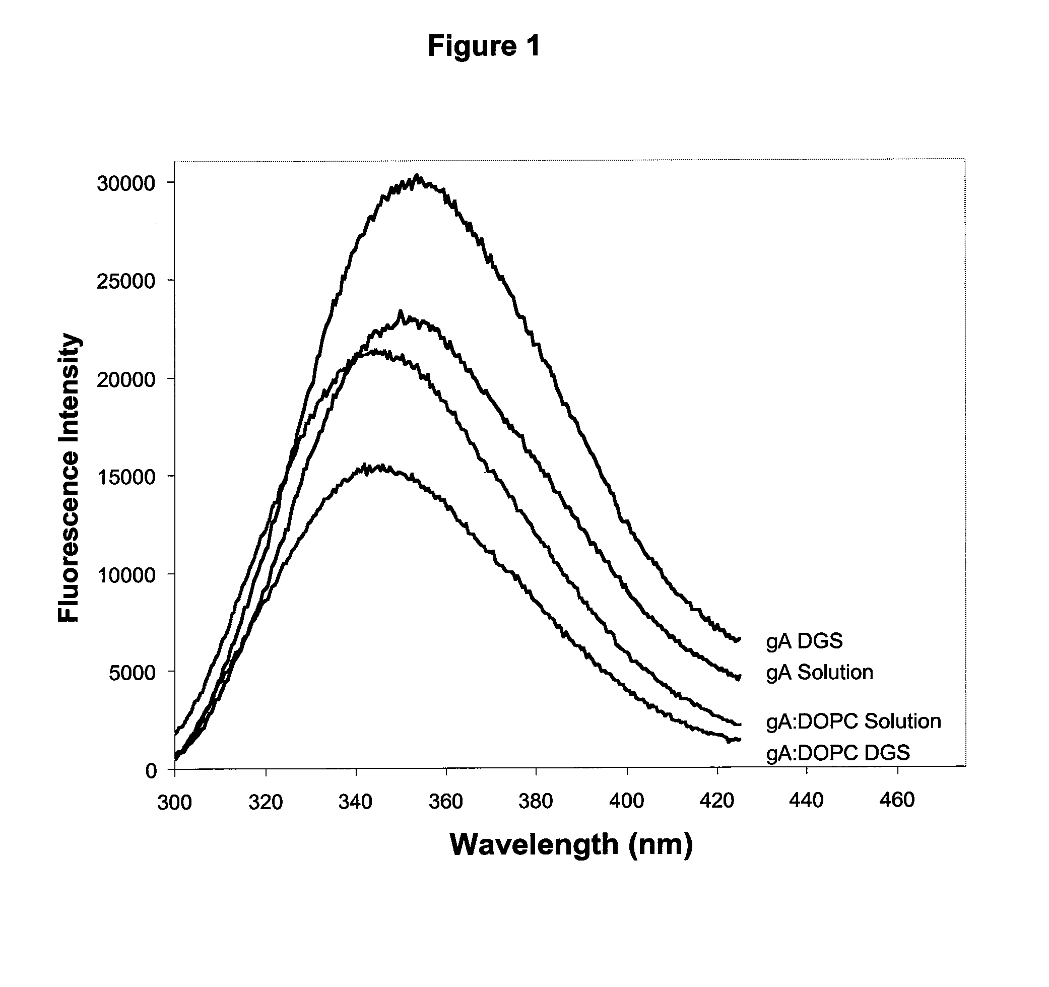
[0135]The emission of Trp residues within proteins has been widely used to probe the conformation and dynamics of proteins within sol-gel derived silica.66,67 In the case of gramicidin A, each homodimeric subunit of the ion channel contains four tryptophan residues, which NMR and crystallographic data have shown to be buried within the lipid bilayer.68 Furthermore, the tryptophan residues of gramicidin have been shown to have distinctly different fluorescence emission spectra when located in the bilayer relative to being in solution. The fluorescence emission properties of gA can therefore be used to indicate if gramicidin has survived the entrapment process and remained in the bilayer.

[0136]FIG. 1 shows the emission spectra of gramicidin A before and after reconstitution into phospholipid vesicles comprised of DOPC, both in solution and after entrapment into DGS derived silicate. The results clearly show that the emission maximum of gramicidin embedded ...

example 2

Ion Channel Activity of gA

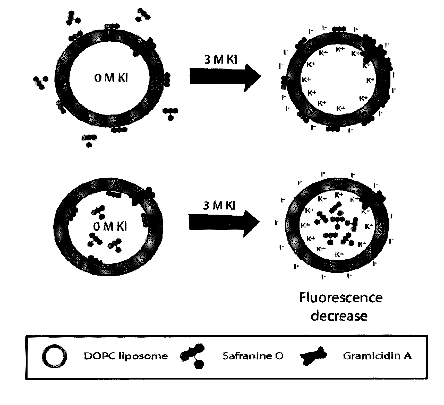
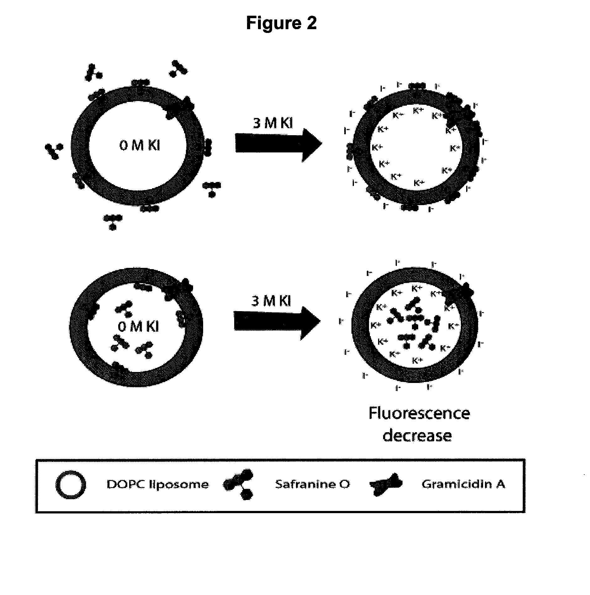
[0138]The lipophilic cationic dye safranine O was used to follow the development of an electrochemical potential of K+ across the phospholipid membrane. As shown in FIG. 2, the changes in emission properties depend on whether the probe is located inside or outside of the membrane. As shown in FIG. 2a, upon addition of KCl or KI to a membrane with the probe in the external solution, the influx of potassium ions through gA into the interior of the liposomes, combined with the exclusion of chloride ions, creates an electrochemical gradient across the membrane that is net positive on the interior and net negative on the exterior. Safranine O responds to development of such a membrane potential by partitioning into the hydrophobic lipid core due to the electrostatic attraction of the dye to the net-negative side of the membrane.69,70,71 The net effect is to produce an increase in both fluorescence intensity and anisotropy as K+ enters the membrane, owing to a re...

example 3

Inhibitors of gA Ion Channel Activity

[0145]A final test of the potential utility of the entrapped gA ion channel was to assess whether the ion channel activity could be inhibited by addition of channel blocking agents. It has been well established that the presence of divalent cations inhibits the flux of potassium and sodium ions through gramicidin by blocking their passage through the channel.76 Inhibition of reconstituted gA entrapped in DGS derived silicate was examined by adding various levels of CaCl2 to the entrapped samples along with 3.0 M KI. As shown in FIG. 6, the presence of calcium ions produces a significant and concentration-dependent decrease in the potential induced fluorescence response to ion flux, consistent with inhibition of the ion-channel activity. The inhibitory effect requires the presence of several hundred millimolar of Ca2+, which in expected given that Ca2+ must compete with molar levels of K+ for access to the ion channel. A benefit of the “inverted” ...


PUM


Abstract

The present invention relates to methods of immobilizing membrane-associated molecules within a sol-gel matrix. The membrane-associated molecule is embedded in the bilayer of a liposome. The molecule-liposome assembly remains functionally intact when it is immobilized within a protein and membrane-compatible sol-gel derived from polyol silane precursors or sodium silicate. The activity and stability of the entrapped membrane-associated molecule was significantly improved in macroporous silica.

Description

FIELD OF THE INVENTION[0001]The present invention relates to methods for the immobilization of membrane-associated molecules, including membrane-associated biomolecules, to composites prepared by such methods and to the use of these composites, in particular for high-throughput drug screening, multianalyte biosensing or bioaffinity chromatography.BACKGROUND TO THE INVENTION[0002]Immobilization of natural cellular receptors, which are mainly membrane associated proteins, is receiving substantial attention in the areas of research, clinical and environmental analysis, and in drug development.1,2,3,4,5,6,7,8,9,10,11 This is a result of increasing demand for robust and portable devices for medical, environmental and bioprocess monitoring. Just as the immobilization of biomolecules such as polynucleotides in the microarray platform has revolutionized the area of genomics, the immobilization of proteins will provide the same advantage to proteomics.12,13,14,15 Furthermore, immobilization ...

Claims


Application Information

Patent Timeline
IPC IPC(8): C40B30/04C40B50/14C40B40/10G01N30/02C40B30/00G01N33/543G01N33/552G01N33/68
CPCG01N33/5432G01N33/552G01N33/6872G01N2333/726
Inventor BRENNAN, JOHN D.BROOK, MICHAEL A.BESANGER, TRAVIS
Owner BRENNAN JOHN D
Learn More