
Fluorescent probes for ribosomes and method of use

a technology of fluorescent probes and ribosomes, applied in the field of fluorescent probes, can solve the problem of not being able to tell the binding site of inhibitors, and achieve the effect of controlling, treating or reducing the progression, severity or effects of nosocomial or non-nosocomial infections

Inactive Publication Date: 2005-06-02
CUMBRE
View PDF1 Cites 11 Cited by
  • Summary
  • Abstract
  • Description
  • Claims
  • Application Information

AI Technical Summary

Benefits of technology

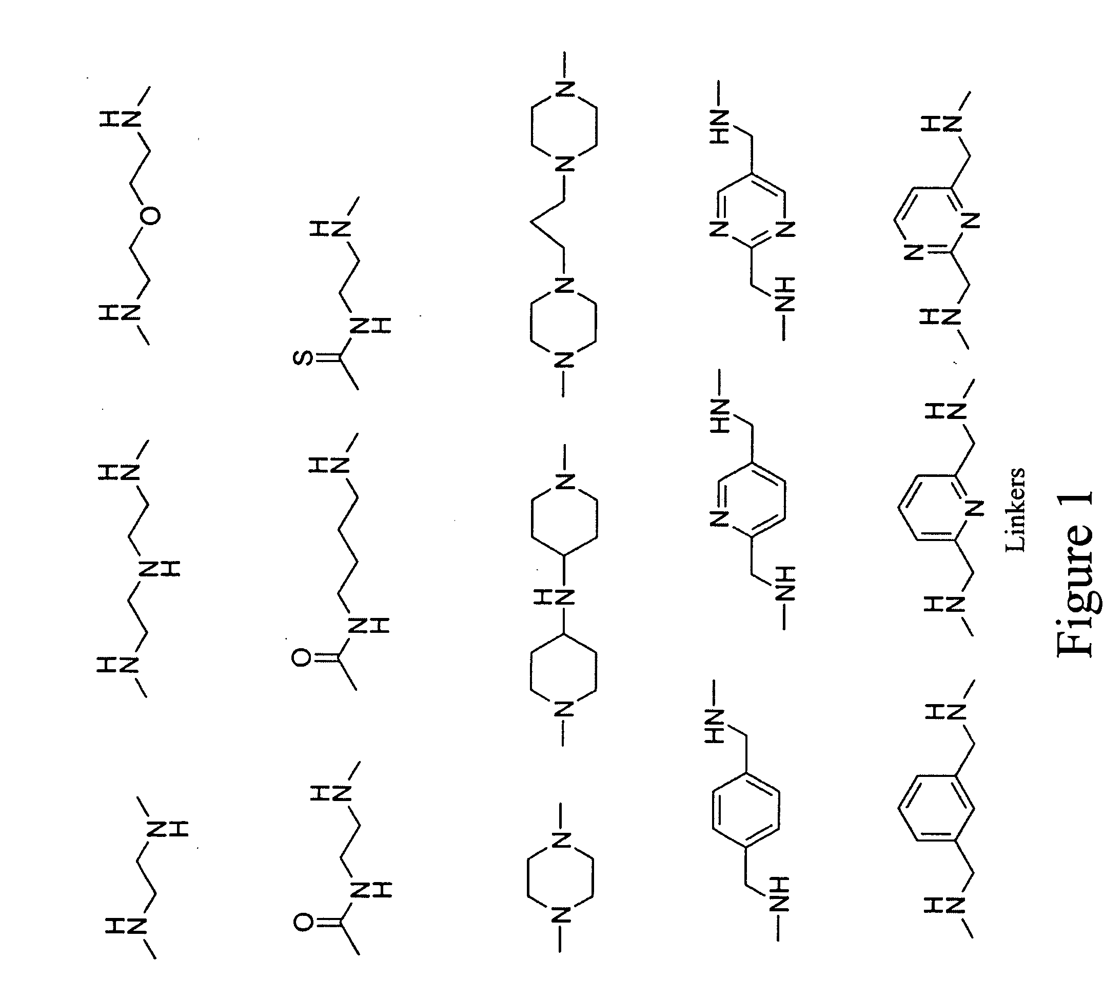
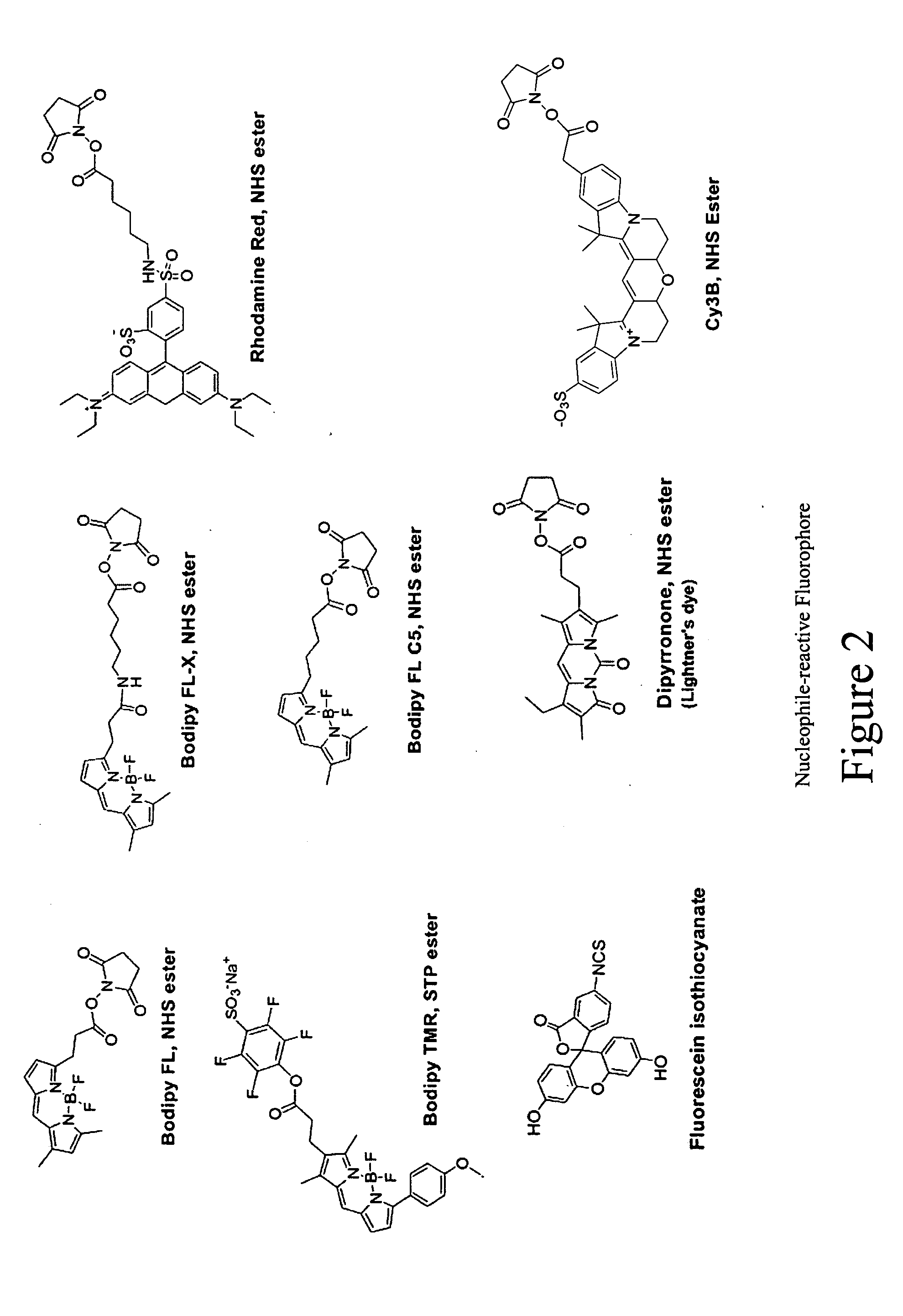
[0015] In a more preferred embodiment, the ribosome ligand is a member of the macrolide family of antibiotics. Examples of macrolide antibiotics are erythromycin, erythromycylamine, clarithromycin, azithromycin, roxithromycin, dirithromycin, flurithromycin, oleandomycin, telithromycin, cethromycin, leucomycin, spiramycin, tylosin, rokitamycin, miokamycin, josamycin, and midecamycin. The linker is a 0 to 16-carbon chain optionally interrupted by 1 to 6 heteroatoms, functional groups, carbocycles and heterocycles. The fluorophore is selected from groups consisting of BODIPY, fluorescein, rhodamine, and dipyranone.
[0016] In another aspect, the fluorescent probes are used for high-throughput screening to identify small molecules that interact with ribosomes and for mechanistic studies of ligand-ribosome interactions. The methods described in this invention are generally applicable for the identification of compounds that selectively modulate the function of ribosomes derived or purified from any organism, and can therefore be applied toward the discovery of novel agents for controlling infections mediated by bacterial, fungal and protozoal organisms. Examples of bacterial organisms that may be controlled by the compositions resulting from the application of the methods of this invention include, but are not limited to the following organisms: Streptococcus pneumoniae, Streptococcus pyogenes, Enterococcus fecalis, Enterococcus faecium, Klebsiella pneumoniae, Enterobacter sps., Proteus sps., Pseudomonas aeruginosa, E. coli, Serratia marcesens, S. aureus, Coag. Neg. Staph., Acinetobacter sps., Salmonella sps, Shigella sps., Helicobacter pylori, Mycobacterium tuberculosis, Mycobacterium avium Mycobacterium intracellulare, Mycobacterium fortuitum, Mycobacterium chelonae, Mycobacterium kansasii, Haemophilus influenzae, Stenotrophomonas maltophilia, and Streptococcus agalactiae. The compositions and methods will therefore be useful for controlling, treating or reducing the advancement, severity or effects of nosocomial or non-nosocomial infections. Examples of nosocomial infection uses include, but are not limited to, urinary tract infections, pneumonia, surgical wound infections, bone and joint infections, and bloodstream infections. Examples of non-nosocomial uses include but are not limited to urinary tract infections, pneumonia, prostatitis, skin and soft tissue infections, bone and joint infections, intra-abdominal infections, meningitis, brain abscess, infectious diarrhea and gastrointestinal infections, surgical prophylaxis, and therapy for febrile neutropenic patients. The term “non-nosocomial infections” is also referred to as community acquired infections. None of the information provided herein is admitted to be prior art to the present invention, but is provided only to aid the understanding of the reader.

Problems solved by technology

This assay monitors a single step of the protein synthesis process but is not informative about the binding sites of the inhibitors.

Method used


Image

  • Fluorescent probes for ribosomes and method of use
  • Fluorescent probes for ribosomes and method of use
  • Fluorescent probes for ribosomes and method of use

Examples


examples

[0079] The invention may be better understood with reference to the following examples, which are representative of some of the embodiments of the invention, and are not intended to limit the invention.

example i

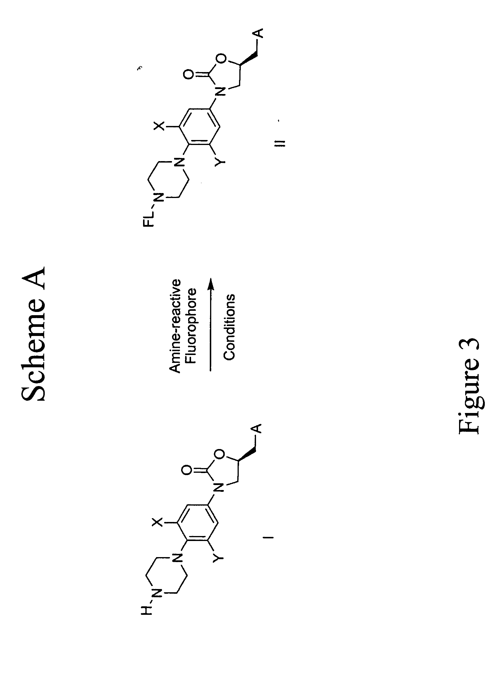
[0080] Oxazolidinone Probes. One series of probes of this invention are based on oxazolidinones. FIG. 27 illustrates the synthesis to prepare the oxazolidinone core compound 112. FIG. 28 illustrates the synthesis comprising compound 112 being reacted with different activated fluorophors to give a variety of oxazolidinone probes under typical coupling conditions. For example, FIG. 27 shows that (1-benzyl-4-(2-fluoro-4-nitro-phenyl)-piperazine) (“103”) was obtained as follows: Step 1, to a solution of difluoronitrobenzene (“101”) (1.08 mL, 9.8 mmol) and benzylpiperazine (“102”) (1.8 mL, 10.4 mmol) in CH3CN (10 mL) was added triethylamine (1.4 mL, 10.0 mmol). The resulting solution was heated at 90° C. for 3.5 h and then diluted with EtOAc and H2O. The organic phase was separated and washed with H2O, brine and dried over Na2SO4. The solvent was evaporated in vacuum to afford a yellow solid 103 (3.68g). Compound 103: TLC (20% EtOAc / Hexane) Rf=0.40. 1H NMR (400 MHz, CDCl3): δ (ppm) 2.64...

example ii

[0091] Macrolide Probes. Another series of probes of this invention are based on Macrolides. FIG. 29 illustrates the preparation of 9N-fluorescein erythromycylamine (“202”). To a stirred solution of erythromycylamine (Timms, G. H. et al. Tetrahedron Lett., 1971, 195-198. 0.10 mmol) and K2CO3 (28 mg, 0.20 mmol) in acetone-water (2 ml) was added 5-fluorescein isothiocyanate (39 mg, 0.10 mmol). The reaction mixture was stirred at r.t. for 20 hrs and the solvent was evaporated. The residue was purified by column chromatography (silica gel, 1% HOAc in ethyl acetate then methanol) to give an orange solid (28 mg, 25%): MS(M+H)+1124.

[0092]FIG. 29 illustrates the synthesis necessary to prepare the 9-BODIPY-amino-erythromycin{9-(4,4-difluoro-5,7-dimethyl-4-bora-3a,4a-diaza-s-indacene-3-propionyl)-amino-erythromycin} (“203”) as follows: To a solution of 9-amino-erythromycin (“201”) in DMF (0.5 mL) was added BODIPY FL SE (4,4-difluoro-5,7-dimethyl-4-bora-3a,4a-diaza-s-indacene-3-propionic acid...


PUM

PropertyMeasurementUnit
temperatureaaaaaaaaaa
pHaaaaaaaaaa
pHaaaaaaaaaa

Abstract

Fluorescent probes that have binding affinity to ribosomes. The fluorescent probes are useful tools for identifying small molecules that bind to the 50S or 30S subunits of the bacterial and other ribosomes and serve as novel ribosome inhibitors. These probes are also useful for determining the interactions between a specific ligand and the ribosome.

Description

[0001] This application claims priority to U.S. Provisional Patent Application, Ser. No. 60 / 508,401, entitled “Fluorescent Probes for Ribosomes and Method of Use” filed on Oct. 3, 2003, the entire content of which is hereby incorporated by reference.BACKGROUND [0002] The present invention is related to fluorescent probes having high binding affinity to ribosomes and their uses. The fluorescent probes of this invention are useful tools for identifying small molecules that bind to the 50S or 30S subunits of the bacterial ribosome and serve as novel ribosome inhibitors. These probes are also useful for determining the interactions between a specific ligand and the ribosome. [0003] Antibiotics are commonly utilized to fight a variety of microbial infections. However, many clinically important strains of bacteria have become resistant to one or more classes of the available antibiotics. Novel antimicrobial agents with activity against these resistant organisms are needed for the effectiv...

Claims


Application Information

Patent Timeline
Patent Type & Authority Applications(United States)
IPC IPC(8): C07H17/08C07H21/04C09B11/08C09B11/24C09B23/01C12Q1/68G01N33/533G01N33/58
CPCC09B11/08C09B11/24G01N2500/00G01N33/533G01N33/582C09B23/0075
Inventor MA, ZHENKUNLI, JINGKIM, INJIN, YAFEILYNCH, ANTHONYROCHE, ERICBEEMAN, DOUG
Owner CUMBRE
Learn More