However, excessive meshing usually results in healed
skin that is more susceptible to infections and which has a basket-like pattern, an undesirable result aesthetically.
However, the autograft method faces a few challenges and limitation in the treatment of patients with large surface area wounds.
Adequate, healthy
skin donor sites are difficult to find in such patients.
There is also a time limitation for harvesting the graft from the same site.
Often, a
delay of several weeks is necessary to wait for healing of the donor sites before harvesting them again, thus delaying healing of the "main" wound-the original wound to be treated and increasing the risk of complication.
Even worse is that harvesting an autograft in fact creates a second wound in the normal healthy
skin, which increases the
risk of infection and fluid /
electrolyte imbalance.
In addition, repeated harvests of autografts from a donor
wound site can result in contour defects or scarring, thereby causing disfigurement of the patient.
This two-step approach suffers a few limitations.
First, growth of cultured epidermal sheets in a laboratory needs at least 3 weeks to be achieved, thus delaying the coverage of wounds.
This limitation is even more prominent in areas where such laboratory and human resources are not available, such as the battle fields and the rural areas of developing countries.
Second, the reconstructed epidermal sheets need to be grafted on a clean wound
bed since they are
highly sensitive to bacterial infection and
toxicity of residual antiseptics.
Since the regeneration of the dermal compartment underneath the epidermis is a lengthy process the skin remains fragile for at least three years and usually
blisters.
In addition, the aesthetic effect is usually not as good as with one obtained with a split-thickness graft.
Unfortunately, even though the allograft is depleted of Langerhans' cells, the rejection of the transplant by the host occurs in mice after about 2 weeks.
Maintaining these stem cells in culture conditions can be challenging.
Inappropriate culture conditions can irreversibly accelerate the clonal conversion and can rapidly cause the disappearance of stem cells, rendering the cultured autograft or allograft
transplantation useless.
Although desirable, an
in vitro embryonic development process is highly unpredictable.
Although the programs of
gene expression in these cells somewhat resemble the differentiation pathways typical of developing animals, the triggering of these programs is
chaotic.
However, question remains unanswered as to whether these multipotent stem cells harvested from specific tissues or differentiated from ESCs
in vitro will make site-specific tissue when transplanted to injured adult tissues.
However, no successful regeneration of a fully functional human organ has been reported by using this approach.
For example, treatment of wounds with in vitro cultivated
keratinocyte stem cells merely closes the wound, not resulting in a full restoration of the physiological
structure and function of the skin.
Moderate successes have been achieved using this approach, with serious limitations on
clinical efficacy and cost in materials and labor.
More significantly, so far there has been no
clinical evidence demonstrating regeneration of organ with a complete restoration of physiological functions by using this approach.
By contrast, wounds that are treated by using other approaches, often if not most of the time, heal pathologically, i.e., with abnormal or impaired functions of the skin.
However, these nascent stem cells are quite fragile and are prone to death caused by cytotoxic effects exerted by various environmental elements, and by uncontrolled cellular responses to injury.
These so-called stem cells, although capable of divide without limit and differentiate into cells of various tissue types, have not been shown to be able to regenerate a fully functional human organ, let alone a live human in vitro.
Unfortunately, a
fully developed human is exposed to a completely different, more hostile environment.
Under the influence of both endogenous and exogenous conditions, spontaneous adult
wound healing and organ generation go through somewhat different pathways and end up with
scars and dysfunction of organs.
This spontaneous healing process is totally passive, uncontrolled by therapeutic interventions by embarking on a course of
chaotic cell proliferation and differentiation and reconstitution of regenerated tissues.
Excessive
drying of the wound leads to
eschar formation and damages viable tissues.
The drawback to the approach of implantation of bionic device is the inability to manufacture
artificial materials that duplicate the durability, strength, form, function, and
biocompatibility of natural tissues.
Although progress in
biology has made it possible for apply the
cell transplantation in the clinic, multiple practical limitations still exist and the clinical results are not physiological or cosmetically satisfactory.
One of the limitations associated with this approach is the difficulties with identification and isolation of multipotent stem cells from various tissues.
Results obtained from experimental animals, although encouraging, still cannot translate functionally into human therapy confidently.
For successful organ regeneration using stem cells cultured in vitro, major obstacles lies in its way.
However, question remains unanswered as to whether these multipotent stem cells harvested from specific tissues or differentiated from ESCs in vitro will make site-specific tissue when transplanted to injured adult tissues.
Such approaches trigger bioethical concerns, a problem even harder to solve.
For example, it remains a major challenge to synthesize scaffolding material for bionic implants that have the requisite
topography, surface properties, and growth and differentiative signals to facilitate
cell migration, adhesion, proliferation and differentiation, as well as being moldable into the shape of various tissues and organs.
However, the prevailing thought is that the skin of an adult with deep partial-thickness burn or full-thickness burn can only be regenerated with
scars and substantial loss in the
structure and function of the appendages.
However, delivery of growth factors made exogenously to the
wound site has been proven an unsatisfactory therapy clinically.
More disappointingly, this marginal
gain occurs only when radical debridement and weight off-loading have been achieved by experienced clinicians.
The environment of human chronic wounds cannot be replicated in experimental animals.
Another challenge is that fibroblasts from chronic wounds are often deficient in response to growth factors.
In addition to
hair loss, abnormally accentuated growth of hair can result from some rare conditions.
Such conditions can lead to profound social consequences for affected individuals.
However, despite such a need, effective prophylaxis and therapy remains elusive.
For example, one method used to combat alopecia,
hair transplant surgery, is not available to many people suffering from alopecia (e.g., patients having undergone
chemotherapy, elderly individuals, etc.).
Moreover,
surgery offers, at best, only a partial remedy.
Electrical stimulus has been suggested as an alternative way to promote
hair growth (see, e.g., U.S. Pat. No. 5,800,477 and references cited therein); however, such methods are of questionable
efficacy.
Tissue repair and organ regeneration through modulation of a single or a limited number of growth factors could likely to result in incomplete restoration of physiological structures and functions because the exquisite balance of life is kept by complex
cellular activity regulated by the body itself, not controlled by just a few growth factors.
The cellular and tissue interactions in the wounds treated by using these traditional methods are
chaotic, leading to
pathological healing of the skin with disfiguring
scars, disablement and dysfunction.
However, when the circumstances of development are more grossly abnormal, the embryonic cells can go wildly out of control.
For example, when a normal early mouse
embryo is grafted into the
kidney or testis of an adult, it rapidly becomes disorganized, and the normal controls on cell proliferation break down.
However, the triggering of these developmental programs is chaotic, yielding a jumbled "grab bag" of tissue types.
Because of such uncontrollable, chaotic development of cultured ES cells in vitro, it remains a challenge for people who attempt to regenerate organs
ex vivo and then transplant them to the patient with complete restoration of physiological
structure and function.
To reconstruct a fully functional organ from ES cells in vitro, an enormous challenge is to sift through a "
galaxy" of environmental signals to determine which "constellations" of cues can selectively "coax" ES cells down a specific lineage pathway at the expense of all others.
Before these signals are deciphered and absent a suitable physiological environment as in the body itself, attempts to reconstruct a fully functional organ in vitro would most likely to fail despite of extensive intervention with cocktails of growth factors.
However, since these regenerative ACS are exposed to a completely different, more hostile environment than those in a
fetus, the fate of the ASCs is not only controlled by endogenous regulatory mechanisms but also by exogenous interference such as bacterial infection and
air pollutants.
Loss or inactivation of the receptors leads to ejection from the basal layer, confirming the decision to differentiate; and ejection from the basal layer through other causes leads to loss of the receptors, forcing the cell to differentiate prematurely.
However, scarring begins to occur at this stage.
In addition, eschars form when wounds are exposed to dry air and are incapable of supporting overlying cells.
The wound is susceptible to adverse effects caused by "normal"
inflammatory response of the body to wounding and by exogenous agents such as
bacteria that causes infection and further
inflammation systemically and on the site.
Overgrowth of fibroblasts leads to
overproduction of collagen which aggregates to form disorderly fibers and eventually causes scarring after closure of the wound.
Treatment of full-thickness burn with conventional methods such as dry therapy and skin grafts results in wound-closure with disfiguring scars and substantial loss of normal functions of appendages of the skin.
However, some cells proliferate continuously through out the body's life, thus demanding a continuous supply of stem cells.
This outcome has been dreamed by scientists and physicians in the art but never achieved clinically.
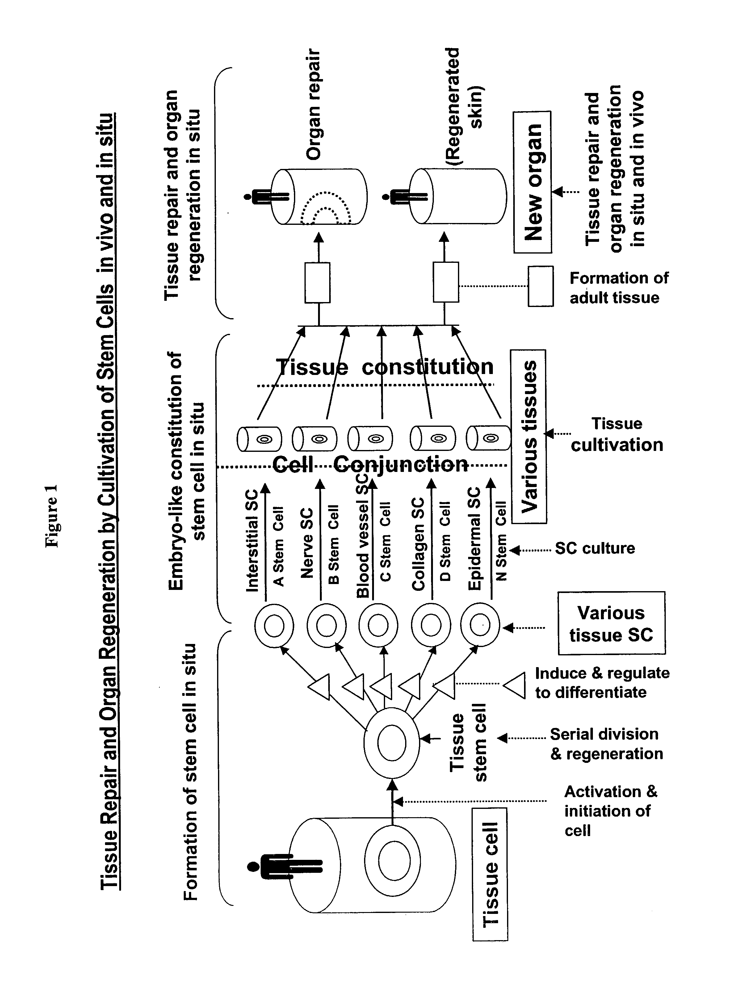
The inventor believes that although
transplantation of stem cells cultivated in vitro has enjoyed limited successes in repairing damaged epidermis and
dermis, the healing of the wounds is not physiological.
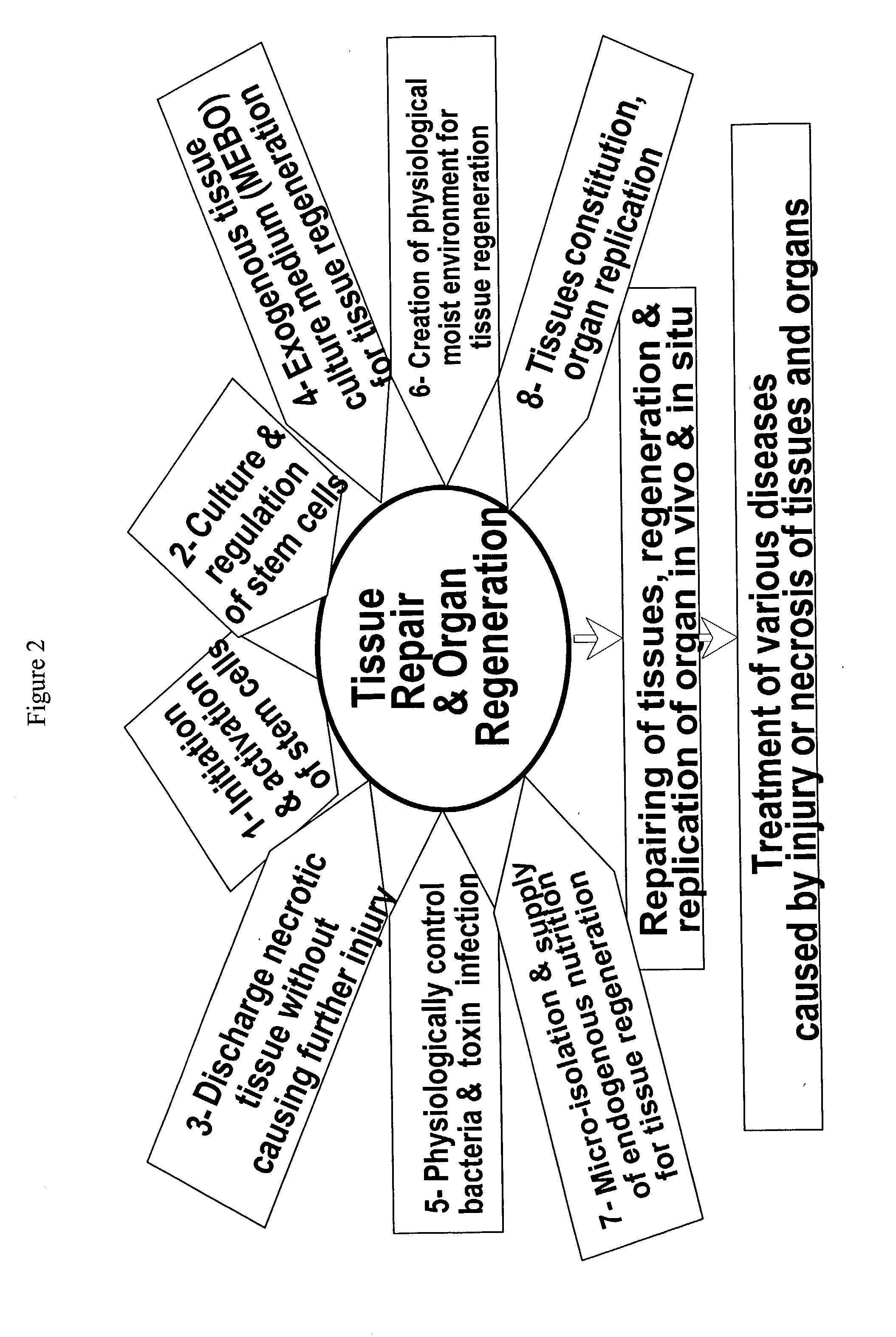
If the necrotic cells remain in the diseased or the wounded site, various biochemical products from these cells will trigger
inflammatory response of the body, which in turn inhibits the tissue regeneration and induces damage to the remaining viable cells.
When the composition is applied to a damaged tissue such the
wound site of a burn patient, a serious of
biochemical reactions occur as a result of the release of the oil from the pigeonholes formed by
beeswax (FIG. 39).
Second, with the effective removal of the necrotic tissues, the conditions favorable for
bacteria growth are destructed, thus dramatically reducing the risk of
bacteria infection.
As shown in FIG. 47, burn wounds of rabbits that were exposed to
open air undergo active
evaporation of water, causing overdrying of the wound.
In contrast, the
burn wound of the rabbit covered by
Vaseline is drenched, showing signs of dislodging of tissues; and the
normal skin surround the wound also suffered excessive drenching.
Instead, bacterial cells are still capable of genetic replication and yet the
toxicity of bacteria is severely inhibited by the inventive composition's intervention with the bacterial
cell division and thus the production of
toxin.
Certain types of bacteria exert their
toxicity to animal through production of bacterial
toxin which triggers the infected host animal's immune response, causing
inflammation and damage of organs.
Bacterial infection, if not controlled timely, can result in severe
organ damage and sometimes death of the infected host.
However, most of the
antibiotics interfere with bacteria growth at the translation stage of
gene replication.
For example,
puromycin can cause premature release of nascent polypeptide chains by its addition to growing chains end.
These alcoholic reagents, however, are not selective in terms of
cell killing and can be too harsh as to injure the nascent, fragile regenerative cells in the wound site.


