Super-resolution stimulated Raman microimaging method and device for realizing near resonance enhancement

A stimulated Raman and microscopic imaging technology, which is applied in the fields of biomedicine and optical imaging, can solve the problems of inability to image fine structures, achieve the effects of reducing the effective spot volume, improving imaging sensitivity, and improving lateral resolution

Active Publication Date: 2021-01-19
HUAZHONG UNIV OF SCI & TECH
View PDF6 Cites 3 Cited by
  • Summary
  • Abstract
  • Description
  • Claims
  • Application Information

AI Technical Summary

Problems solved by technology

[0005] Aiming at the defects of the prior art, the purpose of the present invention is to provide a super-resolution stimulated Raman microscopy imaging method and device for near-resonance enhancement, aiming to solve the problem of non-invasive label-free optical imaging. Limited to around 300 nanometers, the problem of not being able to image fine structures

Method used


Image

  • Super-resolution stimulated Raman microimaging method and device for realizing near resonance enhancement
  • Super-resolution stimulated Raman microimaging method and device for realizing near resonance enhancement
  • Super-resolution stimulated Raman microimaging method and device for realizing near resonance enhancement

Examples


Embodiment 1

[0105] Example 1: High-resolution imaging of unlabeled neurons

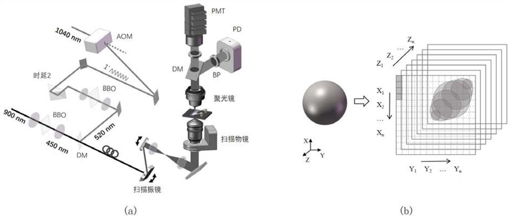
[0106] For stimulated Raman imaging, when the frequency difference Δω of the two laser beams matches the molecular vibration frequency, the molecular transition rate is excited and the Raman signal is stimulated and amplified. By changing the excitation wavelength or spectral focusing method, etc., the imaging of different molecular bonds such as C-H bond and C=C bond can be realized. Taking the laser wavelength of 520nm & 450nm as an example, the spectral focusing technology can achieve 2800~3050cm -1 range of chemical imaging, including C-H 2 Molecular bond, C-H 3 Molecular bond, =C-H molecular bond.

[0107] Figure 8 (a) shows images of cultured osteosarcoma cells obtained by conventional near-infrared laser-based stimulated Raman microscopy. Figure 8 (b) shows the image of osteosarcoma cells obtained by the visible laser-based stimulated Raman microscope of the present invention, and the image has been...

Embodiment 2

[0108] Example 2: In situ three-dimensional hyperspectral imaging of unlabeled cells and tissues

[0109] Based on nonlinear effects, stimulated Raman microscopy has natural optical section imaging capabilities. After collecting one XY plane through the galvanometer scanning, the Z-axis height of the objective lens is adjusted synchronously to image the next plane. Figure 9 (a) is the cross-section display at different heights in the high-resolution 3D image of human cervical cancer cells (HeLa cells). The three-dimensional structure of cell shape, nuclear membrane and nucleolus is clearly presented. Figure 9 (b) is a three-dimensional image of brain tissue, and the invention has the ability of direct tissue imaging.

[0110] The difference between the second embodiment and the first embodiment lies in the application of the three-dimensional imaging technology.

Embodiment 3

[0111] Example 3: Large-scale high-resolution hyperspectral stimulated Raman microscopic imaging of brain tissue

[0112] High-resolution imaging of intact tissue is very challenging for fluorescence microscopy due to the difficulty of eliminating out-of-focus fluorescence background, but this problem does not exist for stimulated Raman microscopy. Figure 10 Imaging results of unprocessed high-resolution C57 mouse brain tissue.

[0113] Figure 10 (b) with Figure 10 (c) Hyperspectral high-resolution SRS imaging of mouse brain tissue slices. Based on the improved resolution, the invention enables chemical analysis of samples in Figure 10 (d), the imaged strip area covers the cortex, hippocampus and other brain regions.

[0114] In visible light stimulated Raman imaging, the axon appears as a closed circle with a diameter of about 1 micron in the cross section, and two parallel curves in the longitudinal section. Such as Figure 10 (d), The neural network is composed of ...


PUM

PropertyMeasurementUnit
wavelengthaaaaaaaaaa

Abstract

The invention discloses a super-resolution stimulated Raman microimaging method and device for realizing near resonance enhancement. The method comprises the following steps: (1) obtaining a first pulse light source lambda 1 and a second pulse light source lambda 2 which are synchronous and phase-locked; (2) carrying out frequency multiplication processing on the first pulse light source lambda 1,and carrying out frequency multiplication processing on the second pulse light source lambda 2 subjected to intensity modulation, so as to halve the wavelength of the second pulse light source lambda2 and obtain third pulse laser lambda 3 and fourth pulse laser lambda 4; (3) carrying out delay processing on the fourth pulse laser lambda4 to realize time domain matching with the third pulse laserlambda3, and carrying out beam combination processing on the third pulse laser lambda3 and the fourth pulse laser lambda4 which are matched in time domain to realize complete matching of a spatial domain; (4) simultaneously coupling the combined third pulse laser lambda 3 and fourth pulse laser lambda 4 into a single-mode polarization maintaining optical fiber; and (5) processing an optical signal generated after the light transmitted by the single-mode polarization maintaining optical fiber and the sample act, and obtaining a microscopic image. According to the invention, the spatial resolution is improved.

Description

technical field [0001] The invention belongs to the technical field of biomedicine and optical imaging, and more specifically relates to a super-resolution stimulated Raman microscopic imaging method and device for realizing near-resonance enhancement. Background technique [0002] Optical microscopy imaging has become a cornerstone of biomedical research, enabling the detection and function of cellular structures with minimal invasiveness to the cell or tissue of interest. At present, the development of fluorescence microscopy is in full swing. Microscopic imaging technology based on fluorescent labels can indirectly realize specific super-resolution imaging of various biomolecules including proteins and nucleic acids through the manipulation of exogenous marker molecules. However, too large fluorescent protein label will affect the activity of biomolecules, too broad fluorescent spectrum limits the development of multicolor labeling, and the bleaching characteristics of fl...

Claims


Application Information

Patent Timeline
Patent Type & Authority Applications(China)
IPC IPC(8): G01N21/65
CPCG01N21/65G01N2021/655
Inventor 王平毕亚丽杨光杨驰
Owner HUAZHONG UNIV OF SCI & TECH
Why Eureka
Try Eureka