Automatic extraction method of cell components

An automatic extraction and cell technology, which is applied to the device for automatic separation of protein and subcellular structure in animal and plant cells, and in the field of multi-sample batch processing, can solve the problems of subcellular structure destruction, excessive homogenization, and manual operation. Achieve the effect of uniform cell crushing, avoiding differences, and saving manpower

Pending Publication Date: 2019-12-31
CHONGQING UNIV
View PDF0 Cites 2 Cited by
  • Summary
  • Abstract
  • Description
  • Claims
  • Application Information

AI Technical Summary

Problems solved by technology

[0003] The traditional method of separating cell proteins is to use RIPA lysate (the main components are 50mM Tris (pH 7.4), 150mM NaCl, 1% Triton X-100, 1% sodium deoxycholate, 0.1% SDS) to lyse the cells, and the protein dissolves until it is lysed. centrifuge at high speed in a centrifuge, and discard the insoluble part. The supernatant is the separated protein solution. The protein that can be separated by this method must be able to be dissolved by the cell lysate. In fact, about 30% of the protein is insoluble in For RIPA lysate, this part of the protein exists in the centrifuge pellet and is discarded, resulting in the loss of the target protein or inaccurate protein quantification
The classic method of separating subcellular structures is to separate the cells by lysing and homogenizing them with differential centrifugation combined with density gradient centrifugation. This method requires the configuration of a replicated density gradient medium and an ultra-high-speed centrifuge, so the experimental skills of researchers High requirements for experimental equipment and cumbersome operation, large sample size, long time-consuming and poor repeatability
In recent years, some subcellular separation methods based on antibody affinity have emerged. This method uses the specificity of immunoaffinity, so it is necessary to prepare a special antibody for each structure. Although the specificity has been improved, the price of the antibody is high, and the experimental cost is high. too high
At the same time, due to the extensive use of RIPA in the traditional method for cell lysis and protein dissolution, although the anionic surfactants such as SDS contained in it can efficiently lyse, it will also cause protein loss and denaturation
The traditional method of physical cell disruption is ultrasonic or glass homogenizer homogenization. Ultrasonic method is not suitable for scientific research with small sample volume, and the thermal effect of ultrasonic will lead to protein denaturation; the homogenization effect is artificially random and prone to homogenization effect Insufficient or excessive homogenization will destroy the subcellular structure and affect downstream separation
At the same time, manual homogenization is difficult to ensure the low temperature environment throughout the process, resulting in protein denaturation or degradation
The genomic nucleic acid released at the same time will make the cell homogenate extremely viscous, which will affect further separation experiments
[0004] In the traditional method, low-temperature high-speed centrifugation or even ultra-high-speed centrifugation is a necessary separation method. The centrifugation operation requires manual completion of sample placement and recovery, which is not suitable for integration with other operations to achieve automation. Therefore, there is currently no automatic cell protein extraction device.
Under the requirement of separation and extraction of multiple samples, the manual operation is intensive and the risk of batch difference increases, which will inevitably affect the scientificity of the experimental results. Therefore, it is necessary to carry out technological innovation and develop an automatic extraction device.

Method used


Image

  • Automatic extraction method of cell components
  • Automatic extraction method of cell components
  • Automatic extraction method of cell components

Examples


Embodiment 1

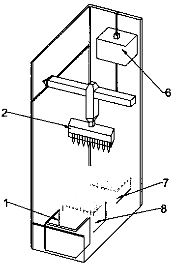
[0041] Such as Figure 1~3 As shown, a method for automatic extraction of cell components, the instrument used in the method includes an automatic extraction device for cell components, and the reagents used include sensitizers and a series of cell protein extracts;

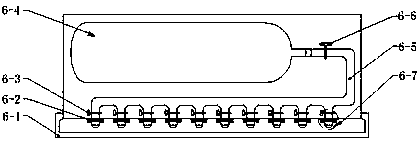
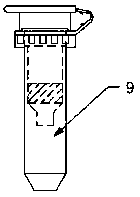
[0042] The automatic cell component extraction device includes: a pipetting system, a filtration system, a cooling system, and an automatic control system. The cooling system provides a low temperature of 0-4°C for the pipetting system and / or the filtration system, wherein the automatic control system is respectively Connect with pipetting system, filter system, cooling system; Described filter system includes: include filter tube placement box 7, ventilation pressure supply system 6, filter tube 9, wherein filter tube 9 includes filter column 9-3, collection tube 9- 1. The filter column 9-3 includes a filter element 9-2. The filter element 9-2 breaks up the cells with cell membrane sensitization and filters out ...

Embodiment 2

[0056] Example 2 Extraction of human liver cancer cell total protein

[0057] Collect 5×10 per well 6 HepG2 cells were washed with cold PBS. Add about 200 μL of cell sensitization solution to each well, add it to the cell sample, mix well, and add it to the sample tank. Put the corresponding filter tube in the hole of the filter tube placement box. The filter element is made of sintered polytetrafluoroethylene particles with an average particle size of 50 μm, a porosity of 45%, and an average pore size of 30 μm. Select the gas injection port and the sample tank.

[0058] Start the automatic extraction program, and after 5 minutes of low-temperature incubation at 0-4°C, the row-type pipette moves to the top of the pipette tip placement box, and moves down to insert the pipette tip. Move to the sampling tank, draw 200 μL of cell suspension, then move to the filter tube placement box, add to the filter tube, and return the pipette to its original position. The ventilation an...

Embodiment 3

[0060] Example 3: Extraction of human liver cancer membrane protein

[0061] Collect 5×10 per well 6 HepG2 cells were washed with cold PBS. Add about 200 μL of membrane protein sensitization solution to each well, add it to the cell sample, mix well, and add it to the sample tank. Put the corresponding filter tube in the hole of the filter tube placement box. Select the air nozzle and the sample tank.

[0062] Start the automatic extraction program, and after 10 minutes of low-temperature incubation at 0-4°C, the row-type pipette moves to the top of the pipette tip placement box, and moves down to insert the tip. Move to the sampling tank, draw 200 μl of the mixed sample, then move to the filter tube placement box, add the sample to the filter tube, and return the pipette to its original position. The ventilation and pressure supply system moves down, the air nozzle and the filter column are closed, and the air nozzle ejects pressurized gas, so that the cell suspension tre...


PUM

PropertyMeasurementUnit
Particle sizeaaaaaaaaaa
Apertureaaaaaaaaaa
Thicknessaaaaaaaaaa

Abstract

The present invention belongs to the technical field of biology and discloses an automatic extraction method of cell components. The automatic extraction method comprises the following steps: (1) after mixing a cell sample and a sensitizer, adding the mixture into a sample adding groove for incubation; (2) after incubation for 5-10 min, adding the sample into a filter tube by a pipettor; (3) conducting air injection pressurization and crushing cells under action of shearing force and inertial impact; and (4) if total cellular protein or proteins with a special distribution of a certain subcellular structure need to be collected, pretreating the product obtained in the step (3) and then adding a series of cell component extracts into the filtered product for extraction. The method is different from the traditional method which completely depends on chemical crushing of lysate or grinding and homogenizing method for crushing, so that cell crushing is more uniform, efficiency is high, andprotein damage is small. The method is suitable for processing with small number of samples and batch processing under condition of large total number of the samples, has small requirements on singlesample amount, can effectively save manpower and avoids difference of manual operation.

Description

technical field [0001] The invention relates to the field of biotechnology, in particular to a device and method for automatically separating proteins and subcellular structures in animal and plant cells, which is suitable for multi-sample batch processing. Background technique [0002] In the post-genome era, the research of functional omics is the main content of life science research, and protein is the main executor of life activities, so it is the focus of research. By analyzing the similarities and differences of proteins in tissues, cells or subcellular structures under different physiological / pathological conditions, it can reveal complex life phenomena and find targets for disease diagnosis and treatment. Due to the large variety of proteins in a cell (more than 100,000), the dynamic distribution range of different proteins is extremely wide (from 10 2 to 10 9 molecules), and their properties are different, these factors bring difficulties to the separation and an...

Claims


Application Information

Patent Timeline
IPC IPC(8): C07K1/34C12N5/04C12M1/33C12M1/12C12M1/04
CPCC07K1/34C12N5/04C12M23/02C12M33/04C12M45/02C12M47/04
Inventor 张兵兵李樑杨云澜周雪毅
Owner CHONGQING UNIV
Why Eureka
Try Eureka