
Plasma biomarker tool for the diagnosis of liver cancer comprising liver carboxylesterase 1 and liver cancer screening method

a liver cancer and carboxylesterase technology, applied in biochemistry equipment and processes, instruments, material analysis, etc., can solve the problems of poor sensitivity and/or specificity, low survival rate after diagnosis, and difficulty in early-stage diagnosis of liver cancer, so as to effectively detect the presence of hcc

Active Publication Date: 2012-06-12
IND ACADEMIC COOP FOUND YONSEI UNIV
View PDF5 Cites 8 Cited by
  • Summary
  • Abstract
  • Description
  • Claims
  • Application Information

AI Technical Summary

Benefits of technology

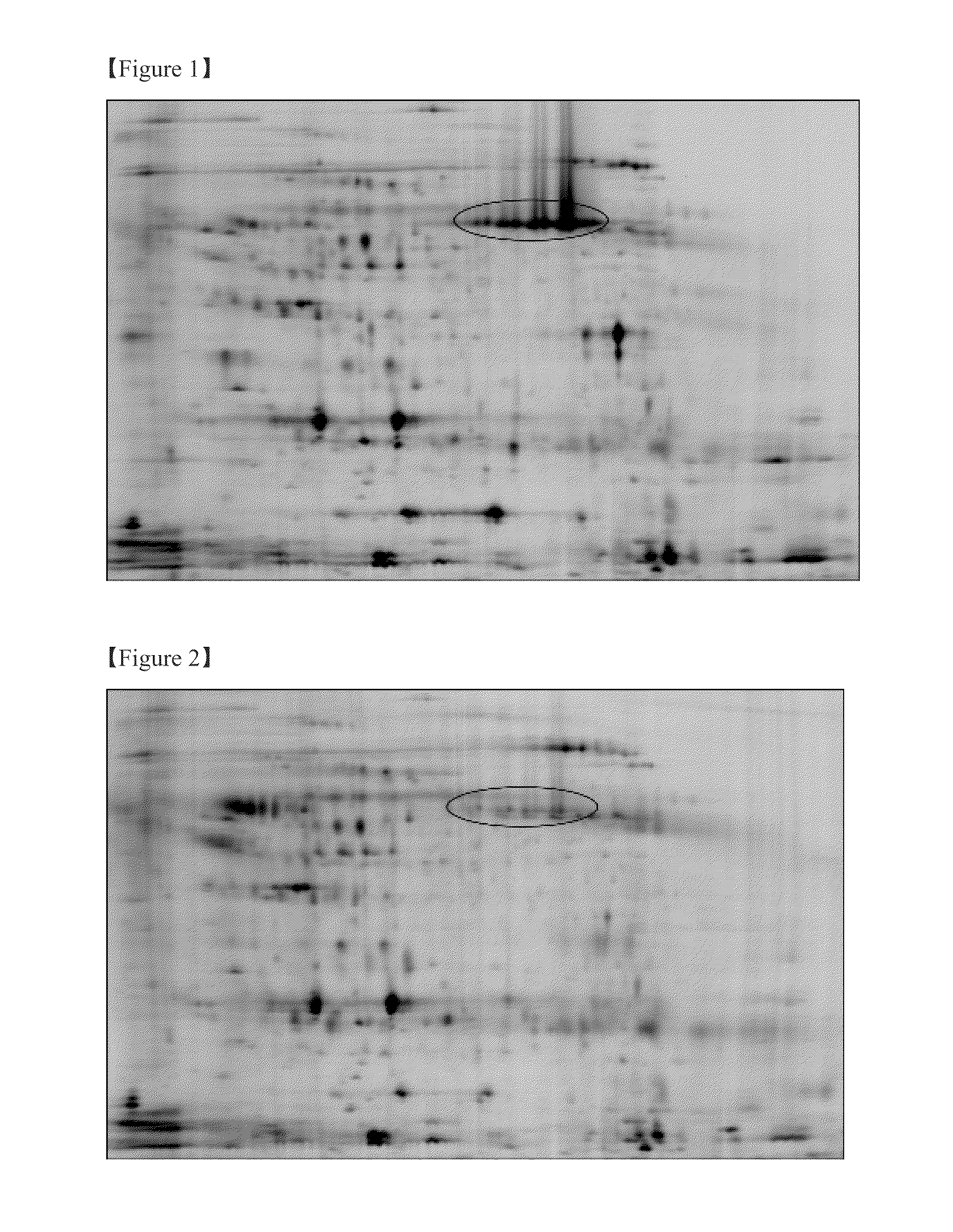
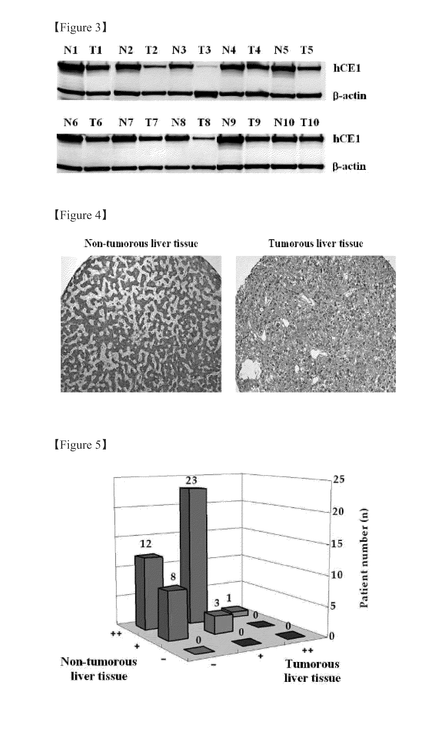
The present invention is about a new biomarker called human liver carboxylesterase 1 (hCE1) that is specifically associated with liver cancer (HCC). The inventors discovered hCE1 in the plasma of patients with HCC and used proteomics techniques to confirm its presence. The invention provides a new method for screening for HCC using patient plasma and a routine detection method. The hCE1 biomarker is an N-linked glycoprotein that binds lectin and is differentially expressed in HCC tissues and plasma. The invention can effectively detect the presence of HCC and discriminate between patients with HCC and healthy volunteers through quantitative measurement of hCE1 in plasma.

Problems solved by technology

It is an especially major problem among Asian and African populations.
Although HCC is the subject of continuing investigation and its symptoms are well known, early-stage diagnosis of this disease remains difficult and the survival rate after diagnosis is very low (3%-5%).
Although these biomarkers are useful for the detection of HCC, they suffer from poor sensitivity and / or specificity.
However, there are no reports on the direct detection of secreted hCE1 protein in human bodily fluid (i.e., plasma).

Method used


Image

  • Plasma biomarker tool for the diagnosis of liver cancer comprising liver carboxylesterase 1 and liver cancer screening method
  • Plasma biomarker tool for the diagnosis of liver cancer comprising liver carboxylesterase 1 and liver cancer screening method
  • Plasma biomarker tool for the diagnosis of liver cancer comprising liver carboxylesterase 1 and liver cancer screening method

Examples


embodiment 1

[0041] Collection of Clinical Tissues and Plasma

[0042]The non-tumorous and tumorous liver tissue from patients with HCC were obtained along with pathological individual information from the Department of Pathology, Yonsei University College of Medicine, Seoul, Korea. The following tests were used to assess the appropriateness of plasma from healthy volunteers to serve as a negative control: HIV-1 and HIV-2 antibodies derived from HIV, which is a liver-cancer indicating standard test element; HIV-1 antigen; hepatitis B surface antigen; hepatitis B core antigen; hepatitis C virus; T-Cell Leukemia Virus (HTLV-I / II) antigen; and Treponema pallidum. Plasma was obtained according to a standardized sample separation method formally adopted by the Human Proteome Organization (HUPO).

[0043]Each blood sample (4 ml) from man or woman was maintained in a tube containing di-potassium ethylenediamine tetraacetic acid (K2EDTA) for 30 minutes at room temperature, followed by centrifugation for 15 mi...

embodiment 2

[0044] Separation of Glycoproteins in Non-Tumorous and Tumorous Liver Tissue

[0045]A 100 mg sample of each tissue was homogenized in RIPA buffer (50 mM Tris, 150 mM NaCl, 1% Nonidet P-40, 0.25% sodium deoxycholate, pH 7.4) at 4° C. Glycoproteins were separated by lectin-affinity chromatography using a mixture of five different agarose-bound lectins: concanavalin A, wheat germ agglutinin, Jacalin, Sambucus nigra, and Aleuria aurantia. The lectin mixture, with binding specificities for glycoproteins with different sugar compositions, was packed into a 2 ml PD-10 column (Pierce). Glycoprotein-containing samples were applied to the column in binding buffer (20 mM Tris, 1 mM MnCl2, 1 mM CaCl2, 0.15 M NaCl, pH 7.4) and allowed to interact with the column for 30 minutes.

[0046]The bound glycoproteins were eluting with a buffer (0.2 M methyl-D-mannopyroside, 0.2 M methyl-D-glucopyroside, 0.2 M N-acetylglucosamine, 0.2 M galactose, 0.1 M lactose, 0.1 M fucose, 20 mM Tris, 0.5 M NaCl, pH 7.0) t...

embodiment 7

[0060] Validation of HCC Biomarker Protein Secretion Level in Human Plasma by Immunoprecipitation and Western Blot Analysis

[0061]Dynabead MyOne™ Tosylactivated (Invitrogen) magnetic beads were coated with anti-hCE1 antibody as described by the manufacturer. Briefly, magnetic beads (10 mg) and anti-hCE1 antibody (400 μg) were mixed and incubated for 16 hours in binding buffer (0.1 M sodium borate, 1 M ammonium sulfate, pH 9.5) at 37° C., and then blocked with TBS-T / 5% skim milk for an additional 16 hours. hCE1 was immunoprecipitated from the plasma of healthy volunteers and patients with HCC by transferring antibody-coated beads (500 μg) to l ml tubes containing 8 mg plasma and incubating for 2 hours.

[0062]Antibody-bound proteins were eluted by adjusting the pH of the TBS-T buffer to pH 2. Differences in hCE1 levels between the plasmas of healthy volunteers and patients with HCC were determined by Western blot analysis using a primary anti-hCE1 antibody (Abcam; 1:1000) and a secondar...


PUM

PropertyMeasurementUnit
pHaaaaaaaaaa
pHaaaaaaaaaa
volumeaaaaaaaaaa

Abstract

The present invention relates to a plasma biomarker for diagnosing hepatocellular carcinoma (HCC), in particular to the discovery of a protein in plasma using 2-D fluorescence differential gel electrophoresis (2-D DIGE), immunoprecipitation and Nano-liquid chromatography mass spectrometry (Nano-LC-MS / MS) system that was unknown on the basis of conventional techniques. By demonstrating the presence of liver carboxylesterase 1 (hCE1) in human plasma and confirming that its secretion level is higher in patients with HCC than in healthy volunteers, this invention may be used as a screening method to diagnose HCC at an early stage.

Description

TECHNICAL FIELD[0001]The present invention pertains to a plasma biomarker (carboxylesterase 1) for the diagnosis of liver cancer (hepatocellular carcinoma; HCC), and the associated screening method for determining differential biomarker expression between healthy volunteers and patients with HCC employing 2-dimensional fluorescence gel electrophoresis (2-D DIGE), immunoprecipitation and nano liquid chromatography mass spectrometry (Nano-LC-MS / MS) system. The amount of biomarker protein secreted into plasma is measured and used as an indicator of the presence of HCC.BACKGROUND ART[0002]Hepatocellular carcinoma (HCC) is the fifth most common cancer worldwide and has the fourth highest mortality rate. It is an especially major problem among Asian and African populations. Unlikely patients with other cancers, such as lung cancer and breast cancers, more than 95% of HCC patients die within five years of being diagnosed with HCC.[0003]Although HCC is the subject of continuing investigatio...

Claims


Application Information

Patent Timeline
Patent Type & Authority Patents(United States)
IPC IPC(8): G01N33/573C12Q1/44
CPCG01N33/57438G01N2333/918C12Q1/44G01N33/574G01N33/576B82Y5/00
Inventor PAIK, YOUNG-KINA, KEUN
Owner IND ACADEMIC COOP FOUND YONSEI UNIV
Learn More