
Microparticle-based diagnostic methods

a technology of microparticles and diagnostic methods, applied in the direction of instruments, biochemical equipment and processes, material analysis, etc., can solve the problems of oral cancer and its lack of early detection, low detection accuracy, and low long-term survival rate, so as to facilitate and accurately detect the surface changes

Inactive Publication Date: 2008-06-24
UNIV OF FLORIDA RES FOUNDATION INC
View PDF14 Cites 10 Cited by
  • Summary
  • Abstract
  • Description
  • Claims
  • Application Information

AI Technical Summary

Benefits of technology

The present invention provides materials and methods for evaluating surfaces for low frequency changes caused by foreign body growth or settlement. This is done by using specifically designed microspheres that can easily and accurately detect these changes. The microspheres are modified to interact with surface markers associated with changes that have occurred, are occurring, or are impending. The microspheres can be incorporated into a solution for use as mouthwash, which can be collected and analyzed for a decrease in microsphere concentration to indicate oral cancer. The invention also provides a composition of microspheres in a carrier that can be used for detecting oral cancers. A kit is also provided for an easy to use, inexpensive home or clinical test for the detection of oral cancers.

Problems solved by technology

Without early detection and subsequent remediation, these surface modifications can lead to deleterious health, economic, environmental, and structural damage.
A serious global health issue concerns oral cancer and its lack of early detection.
In fact, long term survival rates have not improved significantly in the last 20 years.
Early detection is hampered because pain and other symptoms are normally not present during early stages.
In addition, successful treatment of HNSCC frequently involves radiation therapy and leaves the patient with significant deficits in quality of life factors including decreased saliva production.
Surgical removal of deeper lesions, which result from invasion initiated at the surface, also can be very traumatic (e.g., removal of large parts of the tongue or jaw).
Unfortunately, no effective and easily administered screening test is currently available.
Currently, early detection is difficult since early lesions are small, requiring a careful physical examination to detect.
Lighting is difficult, since head mirrors, lamps and penlights lack sufficient intensity and color balance to detect the most common pre-cancerous lesions (Mashberg, S. et al., “Early Diagnosis of Asymptomatic Oral and Oropharyngeal Squamous Cancers,” CA Cancer J. Clin. 45, 328-51 (1995)).
However, toluidine also stains non-malignant lesions, such as ulcers, requiring additional examination after a period of time sufficient for healing to take place.
Hence, folate is required in rapidly growing cell populations, and more rapid uptake is required since it cannot be synthesized by the cell.

Method used


Image

  • Microparticle-based diagnostic methods
  • Microparticle-based diagnostic methods

Examples


example 1

Home Test Kit

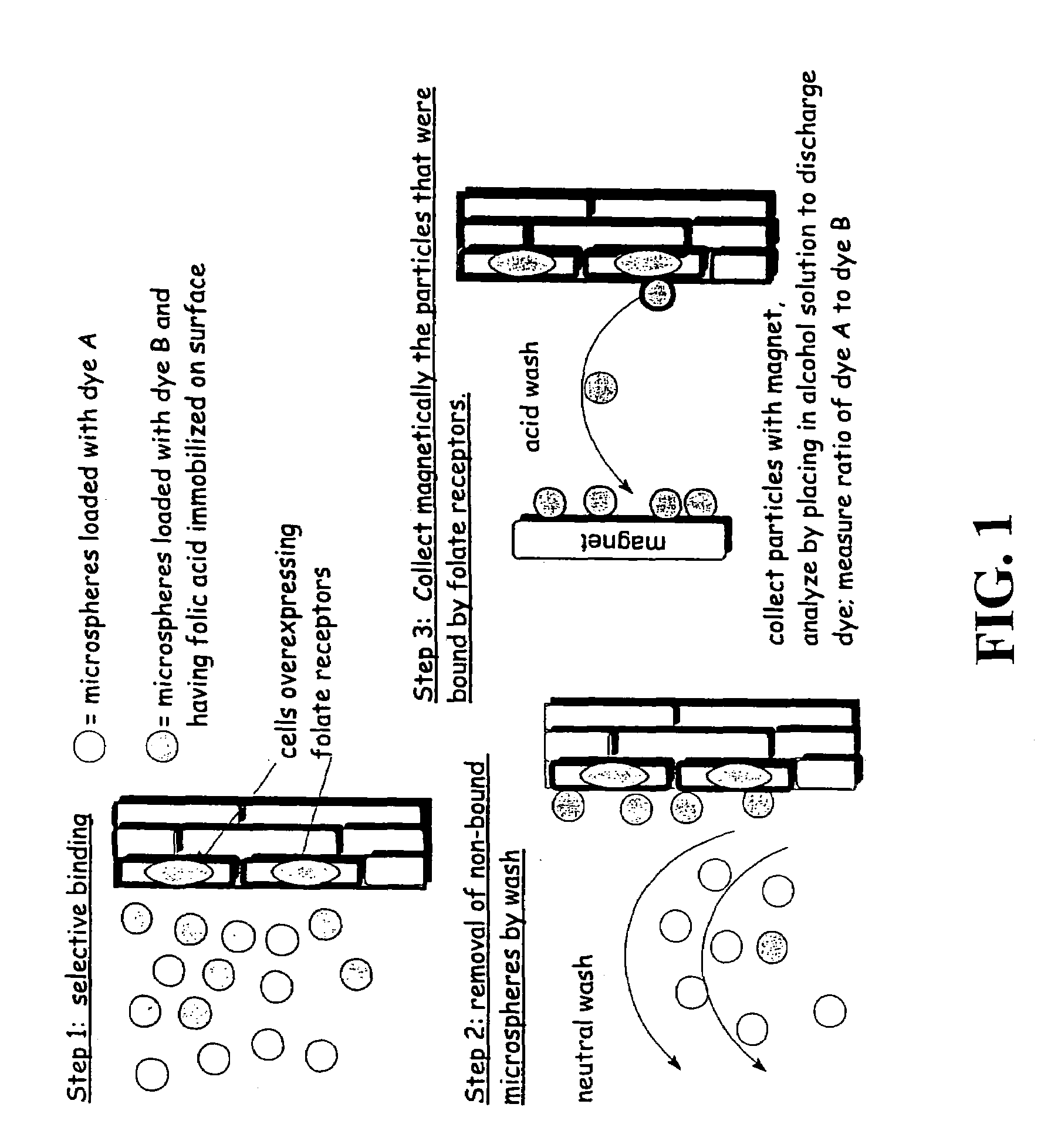
[0062]One embodiment of the present invention involves a kit for a home based test for oral cancer. Optionally, the kit can be administered in a clinical setting such as a physician's or a dentist's office. The kit contains two marked vials each holding a mouthwash—one mouthwash with the first set of folate-modified and unmodified microspheres and one mouthwash with the just an acidic solution—and an empty collection container. The accompanying directions instruct the consumer or test administrator to rinse the oral cavity with the mouthwash containing the first set of microspheres for a sufficient amount of time to ensure complete contact between the mucosal surface and the mouthwash followed by disposal of the expectorated mouthwash. Subsequently, the mucosa should be exposed to the second mouthwash, and the resulting expectorate stored in the collection container and sealed. The collection container can then be submitted to a qualified laboratory for analysis of the ...

example 2

Method of Use

[0063]Microspheres prepared in accordance with the subject invention can be incorporated into a liquid suspension, which can be used as a, for example, a mouthwash delivery system for oral mucosa. Preferably, the properties of this suspension include the ability to prevent significant settling of the heavier particles (less that 25%) over 30 seconds after stirring, but the compositions can simply use microspheres in pH 7.4 phosphate buffered saline (PBS) or culture media.

[0064]In one example, a few cubic millimeters of biopsy tissue is placed in a polyethylene 5 mL centrifuge tube, containing PBS, and a mixture of the two microsphere populations (folate surface with dye “A” and non-folate surface with dye “B”). The tubes are gently shaken for one minute in an automated shaker, and then removed for extraction of microspheres by filtering the PBS mixture through filters with porosity greater than 100 micrometers, or simply removing the tissue with forceps. This removes an...

example 3

Attachment of Folic Acid

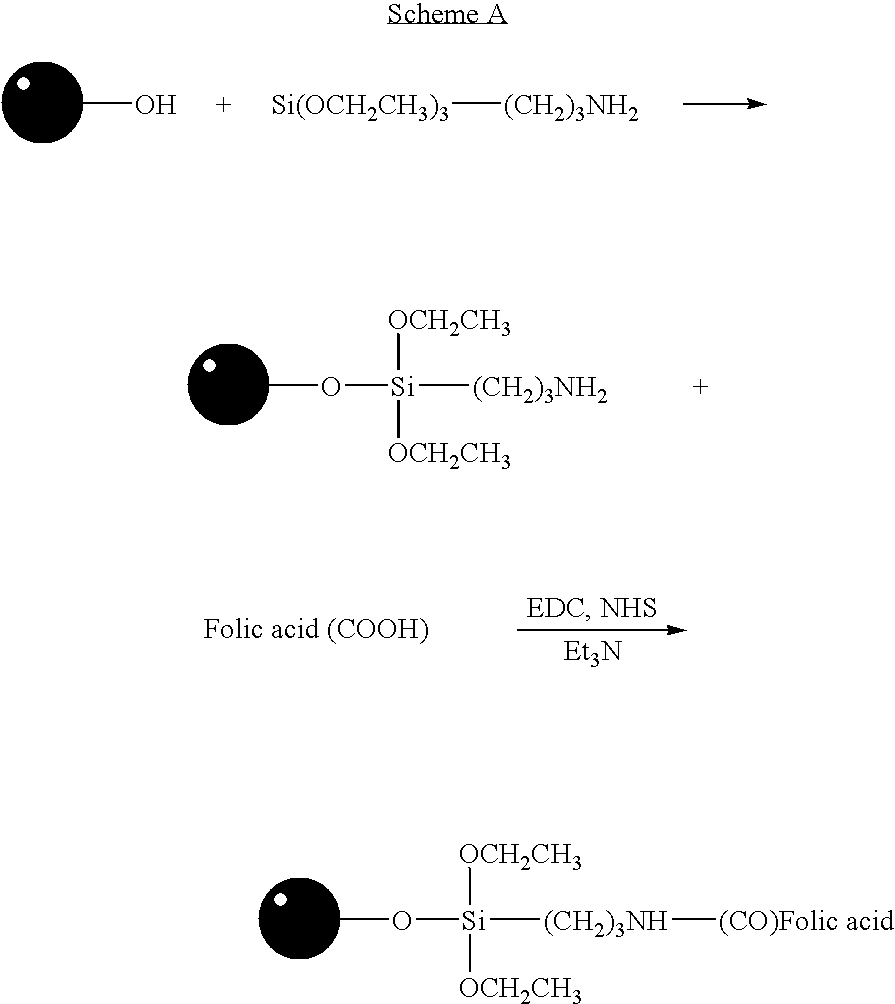
[0065]Folic acid can be attached to the surface of the microspheres by means of a technique described by Zhang et al. (Zhang, Y, N. Kohler, & M. Zhang “Surface modification of superparamagnetic magnetite nanoparticles and their intracellular uptake.”Biomaterials, 2002 April; 23 (7): 1553-61.) The mechanism for attachment of folic acid onto microspheres is shown in Scheme A.

[0066]

[0067]Dried particles are dispersed in 3 mM (3-aminopropyl)-trimethoxysilane in 5 ml toluene, vortexed and sonicated, then incubated at 60° C. for 4 hours. The product is centrifuged, and the precipitate is placed in hexane and sonicated for 10 minutes, then washed with hexane and ethanol. A solution is made of 1.5 ml each of 15 mM NHS (N-hydroxy succinimide) and 75 mM EDC (1-ethyl-3-(3-dimethylamino-propyl) carbodiimide) and 1 ml 10 mM folic acid in DMSO, to which the precipitate is added, and ethylene triamine used as a catalyst. The pH is adjusted to 9 and the mixture incubated at ...


PUM

PropertyMeasurementUnit
sizeaaaaaaaaaa
particle sizeaaaaaaaaaa

Abstract

The present invention concerns novel materials and methods to detect low frequency surface changes utilizing modified microspheres. Upon exposing the surface to the modified microspheres of the subject invention, the microspheres selectively adhere to any targeted surface marker.

Description

CROSS-REFERENCE TO RELATED APPLICATION[0001]This application claims the benefit of provisional patent application Ser. No. 60 / 532,167, filed Dec. 23, 2003, which is hereby incorporated by reference in its entirety.FIELD OF THE INVENTION[0002]The present invention relates to methods for detecting evidence of modifications on surfaces. In a specific embodiment, the subject invention provides diagnostic methods for disease.BACKGROUND OF THE INVENTION[0003]Low frequency changes on the surfaces of substrates are possible indications of contamination and / or abnormalities. Examples include lead in painted surfaces; scaling inside pipes; mold on carpeting, walls, or heating and cooling ducts; bacteria on skin; and cancer cell receptors. Without early detection and subsequent remediation, these surface modifications can lead to deleterious health, economic, environmental, and structural damage. An area wherein surface modification detection is particularly needed involves the early diagnosis...

Claims


Application Information

Patent Timeline
Patent Type & Authority Patents(United States)
IPC IPC(8): G01N33/574A61K9/14A61K49/00G01N33/543G01N33/58
CPCG01N33/54313G01N33/57492G01N33/585
Inventor BATICH, CHRISTOPHER D.
Owner UNIV OF FLORIDA RES FOUNDATION INC
Learn More