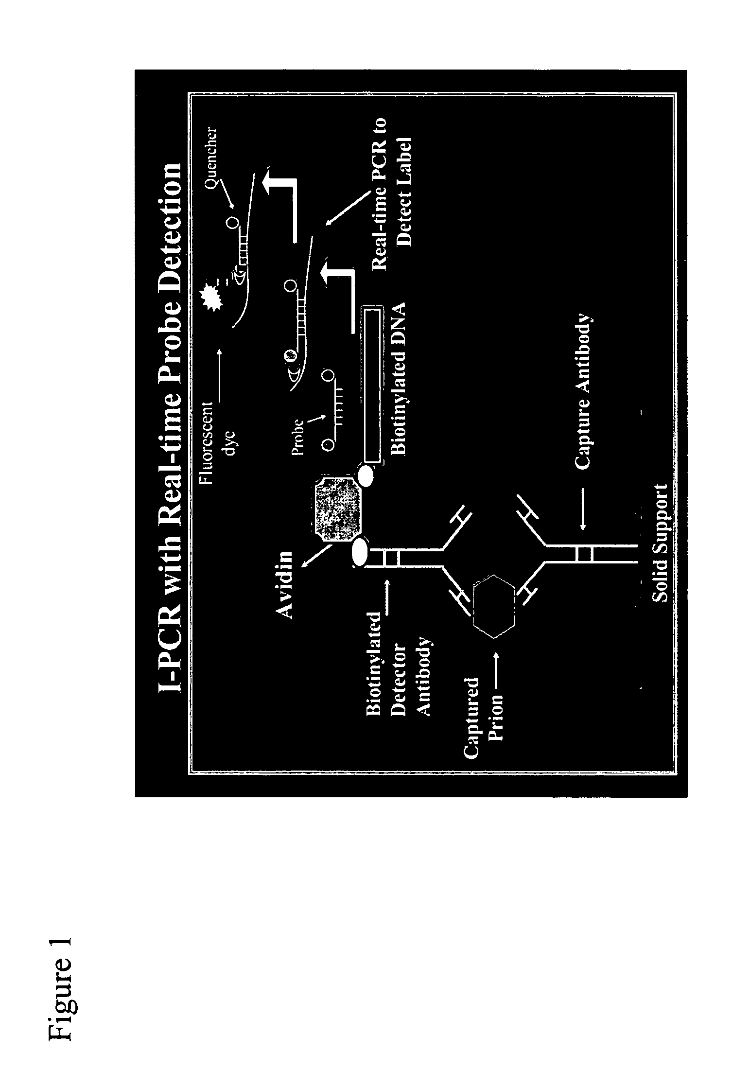
Immuno-PCR method for the detection of a biomolecule in a test sample
a biomolecule and test sample technology, applied in the field of immunopcr method for the detection of a biomolecule in a test sample, can solve the problems of economically devastating food bans and concerns for the safety of blood supply, difficult early disease diagnosis, and a large financial loss due to the ban on feed, so as to achieve simple and highly sensitive results
- Summary
- Abstract
- Description
- Claims
- Application Information
AI Technical Summary
Benefits of technology
Problems solved by technology
Method used
Image

Examples
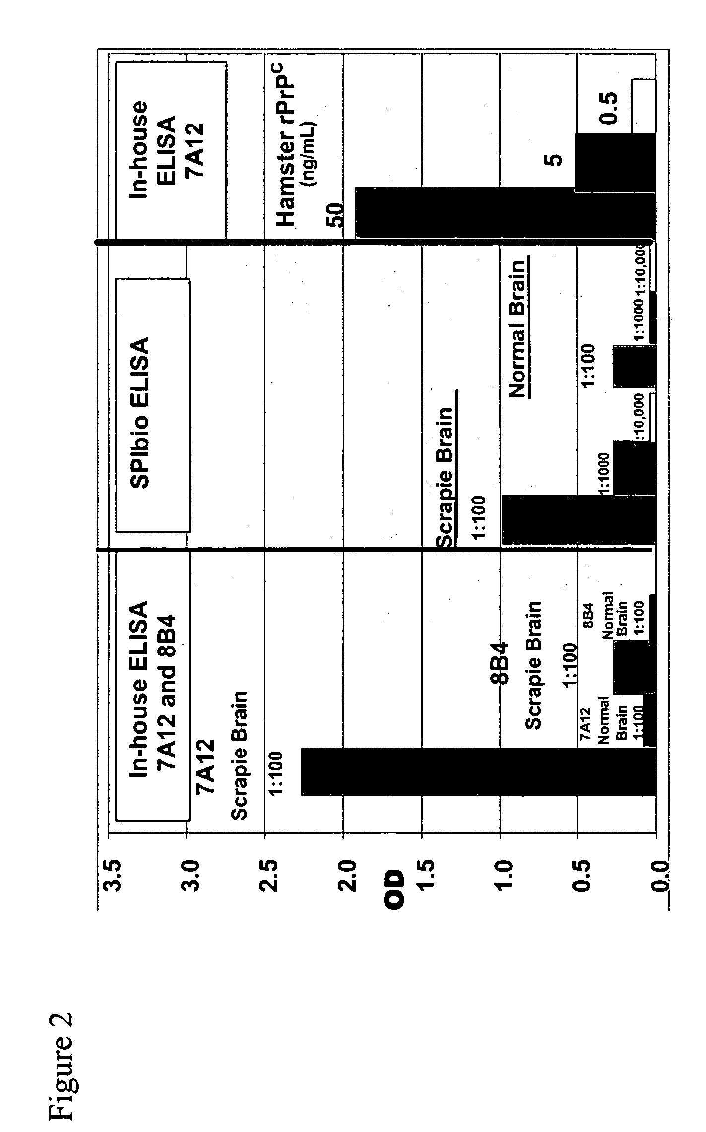
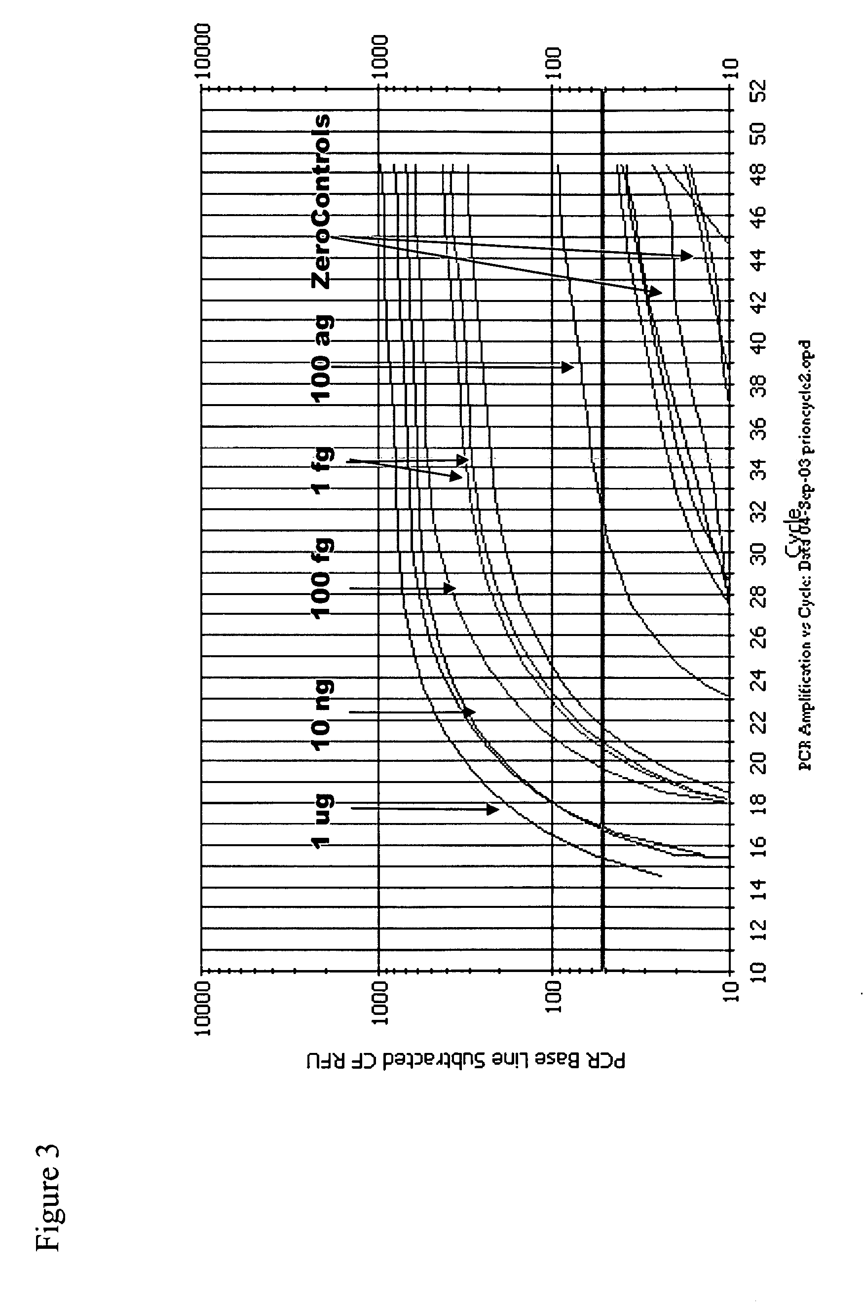
example 1
Protocol
[0260] TopYield stripwell plates (NalgeNunc Corp., Naperville, Ill.) were coated with anti-prion antibody 8B4 (from Dr. Man Sun Sy, Case Western Reserve, Cleveland, Ohio) (capture antibody) in bicarbonate buffer (pH 9.4) for 3 hr, RT, and then blocked with Stabilcoat (Surmodics: Eden Prairie, Minn.) for 1 hr, at room temperature (RT). Varying concentrations of recombinant hamster prion protein (Prionics AG, Schieren, Switzerland) diluted in 0.1% Triton-X / PBS buffer were added to wells for 2 hr, RT. Plates were washed 6 times and then incubated with the detector antibody, a biotinylated 3F4 antibody (Signet Pathology Systems Inc, Dedham Mass.) (2.5 ug / mL) diluted in diluent buffer (Zeptometrix Corp., Buffalo, N.Y.), for 1 hr, RT. Plates were washed 6 times and then incubated with streptavidin (10 ng / mL) diluted in Zeptometrix diluent buffer for 1 hr, RT. Plates were washed 6 times and then blocked for 30 min with DNA Blocking Reagent (Roche Diagnostics; Indianapolis, Ind.)....
example 2
Method for the Detection of a Biomolecule in a Test Sample
[0276] A. Sample Collection, Processing and Storage:
[0277] This method may be used with biological samples such as any human body fluid (e.g., whole blood, plasma, serum, saliva, neuronal tissues, and urine) or environmental samples such as water or soil.
[0278] All specimens used in this assay may be processed on the same day as collected or stored frozen at −20° C. or below until tested. Clear, non-hemolyzed plasma or serum specimens should be used whenever possible.
[0279] B. Test Protocol:
[0280] Note: All steps require optimization depending on the antigen or antibody.
[0281] 1. The solid support (e.g., microwells, microbeads, or membranes) is coated with antibody solution (5-10 ug / mL) for 8-16 hrs at RT.
[0282] 2. The solution is removed by aspiration and the blocking reagent is added for 1 hr at RT.
[0283] 3. The solution is removed by aspiration and the sample (10-100 uL) is added for 10 min to 1 hr at RT.
[0284] 4....
PUM
| Property | Measurement | Unit |
|---|---|---|
| Fraction | aaaaa | aaaaa |
| Mass | aaaaa | aaaaa |
| Concentration | aaaaa | aaaaa |
Abstract
Description
Claims
Application Information
 - R&D
- Intellectual Property
- Life Sciences
- Materials
- Tech Scout
- Unparalleled Data Quality
- Higher Quality Content
- 60% Fewer Hallucinations
Browse by: Latest US Patents, China's latest patents, Technical Efficacy Thesaurus, Application Domain, Technology Topic, Popular Technical Reports.
© 2025 PatSnap. All rights reserved.Legal|Privacy policy|Modern Slavery Act Transparency Statement|Sitemap|About US| Contact US: help@patsnap.com