
Method for preparing conductive composite nanofiber nervous tissue engineering scaffold based on graphene

A technology of composite nanofibers and nerve tissue, applied in the direction of fiber type, fiber processing, animal fiber, etc., to achieve the effect of good biocompatibility, improved biological activity, and high porosity

Inactive Publication Date: 2017-02-15
DONGHUA UNIV
View PDF10 Cites 20 Cited by
  • Summary
  • Abstract
  • Description
  • Claims
  • Application Information

AI Technical Summary

Problems solved by technology

At present, some scholars have made use of the excellent properties of graphene to prepare a variety of graphene and its derivatives reinforced three-dimensional composite scaffolds for bone tissue repair and regeneration, but using the excellent electrical conductivity of graphene to prepare conductive composite nanofibers for neural tissue engineering There are no related documents and patents for stents

Method used


Image

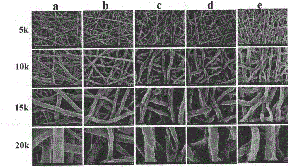
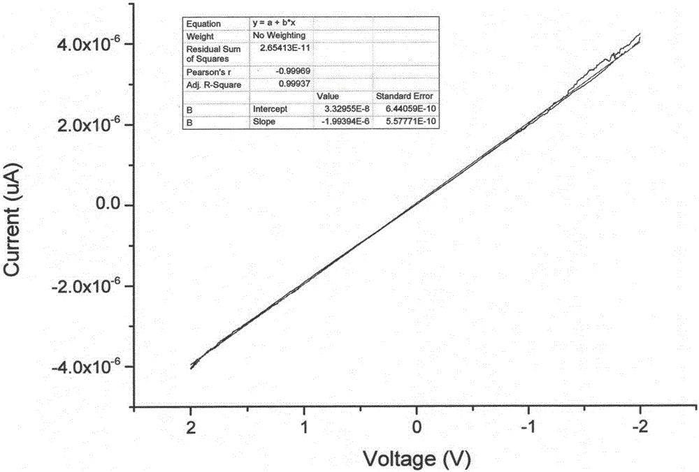
  • Method for preparing conductive composite nanofiber nervous tissue engineering scaffold based on graphene
  • Method for preparing conductive composite nanofiber nervous tissue engineering scaffold based on graphene
  • Method for preparing conductive composite nanofiber nervous tissue engineering scaffold based on graphene

Examples


Embodiment 1

[0044] A preparation method based on graphene conductive composite nanofiber neural tissue engineering scaffold, the specific steps are:

[0045] 1. Place the tussah silk at 95~100℃ 2 CO 3 The degumming solution was degummed 3 times, each time for 30 minutes, and the bath ratio was 1:50. After degumming, the tussah silk fibers are obtained and dried at 60°C. Put the tussah silk fibroin fiber in a saturated LiSCN solution at a bath ratio of 1:10, dissolve it for 70 minutes at 50°C±2°C, put the obtained tussah silk fibroin protein solution into a dialysis bag with a molecular weight cut-off of 8-10KDa, and use Dialyzed with deionized water for 3 days, and freeze-dried to obtain tussah silk fibroin.

[0046] 2. Weigh 0.25g of tussah silk fibroin protein, 0.75g of polylactic acid-polycaprolactone, dissolve in 10ml of hexafluoroisopropanol, and magnetically stir at a certain speed until completely dissolved, and the obtained concentration is 10% (w / v ) spinning solution. Condu...

Embodiment 2

[0049] A preparation method based on graphene conductive composite nanofiber neural tissue engineering scaffold, the specific steps are:

[0050] 1. Place the tussah silk at 95~100℃ 2 CO 3 The degumming solution was degummed 3 times, each time for 30 minutes, and the bath ratio was 1:50. After degumming, the tussah silk fibers are obtained and dried at 60°C. Put the tussah silk fibroin fiber in a saturated LiSCN solution at a bath ratio of 1:10, dissolve it for 70 minutes at 50°C±2°C, put the obtained tussah silk fibroin protein solution into a dialysis bag with a molecular weight cut-off of 8-10KDa, and use Dialyzed with deionized water for 3 days, and freeze-dried to obtain tussah silk fibroin.

[0051] 2. Weigh 0.25g of tussah silk fibroin protein, 0.75g of polylactic acid-polycaprolactone, dissolve in 10ml of hexafluoroisopropanol, and magnetically stir at a certain speed until completely dissolved, and the obtained concentration is 10% (w / v ) spinning solution. Condu...

Embodiment 3

[0054] A preparation method based on graphene conductive composite nanofiber neural tissue engineering scaffold, the specific steps are:

[0055] 1. Put the tussah silk in the environment of 95~100℃, 2 CO 3 The degumming solution was degummed 3 times, each time for 30 minutes, and the bath ratio was 1:50. After degumming, the tussah silk fibers are obtained and dried at 60°C. Put the tussah silk fibroin fiber in a saturated LiSCN solution at a bath ratio of 1:10, dissolve it for 70 minutes at 50°C±2°C, put the obtained tussah silk fibroin protein solution into a dialysis bag with a molecular weight cut-off of 8-10KDa, and use Dialyzed with deionized water for 3 days, and freeze-dried to obtain tussah silk fibroin.

[0056] 2. Weigh 0.25g of tussah silk fibroin protein, 0.75g of polylactic acid-polycaprolactone, dissolve in 10ml of hexafluoroisopropanol, and magnetically stir at a certain speed until completely dissolved, and the obtained concentration is 10% (w / v ) spinnin...


PUM

PropertyMeasurementUnit
concentrationaaaaaaaaaa

Abstract

The invention provides a method for preparing a conductive composite nanofiber nervous tissue engineering scaffold based on graphene. The method includes the following steps: a step 1, dissolving tussur silk fibroin and poly (lactic acid)-poly caprolactone in a solvent with stirring till complete dissolution of the tussur silk fibroin and the poly (lactic acid)-poly caprolactone so as to acquiring a spinning solution; a step 2, performing electrostatic spinning on the spinning solution obtained from the step 1 so as to acquire a nanofiber membrane, performing steam fumigation treatment by using ethyl alcohol, and performing drying so as to acquire tussur silk fibroin / poly (lactic acid)-poly caprolactone composite nanofibers; and a step 3, dipping the tussur silk fibroin / poly (lactic acid)-poly caprolactone composite nanofiber scaffold material obtained from the step 2 in a graphene oxide dispersion liquid, taking out the tussur silk fibroin / poly (lactic acid)-poly caprolactone composite nanofiber scaffold material, cleaning the tussur silk fibroin / poly (lactic acid)-poly caprolactone composite nanofiber scaffold material, soaking the tussur silk fibroin / poly (lactic acid)-poly caprolactone composite nanofiber scaffold material in an ascorbic acid solution, taking out the tussur silk fibroin / poly (lactic acid)-poly caprolactone composite nanofiber scaffold material, and cleaning the tussur silk fibroin / poly (lactic acid)-poly caprolactone composite nanofiber scaffold material so as to acquiring the conductive composite nanofiber nervous tissue engineering scaffold based on the graphene. The method is simple to operate, is excellent in repeatability, and can provide a new thought for nerve defect repairing.

Description

technical field [0001] The invention relates to a preparation method of a nerve tissue engineering scaffold based on graphene conductive composite nanofibers, and belongs to the field of biomedical materials related to nerve tissue engineering repair. Background technique [0002] In recent years, various nerve tissue defects have brought great distress and pain to patients. The regeneration and repair of nerve tissue has always been a hot spot in international research, and technologies such as autologous nerve transplantation, gene therapy, growth factor induction and tissue engineering can be used to treat and repair nerve defects. Among them, the development of tissue engineering and regenerative medicine has provided effective treatment means for reconstructing or repairing nerve tissue, and has brought dawn to clinical nerve defect repair. [0003] The design of nerve scaffold materials should not only biomime the structure and function of human extracellular matrix (...

Claims


Application Information

Patent Timeline
Patent Type & Authority Applications(China)
IPC IPC(8): D04H1/728D04H1/4382D04H1/4266D04H1/435D01D5/00D06M11/74D06M101/12D06M101/32
CPCD04H1/728D01D5/003D01D5/0069D01D5/0092D04H1/4266D04H1/435D04H1/4382D06M11/74D06M2101/12D06M2101/32
Inventor 莫秀梅王娟孙彬彬
Owner DONGHUA UNIV
Learn More