Monoclonal antibody for identifying HPV18 positive cervical epithelial cancer cells, and applications thereof
A technology of antibodies and vectors, applied in applications, antibodies, antiviral agents, etc., can solve the problems of low expression, culture survival, HPV virus failure, etc.
- Summary
- Abstract
- Description
- Claims
- Application Information
AI Technical Summary
Problems solved by technology
Method used
Image


Examples
preparation example Construction
[0176] Preparation of monoclonal antibodies
[0177] Antibodies of the present invention can be prepared by various techniques known to those skilled in the art. For example, an antigen of the invention may be administered to an animal to induce the production of monoclonal antibodies. Monoclonal antibodies can be prepared using hybridoma technology (see Kohler et al., Nature 256; 495, 1975; Kohler et al., Eur.J. Immunol. 6:511, 1976; Kohler et al., Eur.J.Immunol. 6:292,1976; Hammerling et al., In Monoclonal Antibodies and T Cell Hybridomas, Elsevier, N.Y., 1981) or can be prepared by recombinant DNA methods (US Patent No. 4,816,567).
[0178] Representative myeloma cells are those that fuse efficiently, support stable high-level production of antibody by selected antibody-producing cells, and are sensitive to culture medium (HAT medium matrix), including myeloma cell lines, such as murine Myeloma cell lines, including those derived from MOPC-21 and MPC-11 mouse tumors (avai...
Embodiment 1
[0228] 1. Preparation of human papillomavirus HPV18E7 monoclonal antibody
[0229] 1.1 Animal immunity
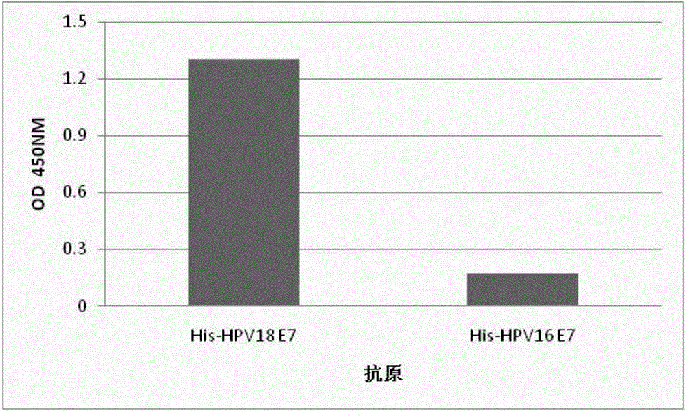
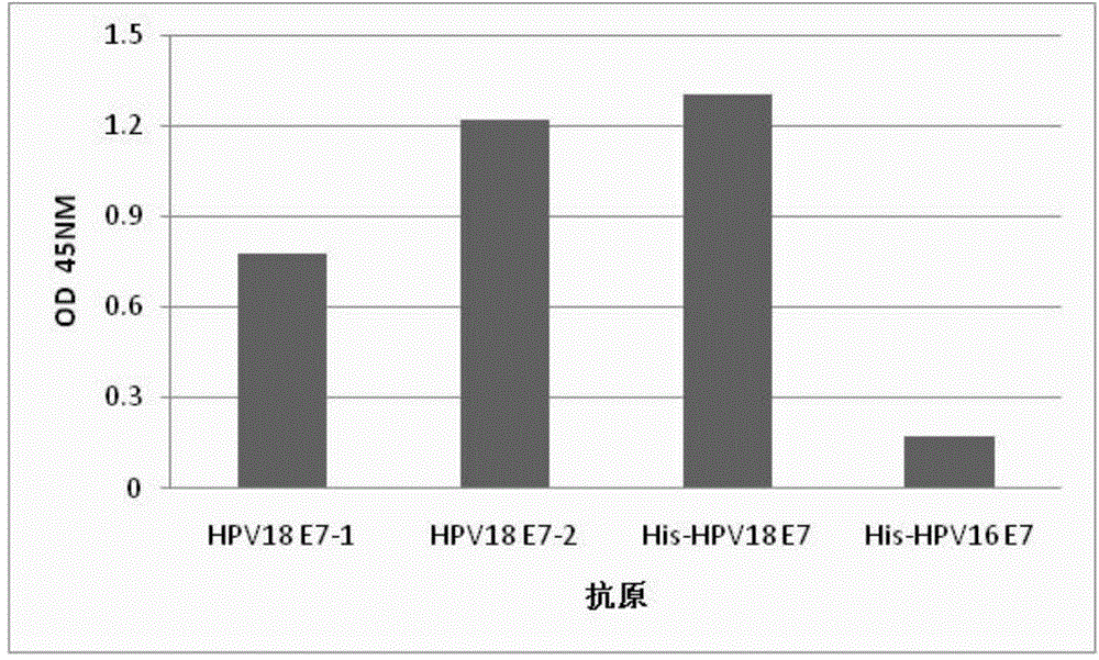
[0230] 6-8 weeks old female BALB / C mice were taken, the immunogen was GST-HPV18E7 protein, and the immunization program was shown in Table 1. Before each immunization, blood was collected by tail docking of mice, and Hi s - HPV18E7 was used as the detection antigen to coat the indirect ELISA method to detect the mouse serum titer. Splenocytes were taken for fusion when the serum titer titer of the immunized mice reached the maximum and no longer increased.
[0231] Table 1 Mouse immunization program
[0232]
[0233]
[0234] 1.2 Cell Fusion and Culture
[0235] The splenocytes of immunized mice were collected and fused with myeloma cells SP2 / 0 according to conventional methods. The fusion ratio of splenocytes: SP2 / 0=5:1. Selective culture in CO2 constant temperature incubator. After the fusion, the HAT medium was used to change the medium three times; when the ...
Embodiment 2
[0261] Immunocytochemical staining to detect the specificity of HPV18E7 monoclonal antibody MAB-F2 in suspended and fixed cervical cancer cell lines:
[0262] The cervical cancer cell line Hela cells expressing HPV18E7 protein and the cervical cancer cell line C-33A cells without HPV DNA were fixed with different cell fixatives and then immunocytochemical staining was performed with monoclonal antibody MAB-F2. The specific experimental method is as follows:
[0263] Collect C-33A cells and Hela cells respectively, fix them with 4% paraformaldehyde or LBC fixative for several days, and take a total of 50,000 cells, in which cervical cancer cell Hela cells and negative control C-33A cells are mixed in a certain proportion, Hela The ratio of the number of cells to C-33A cells was 1:9, 1:19, 1:49, 1:99, 1:499, 1:999. After mixing, put it on poly-L-Lysine-treated coverslip and air-dry at room temperature to fix the cells on the coverslip; add TBS washing solution to wash for 5 min...
PUM
 Abstract
Description
Claims
Application Information
 - R&D
- Intellectual Property
- Life Sciences
- Materials
- Tech Scout
- Unparalleled Data Quality
- Higher Quality Content
- 60% Fewer Hallucinations
Browse by: Latest US Patents, China's latest patents, Technical Efficacy Thesaurus, Application Domain, Technology Topic, Popular Technical Reports.
© 2025 PatSnap. All rights reserved.Legal|Privacy policy|Modern Slavery Act Transparency Statement|Sitemap|About US| Contact US: help@patsnap.com