
Methods for sterilizing biological materials containing non-aqueous solvents

a technology of biological materials and solvents, applied in the field of methods for sterilizing biological materials, can solve the problems of not always reliable, inability to detect the presence of certain viruses, and unwanted and potentially dangerous biological contaminants or pathogens

Inactive Publication Date: 2010-12-07
CLEARANT
View PDF115 Cites 10 Cited by
  • Summary
  • Abstract
  • Description
  • Claims
  • Application Information

AI Technical Summary

Benefits of technology

This approach allows for the effective sterilization of biological materials sensitive to radiation, maintaining their activity and integrity by using stabilizers, reducing solvent content, or lowering temperatures during irradiation, thereby ensuring the material's safety and efficacy for use.

Problems solved by technology

Many biological materials that are prepared for human, veterinary, diagnostic and / or experimental use may contain unwanted and potentially dangerous biological contaminants or pathogens, such as viruses, bacteria (including inter- and intracellular bacteria, such as mycoplasmas, ureaplasmas, nanobacteria, chlamydia, rickettsias), yeasts, molds, fungi, prions or similar agents responsible, alone or in combination, for TSEs and / or single or multicellular parasites.
Such procedures, however, are not always reliable and are not able to detect the presence of certain viruses, particularly in very low numbers.
This reduces the value or certainty of the test in view of the consequences associated with a false negative result.
False negative results can be life threatening in certain cases, for example in the case of Acquired Immune Deficiency Syndrome (AIDS).
Furthermore, in some instances it can take weeks, if not months, to determine whether or not the material is contaminated.
Moreover, to date, there is no reliable test or assay for identifying prions within a biological material that is suitable for screening out potential donors or infected material.
Thus the products of unicellular natural or recombinant organisms or tissues carry a risk of pathogen contamination.
In addition to the risk that the producing cells or cell cultures may be infected, the processing of these and other biological materials creates opportunities for environmental contamination.
Interestingly, even products from species as different from humans as transgenic plants carry risks, both due to processing contamination as described above, and from environmental contamination in the growing facilities, which may be contaminated by pathogens from the environment or infected organisms that co-inhabit the facility along with the desired plants.
Indeed, such rodents are notoriously difficult to control, and may gain access to a crop during sowing, growth, harvest or storage.
Likewise, contamination from overflying or perching birds has to potential to transmit such serious pathogens as the causative agent for psittacosis.
Thus any biological material, regardless of its source, may harbour serious pathogens that must be removed or inactivated prior to the administration of the material to a recipient.
This is a result of safety concerns for the workers conducting the tests, and the difficulty and expense associated with the containment facilities and waste disposal.
According to current standards of the U.S. Food and Drug Administration, heat treatment of biological materials may require healing to approximately 60° C. for a minimum of 10 hours, which can be damaging to sensitive biological materials.
Indeed, heat inactivation can destroy 50% or more of the biological activity of certain biological materials.
Unfortunately, this method may also remove products that have a high molecular weight.
Further, in certain cases, small viruses may not be removed by the filter.
This procedure requires that unbound sensitizer is washed from products since the sensitizers are toxic, if not mutagenic or carcinogenic, and cannot be administered to a patient.
The published literature in this area, however, teaches that gamma radiation can be damaging to radiation sensitive products, such as blood, blood products, protein and protein-containing products.
In particular, it has been shown that high radiation doses are injurious to red cells, platelets and granulocytes (Leitman).
Unfortunately, many sensitive biological materials, such as monoclonal antibodies (Mab), may lose viability and activity if subjected to freezing for irradiation purposes and then thawing prior to administration to a patient.

Method used


Image

  • Methods for sterilizing biological materials containing non-aqueous solvents
  • Methods for sterilizing biological materials containing non-aqueous solvents
  • Methods for sterilizing biological materials containing non-aqueous solvents

Examples


example 1

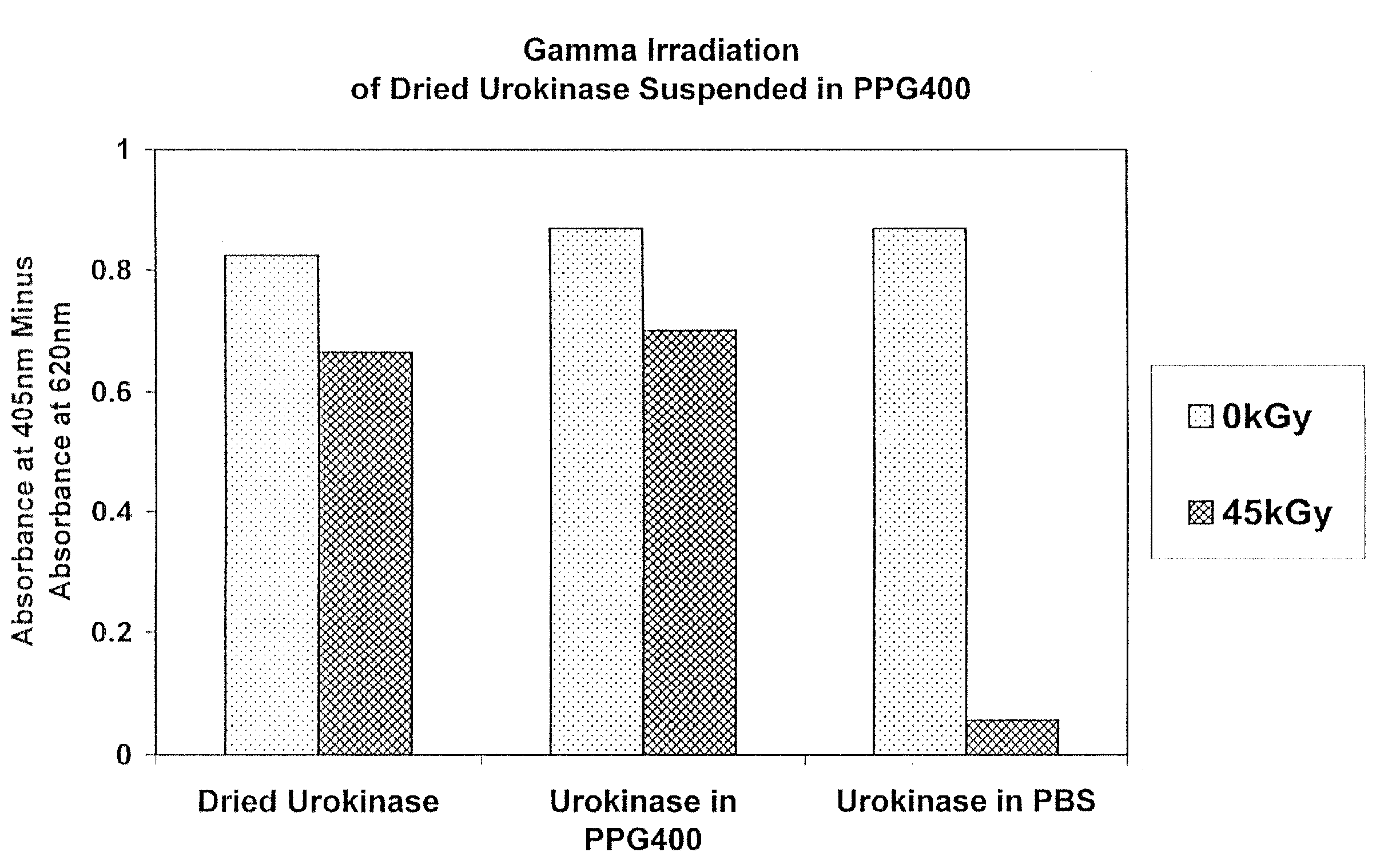
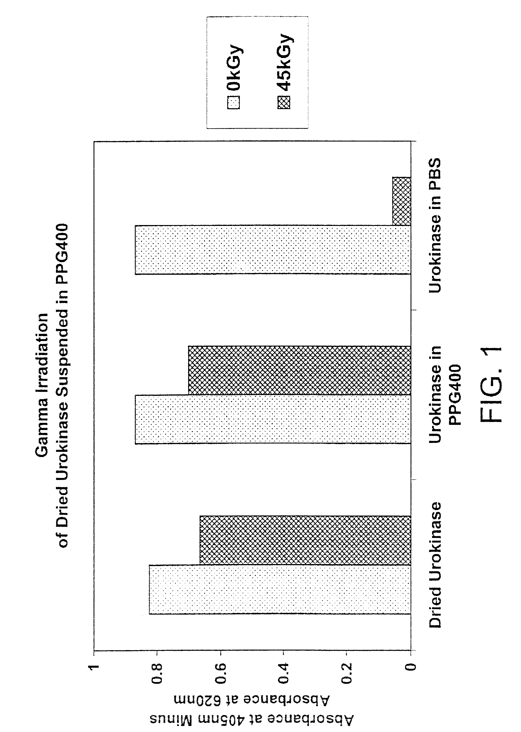
[0101]In this experiment, the effect of gamma radiation on dried urokinase suspended in polypropylene glycol (PPG) 400 or phosphate buffered saline (PBS) was determined.

Method

[0102]Six 1.5 ml polypropylene microfuge tubes containing urokinase and PPG400 (tubes 2 and 5), PBS (tubes 3 and 6) or dry urokinase alone (tubes 1 and 4) were prepared as indicated in the table below. Tubes 4-6 were gamma irradiated at 45 kGy (1.9 kGy / hr) at 4° C. Tubes 1-3 were controls (4° C.).

[0103]

weightof dryvolumeurokinasePPG400volumeTubeSample(mg)(ul)PBS (ul)1dry urokinase alone3.2002urokinase suspended in PPG4003.1612603urokinase suspended in PBS3.0801234dry urokinase alone3.38005urokinase suspended in PPG4003.313206urokinase suspended in PBS3.520141

[0104]After irradiation, the samples were centrifuged at room temperature for 5 minutes at 14 k RPM. PPG400 solvent was removed from tubes 2 and 5 and 120 μl PBS were added to those two tubes. 128 μl and 135 μl PBS were added to tubes 1 and 4, respectively ...

example 2

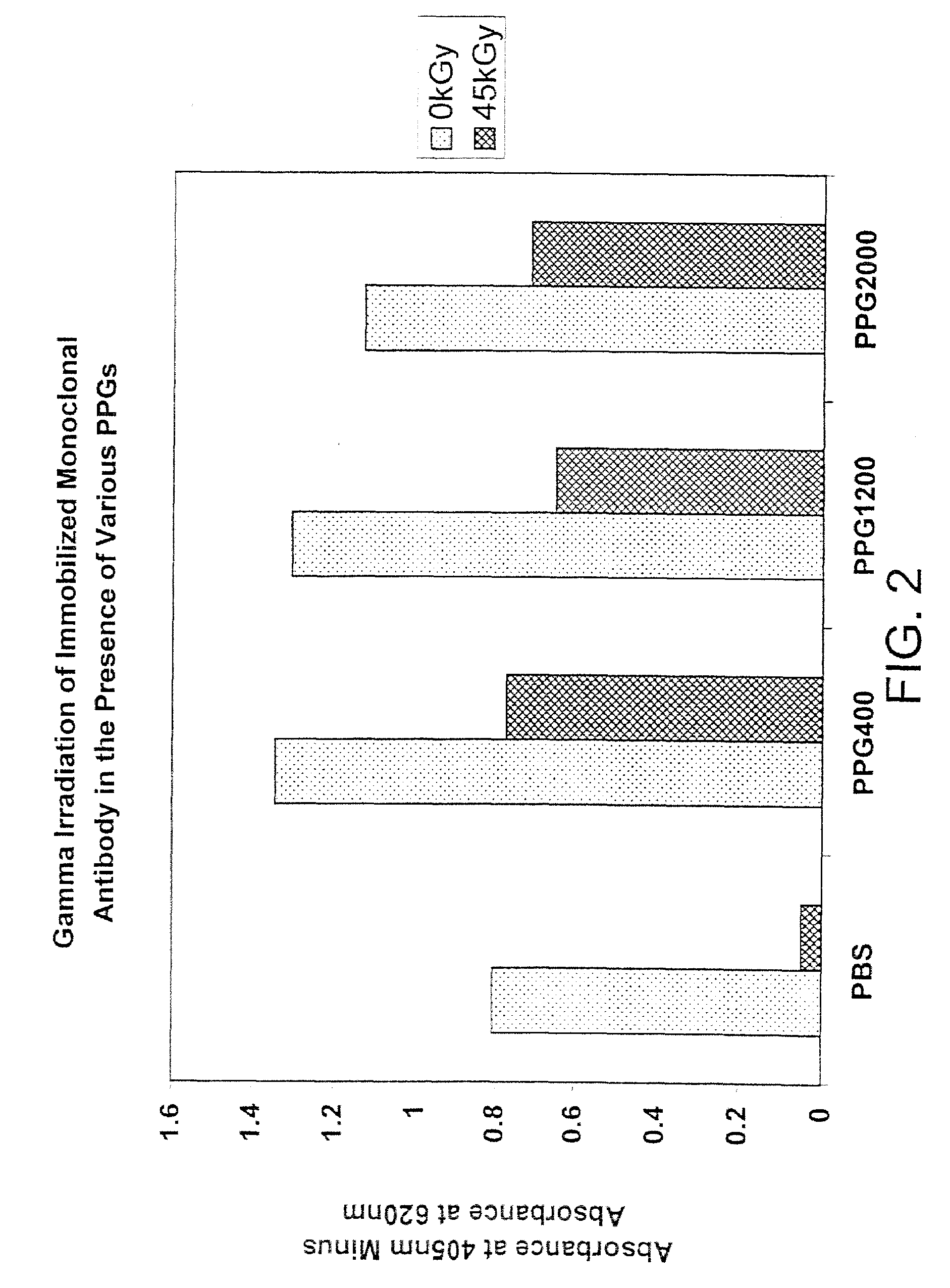
[0110]In this experiment, the activity (as shown by the ability to bind antigen) of immobilized anti-insulin monoclonal antibody was determined after irradiation in the presence of various forms of polypropylene glycol (molecular weights of 400, 1200 and 2000).

Method

[0111]In two 96-well microliter plates (falcon plates—ProBind polystyrene cat. # 353915), the wells were washed four times with full volume PBS (pH 7.4). Once the two plates were prepared as described above, they were coated with 100 μl / well of freshly prepared 2 μg / ml anti-insulin in coating buffer and left overnight at 4° C. The plates were then washed briefly three times with PBS (pH 7.4) and 100 μl of PPG400, PPG1200 or PPG2000 were added to specific wells. Each solution was prepared in a 11, i.e., 2-fold, dilution series with PBS. Both plates were covered tightly with a cap mat (Greiner cap mat cat. # 381070 (USA Scientific)) and irradiated at either 0 kGy / hr or 45 kGy (1.92 kGy / hr), both at 4° C.

[0112]Following irr...

example 3

[0126]In this experiment, liquid thrombin containing 50% glycerol and spiked with porcine parvovirus (PPV) was irradiated to varying total doses of radiation.

Method

[0127]1. Add 100 μl 100% glycerol, 20 μl thrombin (100 U / ml thrombin) spiked with 50 μl PPV and optionally 20 μl (200 mM) sodium ascorbate as a stabilizer (adjusted to a total volume of 1 ml with H2O) to Wheaton 3 ml tubes (in duplicate), and irradiate to a total dose of 10, 30 or 45 kGy at 1.8 kGy / hr at 4° C.[0128]2. Label and seed 96-well cell culture plates to allow at least 4 well per dilution (seeding to be done one day before inoculation). Add 200 μl of cell suspension per well at a concentration of 4×10⁴ / ml. The same cell culture medium is used for cell growth and maintenance after virus inoculation.[0129]3. Perform virus inoculation when the cells sheets are 70-90% confluent. In this experiment 800 μl PK-13 growth media was added to 200 μl samples first.[0130]4. Make appropriate dilution (1:5) of samples with PK-1...


PUM


Abstract

Methods are disclosed for sterilizing biological materials to reduce the level of one or more active biological contaminants or pathogens therein, such as viruses, bacteria (including inter- and intracellular bacteria, such as mycoplasmas, ureaplasmas, nanobacteria, chlamydia, rickettsias), yeasts, molds, fungi, prions or similar agents responsible, alone or in combination, for TSEs and / or single or multicellular parasites. The methods involve sterilizing biological materials containing one or more non-aqueous solvents with irradiation.

Description

[0001]This is a continuation application of U.S. patent application Ser. No. 11 / 812,353, filed Jun. 18, 2007, now abandoned which is a continuation application of U.S. application Ser. No. 09 / 960,703, filed Sep. 24, 2001, now abandoned both of which are herein incorporated by reference in their entireties.BACKGROUND OF THE INVENTION[0002]1. Field of the Invention[0003]The present invention relates to methods for sterilizing biological materials to reduce the level of one or more active biological contaminants or pathogens therein, such as viruses, bacteria (including inter- and intracellular bacteria, such as mycoplasmas, ureaplasmas, nanobacteria, chlamydia, rickettsias), yeasts, molds, fungi, prions or similar agents responsible, alone or in combination, for TSEs and / or single or multicellular parasites. The present invention particularly relates to methods of sterilizing biological materials containing one or more non-aqueous solvents with irradiation.[0004]2. Background of the R...

Claims


Application Information

Patent Timeline
Patent Type & Authority Patents(United States)
IPC IPC(8): G21K5/00A61K39/395A61L2/00A61L2/08A61L2/10A61L2/16C07K16/26
CPCA61K39/39591A61L2/0011A61L2/0035A61L2/0082A61L2/10A61L2/16C07K16/26A61L2/08
Inventor MIEKKA, SHIRLEYMACPHEE, MARTIN J.DROHAN, WILLIAM N.MANN, DAVIDBURGESS, WILSON
Owner CLEARANT
Learn More