
Rapid and non-invasive method to evaluate immunization status of a patient

Inactive Publication Date: 2005-08-09
THE UNITED STATES OF AMERICA AS REPRESENTED BY THE SECRETARY OF THE NAVY
View PDF6 Cites 27 Cited by
  • Summary
  • Abstract
  • Description
  • Claims
  • Application Information

AI Technical Summary

Benefits of technology

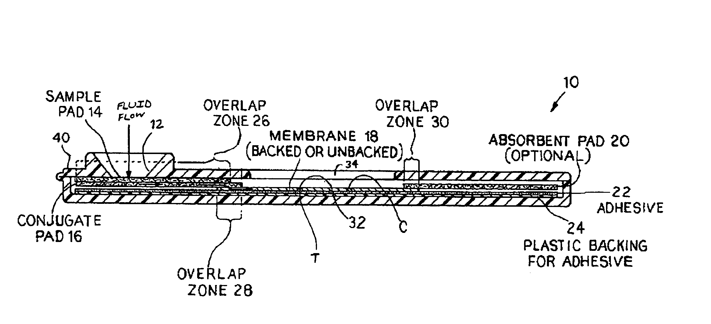
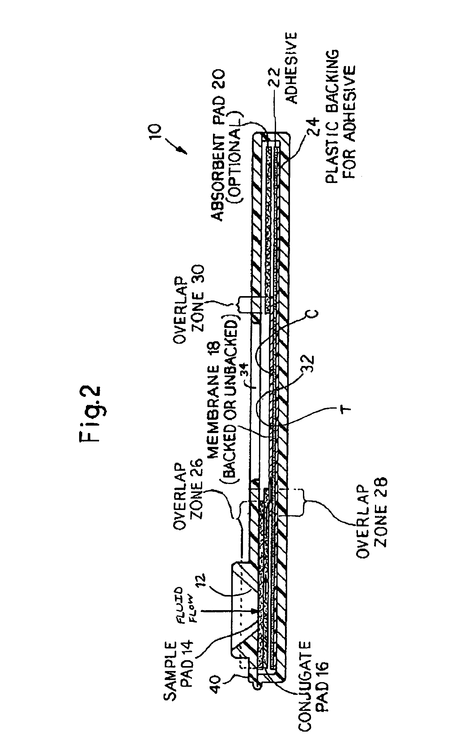
[0051]The present invention comprises an immunodiagnostic assay method and kit to rapidly evaluate immunization status of a patient, or alternatively to verify exposure to a biowarfare agent, such as anthrax, in a patient. The method and kit of the present invention provides rapid, reliable, non-invasive and safe testing procedures.
[0059]Thus, the immunoassays of the present invention provide markers of vaccine effectiveness by detecting specific IgG protective antibodies in patient bodily fluids, such as saliva. The present invention includes in vitro immunological techniques of antibody detection to validate the test by analyses of serum and saliva samples in a population of immunized military personnel. This approach has widespread future potential for monitoring exposure to toxins or disease and for assessing immunization status against various infectious diseases, including anthrax. Possible long-term applications of this hand-held, diagnostic tool include continuing field surveillance of vaccinated personnel to verify protective immunization and to assist in the determination of the optimum dose schedule for anthrax vaccine administrations. This test method may also be employed to monitor personnel and to follow the progression of other infectious diseases for which specific antibodies are known.

Problems solved by technology

Anthrax is considered one of the most dangerous of biowarfare threats because of its potency and very high mortality rate.
Although the mandatory Anthrax Vaccine Immunization Program (AVIP) was initiated in early 1998, there have been no known follow-up studies in immunized personnel to verify antibody production against the antigenic component of the anthrax toxin.
Current testing methods measure serum antibody levels in response to vaccines by time-consuming, labor-intensive processes.
Highly trained laboratory personnel are needed to perform these current tests, and the instrumentation required for current tests is relatively sophisticated and expensive.
While Enzyme-Linked Immunosorbent Assay (ELISA) has been the standard for measuring antibody titers, it is more problematic to perform this diagnostic test in forward-deployed regions where military personnel are at greater potential risk for anthrax toxin exposure.
However, bacterial resistance to third-generation cephalosporins has been documented, further thwarting attempts to quickly treat this infection.
However, outbreaks of the disease still occur in countries that do not have active livestock vaccination programs or effective patient vaccines.
Therefore, anthrax is still a danger to public health in less-developed countries such as India, where a deadly outbreak occurred in 1999-2000 in Pondicherry territory.
It is the PA-LF bound form of the toxin that results in serious illness or death if not promptly treated.
Once in the cytoplasm, these toxins disrupt normal cellular functions, eventually causing cell death.
In the spore form, Bacillus anthracis may easily be dispersed over a wide area and poses a lethal challenge when inhaled.
Further complications result in that exposure to inhalational anthrax is not easily diagnosed because the infection initially presents with nonspecific influenza-like symptoms.
The possibility of chemical or biological agent exposure may not be so unimaginable in that a 1995 terrorist release of sarin nerve gas in a crowded Tokyo subway had a devastating outcome, affecting almost 5,000 people.
Presently, there is little direct scientific evidence that patients vaccinated with AVA are protected against anthrax.
Also, AVA did not provide good protection in the guinea pig against an aerosol spore challenge, with only 20%-26% of the animals surviving.
It was also proposed that this animal model may respond poorly to the AVA vaccine itself.
However, it should also be noted in studies conducted before 1990, the patient vaccine used was not optimized for PA expression.
Therefore, the results are inconclusive because vaccine efficacy in one animal model cannot be compared to the protection afforded other animals immunized with the same vaccines or challenged with the same anthrax strains.
This also clearly points out the inherent difficulty in extrapolating results of anthrax vaccine protection in animals to that in patients.

Method used


Image

  • Rapid and non-invasive method to evaluate immunization status of a patient
  • Rapid and non-invasive method to evaluate immunization status of a patient
  • Rapid and non-invasive method to evaluate immunization status of a patient

Examples


example 1

[0090]General:

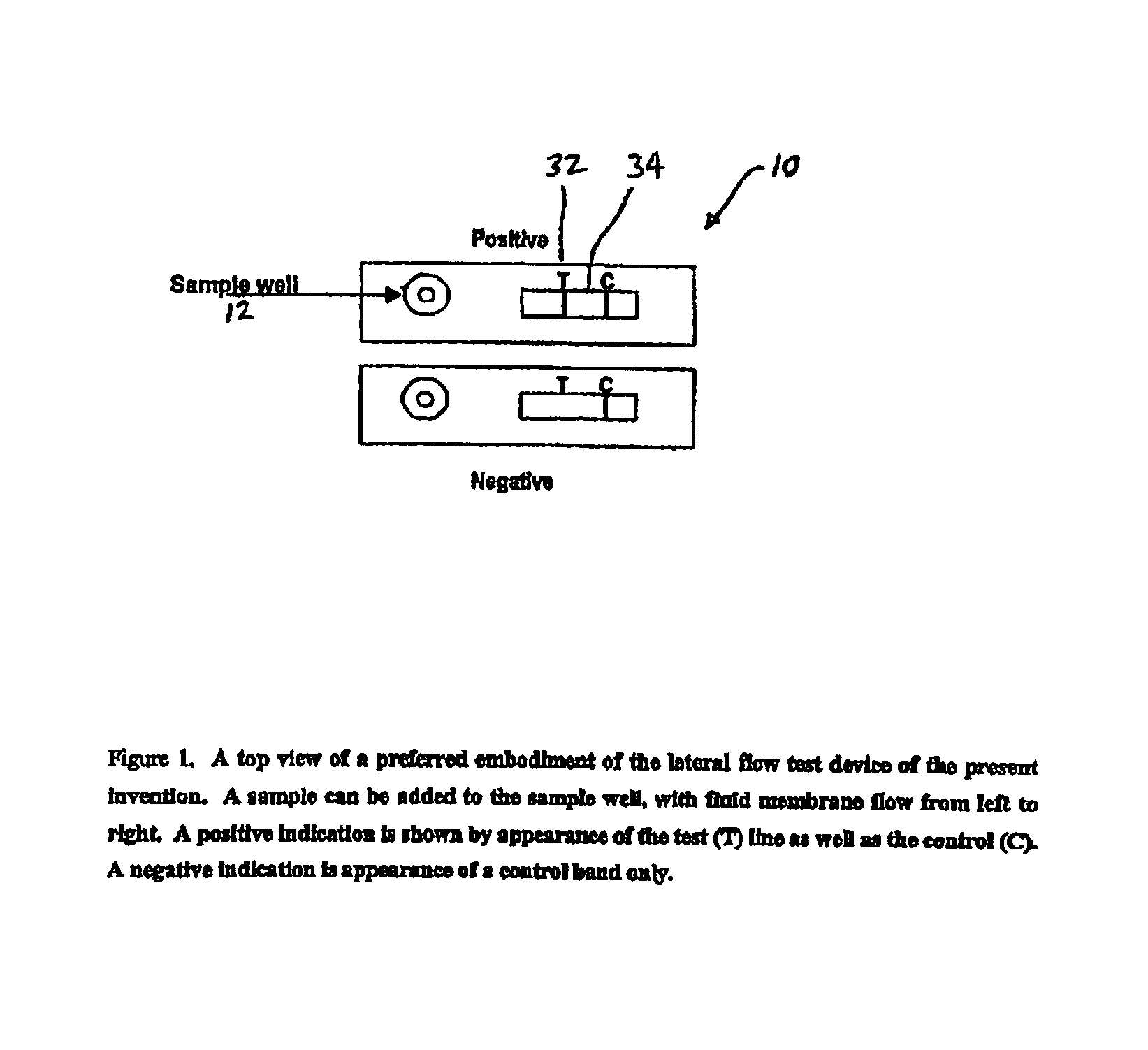
[0091]The design and rationale for this study rest on the importance of using saliva as the test medium to measure anthrax vaccine effectiveness. This rapid immunoassay avoids venipuncture and the necessity of having trained corpsmen to collect samples for measurement of anti-PA antibodies. Sample collection is non-invasive with no perceivable discomfort or risk to the individual. A positive confirmation of anthrax antibodies is readily observed within 5-10 minutes by the appearance of the test band. The assay is straight-forward with no other chemicals or reagents needed to perform the test.

[0092]The following sections describe the experimental approach designed to address three specific aims: (1) To optimize the assay of the present invention by signal amplification methods to achieve the most sensitive results; (2) To determine assay specificity by studying false positive and false negative results in the small pilot study; and (3) To validate the assay with paralle...


PUM


Abstract

An assay method and kit for detecting the presence of a predesignated, target IgG antibody in a sample selected from one or more patient bodily fluids. The method comprises the following steps: (a) contacting the sample of one or more patient bodily fluids with a membrane-bound recombinant protective antigen to bind to the target IgG antibody in the sample; (b) previously, simultaneously or subsequently to step (a), binding the protective antigen (PA) with a conjugated label producing a detectable signal; and (c) detecting the signal whereby the presence of the target IgG antibody is determined in the sample by the intensity of the signal. The method can further comprise the step of evaluating immunization status of the patient from whom the sample came by comparing the signal or lack thereof with immunizations previously received by the patient. In a preferred embodiment, the recombinant protective antigen (PA) specifically binds to anthrax protective antigen-specific IgG antibodies. Preferably, the immunoassay of the present invention comprises a lateral-flow assay comprising a membrane, a conjugated label pad, and a recombinant protective antigen (PA) bound to the membrane.

Description

STATEMENT REGARDING FEDERALLY SPONSORED RESEARCH OR DEVELOPMENT[0001]The invention was made by the U.S. Navy, an agency of the United States Government. The U.S. Government has a paid-up license in this invention.BACKGROUND OF THE INVENTION[0002]1. Field of the Invention[0003]The present invention relates to an immunodiagnostic assay method and kit to rapidly evaluate immunization status of a patient, or alternatively, to verify exposure to a biowarfare agent, such as anthrax, in a patient.[0004]2. Description of the Related Art[0005]A critical need exists for rapid, reliable, non-invasive and safe testing procedures to evaluate immunization status, such as anthrax immunization status, of a patient.[0006]The terrorist attacks of Sep. 11, 2001 in the United States and the outbreak of anthrax in Florida, New York, Washington D.C., etc. shortly thereafter has brought the threat of biowarfare into sharp focus for the general public. Biowarfare agents have been of significant concern for...

Claims


Application Information

Patent Timeline
IPC IPC(8): G01N33/558
CPCG01N33/558G01N2333/32Y10S435/97Y10S436/81
Inventor SIMONSON, LLOYD G.KELLY, JOHN R.
Owner THE UNITED STATES OF AMERICA AS REPRESENTED BY THE SECRETARY OF THE NAVY
Learn More