
Eureka
For R&D, Eureka makes reading and utilizing patents & technical documents easy.
Eureka AIR
Designed for self-driven R&D workflows. Generate viable solutions, solve complex R&D challenges, empower your innovation with AI.

Eureka Materials
Designed for material experts only. Revolutionize your material R&D, from search, analyze, to developing new materials.

TechResearch
Generate reliable direction feasibility study reports for your R&D in just a few steps.

TechSeek
Discover and master advanced knowledge NOW. Basics, ideas, possibilities, all at once.

TechMind
As an expert in R&D Theories, TechMind can generates customized viable solutions instantly.

TechRisk
Analyze your overall solution with one click, know your potential R&D risks in advance.

TechMonitor
Get weekly tech updates, stay abreast of the latest tech innovations and key insights.
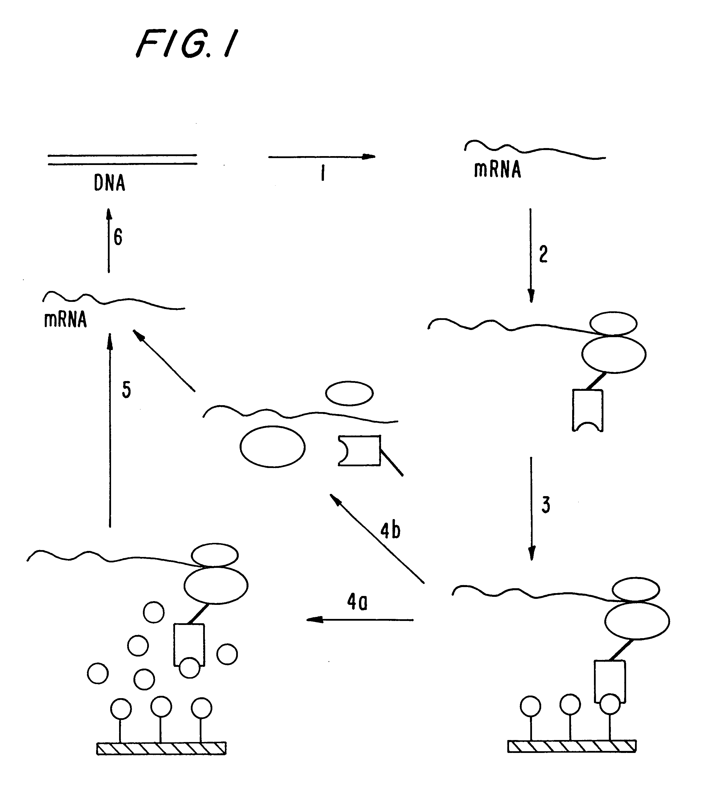
Polysome display in the absence of functional ssrA-RNA
a polysome and functional technology, applied in the field of polysome display in the absence of functional ssra-rna, can solve the problems of severe limitations in the diversity available, the efficiency of the ribosome display of the nascent (poly)peptide, and the power and accuracy of rational design today, so as to increase the stability of mrna, efficiently inhibit the effect of nucleases and stable against nucleases
- Summary
- Abstract
- Description
- Claims
- Application Information
AI Technical Summary
Benefits of technology
Problems solved by technology
Method used
Image

Examples
example 2
RT-PCR and in vitro Transcription
Reverse transcription was performed using Superscript reverse transcriptase (Gibco BRL) according to the manufacturer's recommendation. PCR was performed using Taq polymerase (Gibco BRL) in the presence of 5% DMSO (4 min at 94.degree. C., followed by 3 cycles of 30 sec at 94.degree. C., 30 sec at 37.degree. C., 2 min at 72.degree. C., followed by 10 similar cycles at 60.degree. C. instead of 37.degree. C., 20 similar cycles at 60.degree. C. instead of 37.degree. C. with elongation at 72.degree. C. prolonged by 15 sec per cycle and finished by 10 min at 72.degree. C.). PCR products were analyzed by agarose gel electrophoresis and purified from the gel and reamplified, if the amount and quality was not sufficient, or directly used for transcription without additional purification. In vitro transcription was performed as described (Pokrovskaya, I. D. & Gurevich, V. V. (1994) Anal. Biochem. 220, 420-423).
example 3
Model System and Quantification of Yields of Affinity Selection
As a model system, we used single-chain Fv (scFv) fragments of antibodies (Huston, J. S., Levinson, D., Mudgett-Hunter, M., Tai, M. S., Novotny, J., Margolies, M. N., Ridge, R. J., Bruccolleri, R. E., Haber, E., Crea, R. & Oppermann, H. (1988) Proc. Natl. Acad. Sci. U.S.A. 85, 5879-5883), in which the variable domain of the light chain (V.sub.L) is connected via a flexible linker to the variable domain of the heavy chain (V.sub.H). To tether the folded protein to the ribosome and not interfere with folding we fused a spacer to the C-terminus of the scFv fragment. Since the antibody domains form disulfide bonds, and the RNA polymerase requires .beta.-mercaptoethanol for maximal stability, the effect of performing transcription in a separate reaction was investigated. Further, the conditions for oxidative protein folding during translation (Ryabova, L., Desplancq, D., Spirin, A. & Pluckthun, A. (1997) Nature Biotechnology,...
example 4
Effect of mRNA Structure on Yield
In designing the ribosome display system, we first engineered the flanking regions of the scFv gene (FIG. 2A). The gene should be transcribed very efficiently from the PCR product, and its mRNA should be stable against nucleases. For the 5' end, we used the T7 promoter and the natural T7-gene10 upstream region, which encodes a stemloop structure directly at the beginning of the mRNA (Studier, F. W., Rosenberg, A. H., Dunn, J. J. & Dubendorff, J. W. (1990) Methods Enzymol. 185, 60-89). At the 3' end, we fused a spacer region of 57-116 amino acids to the reading frame of the scFv to tether the emerging, folded polypeptide to the putative polypeptide channel of the ribosome and to give it enough distance to not interfere with folding. This spacer encodes at the RNA level a 3' stemloop, either the terminator of the E. coli lipoprotein (Studier, F. W., Rosenberg, A. H., Dunn, J. J. & Dubendorff, J. W. (1990) Methods Enzymol. 185, 60-89) (lpp term) or the ...
PUM
| Property | Measurement | Unit |
|---|---|---|
| temperature | aaaaa | aaaaa |
| pH | aaaaa | aaaaa |
| physical properties | aaaaa | aaaaa |
Abstract
Description
Claims
Application Information
 - R&D Engineer
- R&D Manager
- IP Professional
- Industry Leading Data Capabilities
- Powerful AI technology
- Patent DNA Extraction
Browse by: Latest US Patents, China's latest patents, Technical Efficacy Thesaurus, Application Domain, Technology Topic, Popular Technical Reports.
© 2024 PatSnap. All rights reserved.Legal|Privacy policy|Modern Slavery Act Transparency Statement|Sitemap|About US| Contact US: help@patsnap.com