
Recombinant Bacterium and Uses Thereof

a bacterium and recombinant technology, applied in the field of recombinant bacterium, can solve the problems of low protective value of mechanisms, high labor intensity, high attenuation of responses or the organism itself, etc., and achieve rapid, potent cd8+ t cell response, high efficacy, safe and cost-effective

Active Publication Date: 2015-05-21
NAT RES COUNCIL OF CANADA
View PDF2 Cites 2 Cited by
  • Summary
  • Abstract
  • Description
  • Claims
  • Application Information

AI Technical Summary

Benefits of technology

The patent text describes how researchers have discovered how pathogens like Salmonella enter the body and how the immune system struggles to control them. They have also developed a new vaccine vector that can quickly and effectively control the bacterium by modifying its cell biology. The vaccine vector uses a key modification to generate a rapid and potent CD8+ T cell response, leading to self-destruction of the vaccine in the body. The approach can be used with other antigens from pathogens or tumors by cloning them into Salmonella and using the YopE / SycE system. Overall, the patent provides a novel tool for fighting against infections and tumors with a safe and cost-effective vaccine vector.

Problems solved by technology

Intracellular pathogens induce CD8+ T cell responses; however, the responses are either highly attenuated or the organism itself is highly toxic.
While the CD8+ T cells play a key role against various diseases, their induction is highly tedious.
However, these mechanisms are of little protective value since rapid pathogen elimination is not observed.
Subunit vaccines are difficult to mass-produce and are faced with numerous technical difficulties including batch to batch variability, quantitation of the antigen-adjuvant ratio, and extensively laborious procedures.
However, live vaccines can be either over- or under-attenuated and it is difficult to find the right balance (Raupach and Kaufmann, 2001).
However, it is difficult to justify the use of viral vectors as a live vaccine due to the lack of availability of reagents to control the virus, particularly in immunocompromised hosts.
However, extracellular bacteria do not gain access to the cytosol of infected cells, hence fail to induce CD8+ T cell response (Bevan, 1995).

Method used


Image

  • Recombinant Bacterium and Uses Thereof
  • Recombinant Bacterium and Uses Thereof
  • Recombinant Bacterium and Uses Thereof

Examples


example 1

Preparation of Recombinant Bacteria

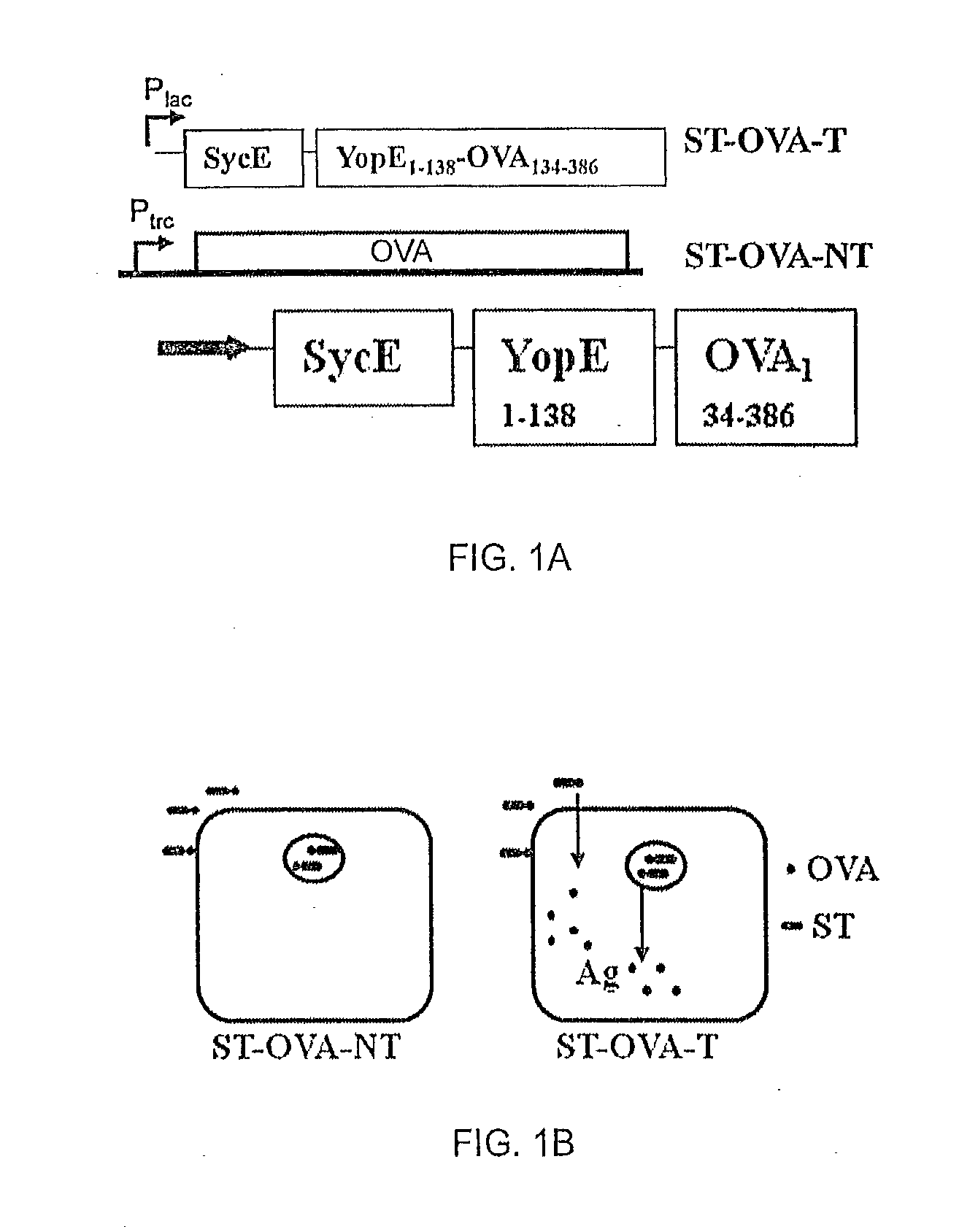
[0060]Recombinant bacteria comprising Salmonella enterica, serovar Typhimurium (ST) expressing ovalbumin (OVA) were prepared. Construct ST-OVA-NT, which does not translocate antigen to the cytosol, was prepared as previously described (Luu et al., 2006). A recombinant construct, ST-OVA-T, that produces an OVA fusion protein that is translocated to the cytosol; FIG. 1A shows a schematic of the fusion protein, where OVA is fused to YopE and SycE.

[0061]YopE is a 23 kDa protein comprising a N-terminal secretion domain (˜11 aa) and a translocation domain (at least 50 aa); the latter domain provides the binding site for the YopE-specific chaperone (SycE) that is required for YopE-mediated translocation of fused proteins to the cytosol (R). SycE is a chaperone necessary for translocation of the fused protein into the cytosol of infected cells through the type III secretion system of ST. A schematic of both ST-OVA-NT and ST-OVA-T constructs and their propo...

example 2

Detection of Antigen

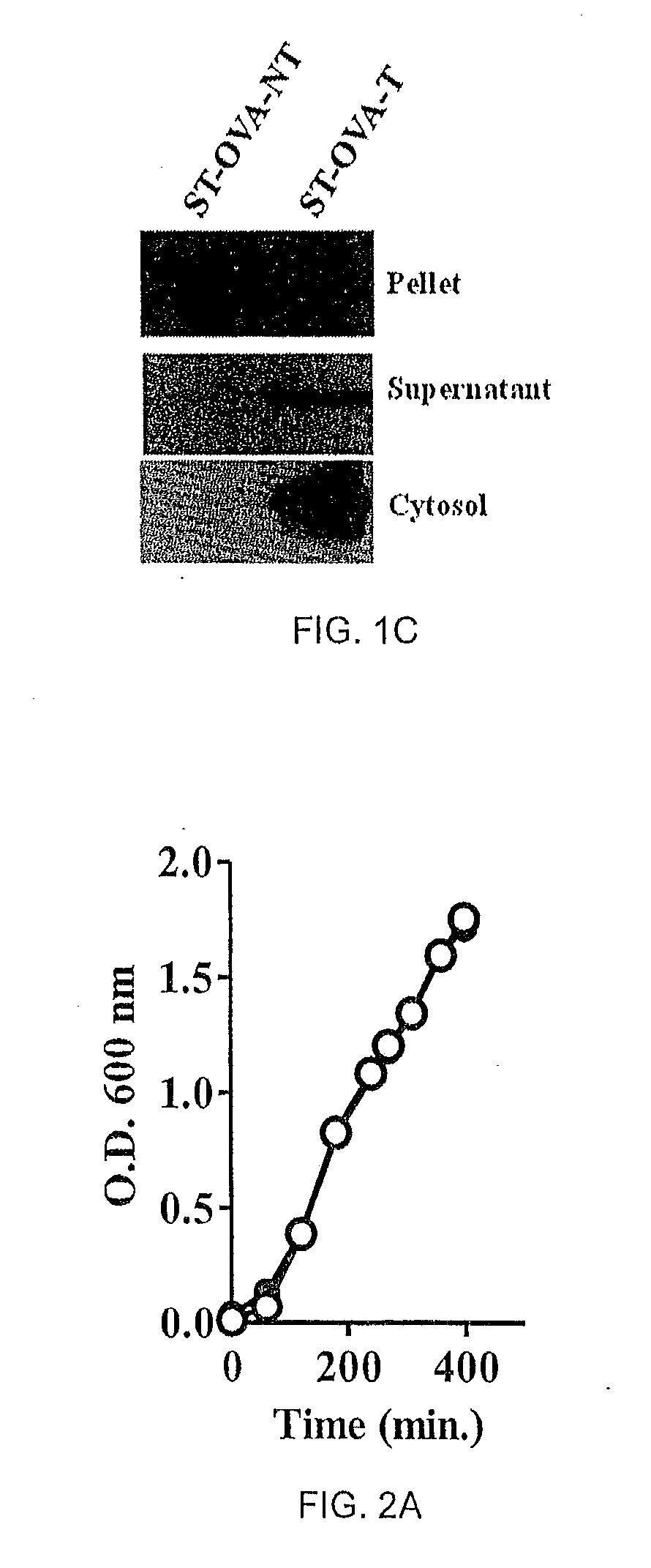
[0063]ST-OVA-NT and ST-OVA-T constructs of Example 1 were grown and expression and translocation of ovalbumin was evaluated. Pellet and supernatant of ST-OVA-NT and ST-OVA-T growing in liquid cultures were tested for the presence of OVA.

[0064]C57BL / 6J mice were injected intravenously with 106 ST-OVA-NT or ST-OVA-T reconstituted in 200 microlitres normal saline. Two days later, spleens were obtained from infected mice; spleen cells were isolated and lysed with TRITON X-100 in the presence of protease inhibitor, phenylmethylsulfonyl fluoride. The soluble lysate containing cytosolic proteins was tested for OVA expression by western blotting. Samples were normalized for cell number and were loaded on SDS-10% polyacrylamide gels. SDS-PAGE was performed and proteins were transferred to membranes, which were then blocked with 5% skim milk powder in PBS-TWEEN. OVA expression was detected using a 1 / 10,000 dilution of polyclonal anti-OVA antibody (Sigma-Aldrich), followed ...

example 3

Proliferation of ST-OVA-T and ST-OVA-NT

[0065]The ability of ST-OVA to proliferate extra- and intra-cellularly was also analyzed.

[0066]Liquid cultures of ST-OVA-NT and ST-OVA-T were set up in flasks to enumerate extracellular proliferation. At various time intervals (eg, 60 min., 120 min., 240 min., etc), aliquots were removed for measurement of OD at 600 nm. Both ST-OVA-NT and ST-OVA-T displayed similar proliferation and doubling time (FIG. 2A).

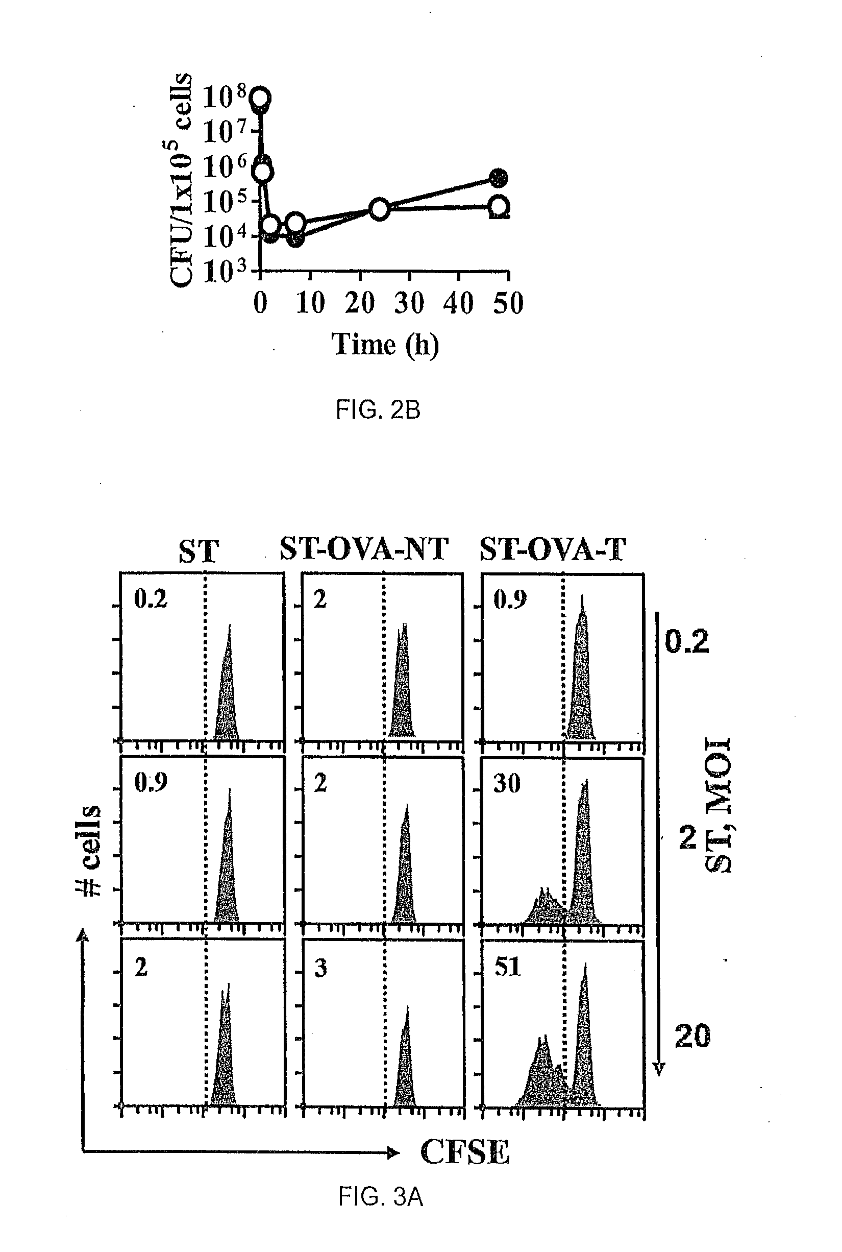
[0067]The influence of antigenic translocation on the ability of ST-OVA to proliferate within the intracellular compartment was evaluated. IC-21 macrophages (H-2b) (5×10⁴ / well) were infected with ST-OVA-NT or ST-OVA-T (MOI=10). After 30 min, cells were washed and cultured in media containing gentamicin (50 μg / ml) to remove extracellular bacteria. After 2 h, cells were washed again and cultured in media containing reduced levels of gentamicin (10 μg / ml). At various time intervals cells were lysed and bacterial burden in the cells determined. N...


PUM

PropertyMeasurementUnit
timeaaaaaaaaaa
ODaaaaaaaaaa
frequencyaaaaaaaaaa

Abstract

The present invention relates to a recombinant bacterium expressing an antigen that is translocated to the cytosol of a host organism, and uses thereof. To this end, the present invention provides a recombinant bacterium comprising a nucleic acid encoding an antigen that is translocated to the cytosol of a host cell utilizing Type III secretion system. The recombinant bacterium is generally chosen from intracellular pathogens that reside in the phagosome and fail to induce rapid T cell activation. The translocated antigen may be a viral antigen, a bacterial antigen, or a tumour antigen. Methods of imparting immunity using the recombinant bacterium are also provided.

Description

CROSS REFERENCE[0001]This application is a continuation of U.S. application Ser. No. 13 / 811,690 filed Jan. 23, 2013, which is a national stage entry from International Patent Application No. PCT / CA2011 / 00848 filed Jul. 28, 2011, which in turn claims priority to U.S. Provisional Patent Application No. 61 / 368,346 filed Jul. 28, 2010, each of which is incorporated herein by reference.FIELD OF THE INVENTION[0002]The present invention relates to recombinant bacterium and uses thereof. More specifically, the invention relates to recombinant bacterium expressing an antigen that is translocated to the cytosol of a host organism, and uses thereof.BACKGROUND OF THE INVENTION[0003]Various vaccine vectors or adjuvants that induce potent T cell responses are known in the art (Kaufmann and Hess, 1997). However, very few vaccine vectors exist that induce rapid and potent memory CD8+ cytolytic T cell responses, and that are safe and cost-effective (Raupach and Kaufmann, 2001). Unlike other T cells,...

Claims


Application Information

Patent Timeline
Patent Type & Authority Applications(United States)
IPC IPC(8): A61K39/00
CPCA61K2039/523A61K39/0005A61K39/0011A61K39/12A61K2039/522C07K14/195C07K14/4713C07K14/4748C07K14/70596C07K14/71C07K2319/036C07K2319/35C12N1/20C12N9/2462C12N9/6494C12N2760/10034C12N9/0059C12Y110/03001C12Y304/24011C07K14/77A61P31/04A61P31/12A61P37/04Y02A50/30A61K39/001191A61K39/001106A61K39/001156A61K39/001192A61K39/001186A61K39/4644A61K2239/31A61K2239/38A61K39/4611A61K39/464832A61K39/464838
Inventor SAD, SUBASHKRISHNAN, LAKSHMITZELEPIS, FANNYALCON, VALERIAYOUNG, KEVIN G.
Owner NAT RES COUNCIL OF CANADA
Learn More