
Reagents and Methods for Engaging Unique Clonotypic Lymphocyte Receptors

Inactive Publication Date: 2010-01-14
THE JOHN HOPKINS UNIV SCHOOL OF MEDICINE
View PDF28 Cites 82 Cited by
  • Summary
  • Abstract
  • Description
  • Claims
  • Application Information

AI Technical Summary

Benefits of technology

[0013]Yet another embodiment of the invention provides a method of increasing the number or percentage of antigen-specific T cells in a population of cells. A first cell population comprising antigen-specific T cells is incubated with at least one first solid support. The solid support comprises at least one T cell affecting molecule and at least one antigen presenting complex that comprises at least one antigen binding cleft. An antigen is bound to the antigenic binding cleft. The step of incubating is carried out for a period of time sufficient to form a second cell population comprising an increased number or percentage of antigen-specific T cells relative to the number or percentage of antigen-specific T cells in the first cell population. The antigen-specific T cells can be administered to a patient.
[0019]Still another embodiment of the invention provides a method of increasing the number or percentage of antigen-specific T cells in a population of cells. The cells comprise at least one T cell affecting molecule and at least one antigen presenting complex. The antigen presenting complex is either an MHC class I molecular complex or an MHC class II molecular complex. The MHC class I molecular complex comprises at least two fusion proteins. A first fusion protein comprises a first MHC class I α chain and a first immunoglobulin heavy chain and a second fusion protein comprises a second MHC class I α chain and a second immunoglobulin heavy chain. The first and second immunoglobulin heavy chains associate to form the MHC class I molecular complex. The MHC class I molecular complex comprises a first MHC class I peptide binding cleft and a second MHC class I peptide binding cleft. The MHC class II molecular complex comprises at least four fusion proteins. Two first fusion proteins comprise (i) an immunoglobulin heavy chain and (ii) an extracellular domain of an MHC class IIβ chain. Two second fusion proteins comprise (i) an immunoglobulin light chain and (ii) an extracellular domain of an MHC class IIα chain. The two first and the two second fusion proteins associate to form the MHC class II molecular complex. The extracellular domain of the MHC class IIβ chain of each first fusion protein and the extracellular domain of the MHC class IIα chain of each second fusion protein form an MHC class II peptide binding cleft. Antigenic peptides are bound to the peptide binding clefts. The step of incubating is carried out for a period of time sufficient to form a second cell population comprising an increased number or percentage of antigen-specific T cells relative to the number or percentage of antigen-specific T cells in the first cell population. The antigen-specific T cells can be administered to a patient.
[0021]Yet another embodiment of the invention provides a method of increasing the number or percentage of antibody-producing B cells in a population. An isolated preparation comprising a plurality of precursor B cells is contacted with at least one first solid support. The solid support comprises at least one B cell affecting molecule and at least one molecular complex that engages B cell surface immunoglobulins or MHC-antigen complexes on a B cell surface. Members of the plurality of precursor B cells are thereby induced to form a first cell population comprising B cells that produce antibodies that specifically bind to the antigenic peptide.
[0022]Another embodiment of the invention provides a method of increasing the number or percentage of antibody-producing B cells in a population. A first cell population comprising antibody-producing B cells is incubated with at least one first solid support. The solid support comprises at least one B cell affecting molecule and at least one molecular complex that engages B cell surface immunoglobulins or MHC-antigen complexes on a B cell surface. The step of incubating is carried out for a period of time sufficient to form a second cell population comprising an increased number or percentage of antibody-producing B cells relative to the number or percentage of antibody-producing B cells in the first cell population.

Problems solved by technology

Development of immunotherapy, both adoptive and active, has been impeded by the lack of a reproducible, economically viable method to generate therapeutic numbers of specific T or B lymphocytes.
This step is both time consuming and expensive.
For these reasons, use of DC has been a limiting step in ex vivo expansion of T cells.
However two problems arise.
First, anti-CD3 / anti-CD28 beads support long-term growth of CD4 T cells, but do not sustain long term growth of CD8 T cells.
In addition, approaches using anti-CD3 based stimulation are associated with a decrease in antigenic specificity even when starting with highly enriched antigen-specific CTL populations.
These problems substantially limit the delivery of therapeutically relevant lymphocytes.

Method used


Image

  • Reagents and Methods for Engaging Unique Clonotypic Lymphocyte Receptors
  • Reagents and Methods for Engaging Unique Clonotypic Lymphocyte Receptors
  • Reagents and Methods for Engaging Unique Clonotypic Lymphocyte Receptors

Examples


example 1

Materials and Methods

[0197]Cell Lines. TAP-deficient 174CEM.T2 (T2) cells and melanoma cell lines were maintained in M′ medium (Oelke et al., Scand. J. Immunol. 52, 544-49, 2000) supplemented with 10% FCS.

[0198]Peptides. Peptides (Mart-1, ELAGIGILTV, SEQ ID NO:3; CMVpp65, NLVPMVATV, SEQ ID NO:4) used in this study were prepared by the JHU core facility. The purity (>98%) of each peptide was confirmed by mass-spectral analysis and HPLC.

[0199]HLA-A2.1+ Lymphocytes. The Institutional Ethics Committee at The Johns Hopkins University approved the studies discussed in the examples below. All donors gave written informed consent before enrolling in the study. Healthy volunteers and a melanoma patient, donor #7, were phenotyped HLA-A2.1 by flow cytometry. The melanoma patient had extensive metastatic disease with lung, liver, and lymph node metastases. PBMC were isolated by Ficoll-Hypaque density gradient centrifugation.

[0200]Generation of aAPC. aAPC were generated by coupling “HLA-Ig” (des...

example 2

Induction and Expansion of Mart-1- and CMV-Specific CTL by aAPC

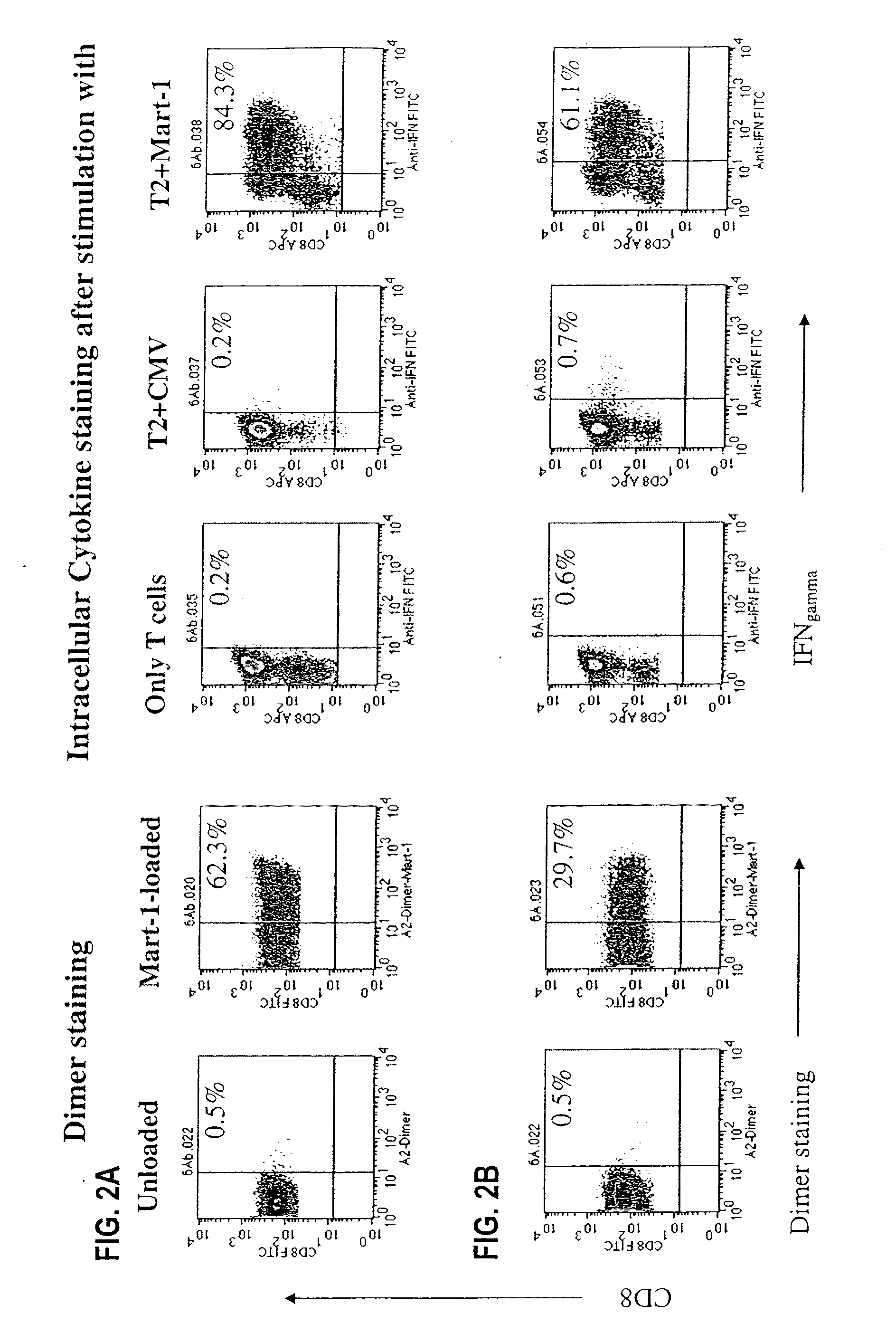
[0206]This example demonstrates the induction and expansion of antigen-specific CTL by two clinically relevant targets, CMV-peptide pp65 and Mart-1. These peptides have widely varying affinities for their cognate TCR. The CMV-peptide pp65 is known to be a high affinity peptide, whereas the modified Mart-1 peptide, derived from a melanocyte self antigen, is a low affinity peptide (Valmori et al., Int. Immunol. 11, 1971-80, 1999).

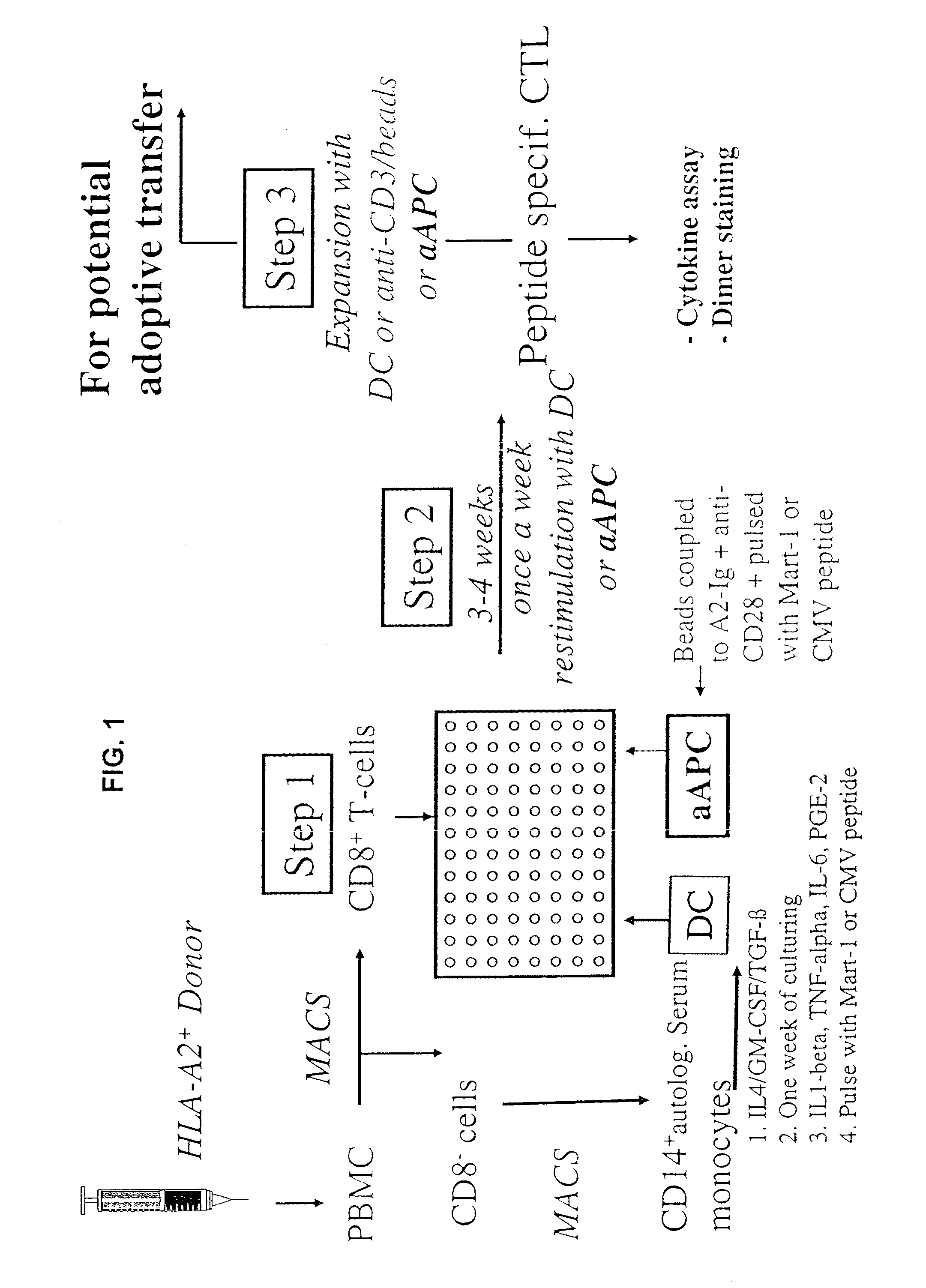
[0207]Current approaches use autologous peptide-pulsed DC to induce antigen-specific CTL from normal PBMC (FIG. 1). These approaches often use DC- or CD40L-stimulated autologous B cells to induce antigen-specific CTL over 2-4 stimulation cycles (FIG. 1, Step 2) until the antigen-specific CTL become a prominent part of the culture. We, therefore, compared aAPC induction to induction by DC. T cells were isolated, purified, and induced with either Mart-1-loaded aAPC or monocyte-derived autologous DC-...

example 3

Recognition of Endogenously Processed Antigen by aAPC-Induced PBMC

[0216]A useful criterion in evaluating CTL function is the recognition of targets expressing endogenous antigen-HLA complexes. Initial work using peptide-pulsed DC for expansion of melanoma-specific CTL resulted in low affinity CTL that mediated lysis of targets pulsed by the cognate antigen but often did not recognize melanoma targets that endogenously expressed antigen-HLA complexes. Yee et al., J. Immunol. 162, 2227-34, 1999. We therefore studied the ability of aAPC-induced CTL to recognize endogenous Mart-1 or pp65 CMV antigen (FIG. 3).

[0217]For the ICS staining the cells were incubated with target cells in regular medium without cytokines. To increase the sensitivity of the ICS assay, a low dose of PMA and ionomycin was added to the medium. As described in Perez-Diez et al., Cancer Res. 58, 5305-09, 1998, this approach enabled us to detect more antigen specific T cells in the population. Differences in the result...


PUM

PropertyMeasurementUnit
Timeaaaaaaaaaa
Timeaaaaaaaaaa
Timeaaaaaaaaaa

Abstract

Platforms comprising at least one lymphocyte affecting molecule and at least one molecular complex that, when bound to an antigen, engages a unique clonotypic lymphocyte receptor can be used to induce and expand therapeutically useful numbers of specific lymphocyte populations. Antigen presenting platforms comprising a T cell affecting molecule and an antigen presenting complex can induce and expand antigen-specific T cells in the presence of relevant peptides, providing reproducible and economical methods for generating therapeutic numbers of such cells. Antibody inducing platforms comprising a B cell affecting molecule and a molecular complex that engages MHC-antigen complexes on a B cell surface can be used to induce and expand B cells that produce antibodies with particular specificities.

Description

[0001]This application is a division of Ser. No. 10 / 618,267 filed on Jul. 13, 2003, which claims the benefit of Ser. No. 60 / 395,781 filed Jul. 12, 2002. Each of these applications is incorporated herein by reference in its entirety.[0002]This invention resulted from research funded in part by National Institutes of Health Grant Nos. AI-29575 and AI-44129. The Federal Government has certain rights in this invention.FIELD OF THE INVENTION[0003]The invention relates to reagents and methods for engaging unique clonotypic lymphocyte receptors.BACKGROUND OF THE INVENTION[0004]Development of immunotherapy, both adoptive and active, has been impeded by the lack of a reproducible, economically viable method to generate therapeutic numbers of specific T or B lymphocytes. For example, the current standard approach to generating antigen-specific cytotoxic T lymphocytes (CTL) for adoptive immunotherapy entails generating monocyte-derived dendritic cells (DC) for expansion of CTL. This step is bo...

Claims


Application Information

Patent Timeline
IPC IPC(8): A61K39/395C12N5/06A61K35/12A61K35/14A61K39/00A61K39/385A61P37/02A61P37/06C07K7/06C07K7/08C07K14/47C07K17/00C07K17/02C12N5/00C12N5/0781C12N5/0783G01N33/50
CPCA61K39/385G01N33/5091A61K2035/124A61K2039/5154A61K2039/605C07K17/00C07K2319/30C12N5/0068C12N5/0635C12N5/0636C12N2501/23C12N2501/24C12N2501/51C12N2501/52C12N2501/58C12N2501/599C12N2533/50A61K2035/122A61P31/04A61P31/12A61P33/00A61P35/00A61P37/02A61P37/06A61K39/4622A61K39/464491A61K39/4611A61K39/464417A61K39/4614A61K39/46449A61K39/4615A61K39/464838A61K39/4612A61K39/464488
Inventor SCHNECK, JONATHANOELKE, MATHIAS
Owner THE JOHN HOPKINS UNIV SCHOOL OF MEDICINE
Learn More