
Buffer solution for electroporation and a method comprising the use of the same

a buffer solution and electroporation technology, applied in the direction of peptide/protein ingredients, genetic material ingredients, enzymology, etc., can solve the problems of not being able to use corresponding methods, at least very tedious, and particular problems frequently occurring, etc., to achieve non-buffered or weakly buffered

Inactive Publication Date: 2005-03-24
LONZA COLOGNE
View PDF6 Cites 108 Cited by
  • Summary
  • Abstract
  • Description
  • Claims
  • Application Information

AI Technical Summary

Benefits of technology

[0010] The object of the invention is thus to provide a buffer solution for electroporation which makes it possible to achieve higher transfection efficiencies with a low cell mortality rate and to develop a corresponding method.
[0014] In an especially advantageous embodiment it is provided that the buffer according to the invention has a lower concentration of potassium ions (K+), preferably 2 to 6 mmol*I−1 K+, and a higher concentration of sodium ions (Na+), preferably 100 to 150 mmol*I−1 Na+, if compared with the cytoplasm of the cells. Thus, the buffer according to the invention is not matched to the intracellular Na+ / K+ ratio but rather “extracellular” ratios are present in this respect. Surprisingly, however, this has no negative effects on the cells but nevertheless results in a significant increase in the transfection and cell survival rate.
[0023] The object is furthermore solved by a method for introducing biologically active molecules into animal or human cells by means of electric current which comprises suspending the cells and dissolving the biologically active molecules in a buffer solution, which has a buffer capacity of at least 20 mmol *11*pH-1 and an ionic strength of at least 200 mmol*I−1 with a change in the pH from pH 7 to pH 8 and at a temperature of 25° C., and applying an electric voltage to the suspension. With the aid of this method, biologically active molecules can be introduced into animal and human cells by means of electroporation with transfection efficiencies up to 93% and at the same time with low cell mortality. Various cell types, especially quiescent and weakly dividing cells can be successfully transfected.
[0025] The buffer solution according to the invention is especially suitable for carrying out an electroporation method in which biologically active molecules are introduced into the cells by a voltage pulse having a field strength of up to 2 to 10 kV*cm−1 and a duration of 10 to 200 μs and a current density of at least 2 A*cm−2. As a result of the high voltage pulse, it is possible for DNA to be transfected directly into the cell nucleus of animal and human cells with irreversible membrane damage being avoided by the shortness of the pulse. As a result of the shortness of the high-voltage pulse, the high ionic strength or conductivity of the buffer also does not result in any disadvantageous heating of the solution so that in addition to dividing cells, quiescent or weakly dividing cells can also be transfected with high efficiency and low mortality. The interaction of the high ionic strength of the buffer and short high-voltage pulses rather brings about efficient pore opening, wherein the strong buffer effect can also compensate for extreme fluctuations of the pH.

Problems solved by technology

In studies of quiescent or weakly dividing cells, for example, primary animal cells, however, the DNA does not enter into the cell nucleus in this way so that corresponding methods cannot be used here or at least are very tedious.
Moreover, especially when DNA is introduced into animal cells, so-called transfection, particular problems frequently occur as a result of the lability of the cells, since the survival rate of the cells influences the efficiency of the transfection as an important parameter.
However, non-buffered or weakly buffered mannitol or saccharose solutions were also used during the electroporation of animal cells (Shimizu et al.
Such non-buffered or weakly buffered solutions have the disadvantage that their use leads to an increased cell mortality and significantly reduced transfection efficiency (Yan et al.
Furthermore, the buffer used by Yan et al., is only optimised for voltage pulses up to 500 V .so that no indication is given as to whether this buffer can also be used for higher voltage pulses such as are required for the direct transfection into the cell nucleus.
In any case, however, the transfection efficiencies achieved in this buffer are too low to meet higher demands.
This change in the pH causes the release of cytotoxic Al3+ ions from the aluminium electrodes of the cuvettes and therefore causes increased cell mortality.
However, a reduction in the transfection efficiency was detected for pulses having a field strength higher than 1300 V / cm.
All the buffer solutions described so far however have the disadvantage that the transfection efficiencies achieved when using them are relatively low and / or the buffers are not suitable for application during the electroporation of quiescent or weakly dividing cells.

Method used


Image

  • Buffer solution for electroporation and a method comprising the use of the same
  • Buffer solution for electroporation and a method comprising the use of the same
  • Buffer solution for electroporation and a method comprising the use of the same

Examples


example 1

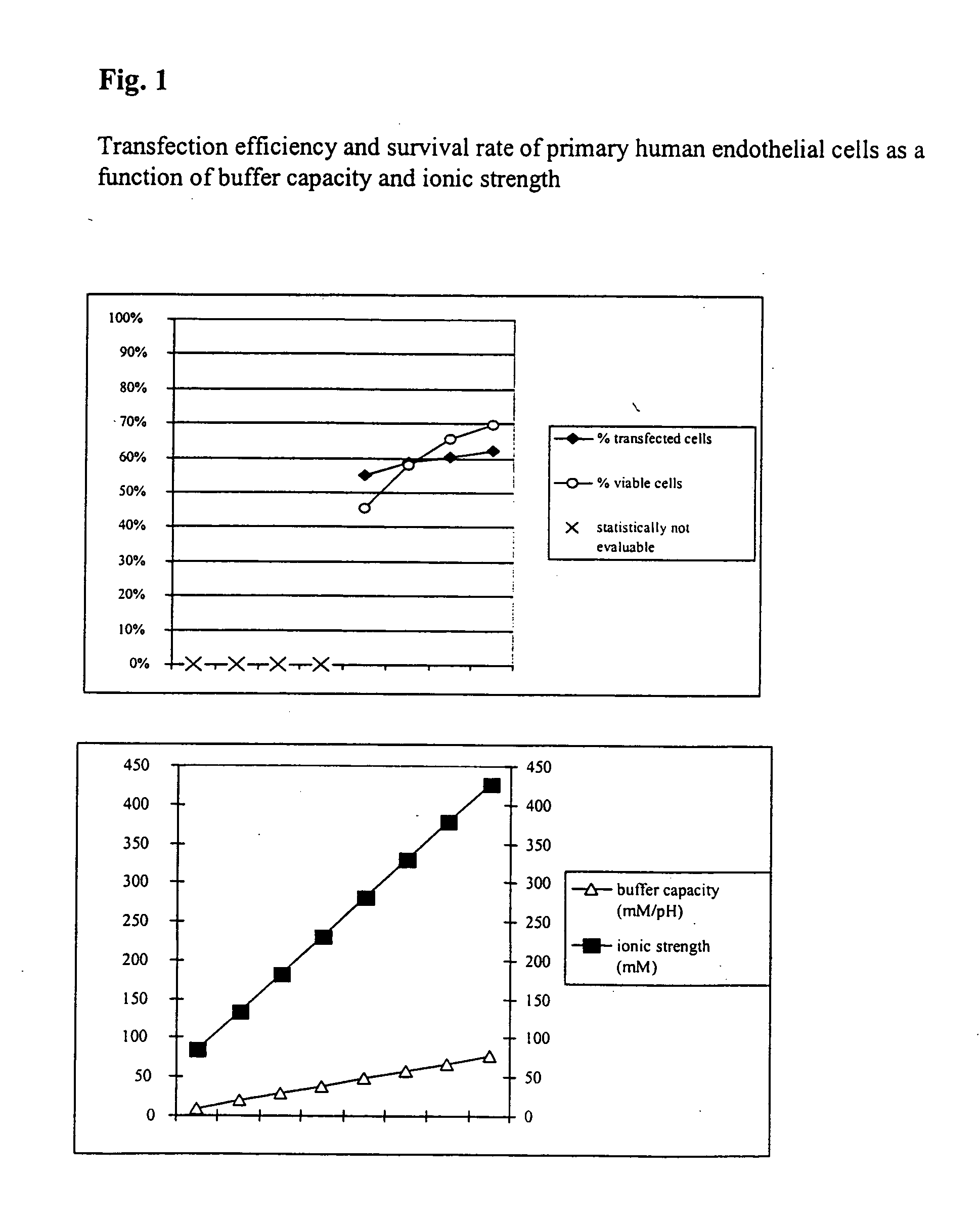
[0042] Transfection Efficiency and Survival Rate of Primary Human Endothelial Cells

[0043] In order to study the transfection and survival rate of cells during electrotransfection as a function of the buffer capacity and the ionic strength, primary human endothelial cells were transfected with a vector coding for the heavy chain of the mouse MHC class 1 protein H-2Kk.

[0044] Respectively 7×10⁵ cells with respectively 5 μg vector DNA and the respective electrotransfection buffer were placed at room temperature in a cuvette having an interelectrode gap of 2 mm and transfected by a 1000 V pulse of 100 μs duration.

[0045] In addition, comparative values were determined using PBS (Phosphate Buffered Saline: 10 mM sodium phosphate, pH 7.2+137 mM NaCl, ionic strength =161.5 mM, buffer capacity =4.8 mM / pH). Directly after the electrotransfection the cells were washed out of the cuvette using 400 μl of culture medium, incubated for 10 minutes at 37° C. and then transferred to a culture dish ...

example 2

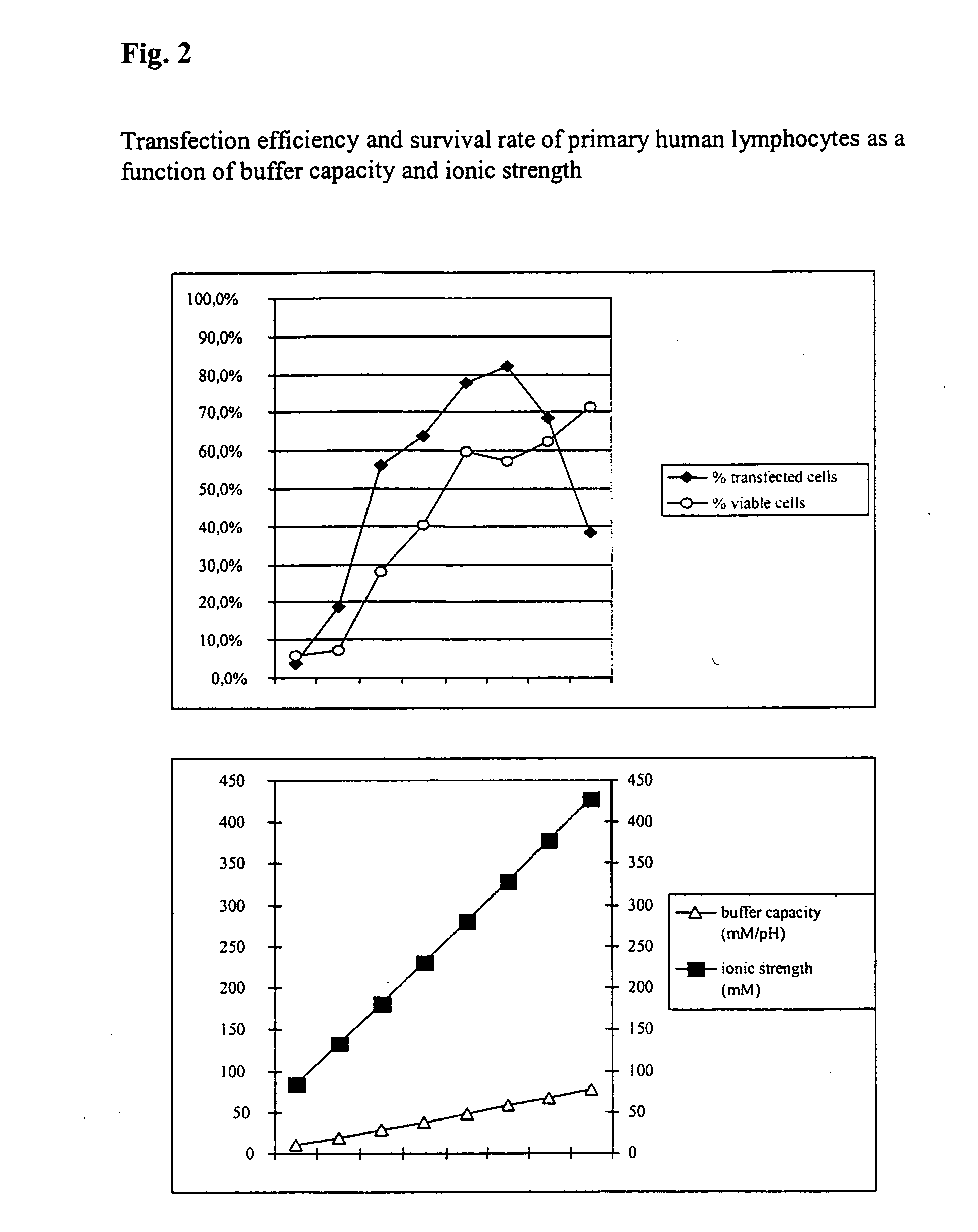
[0048] Transfection Efficiency and Survival Rate of Primary Human Lymphocytes

[0049] Furthermore, the transfection and survival rate of primary human lymphocytes during electrotransfection was studied as a function of the buffer capacity and the ionic strength.

[0050] For this purpose respectively 5×10⁶ cells with respectively 5 μg H-2Kk-expression vector-DNA in the respective buffers were placed at room temperature in a cuvette having a 2 mm interelectrode gap and electrotransfected by a 1000 V, 100 μs pulse followed by a current flow having a current density of 4 A*cm2 and 40 ms. In addition, comparative values were determined using PBS (ionic strength =161.5 mM, buffer capacity =4.8 mM / PH). The analysis was made after 16 hours.

[0051] As can be seen from FIG. 2, it is clear that the survival rates of the cells increase as the buffer capacity and ionic strength of the buffer solutions used increases. In some cases significantly better values could be achieved compared with the com...

example 3

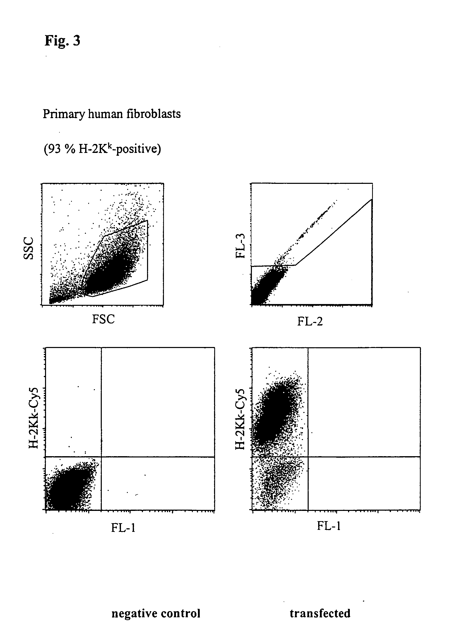
[0052] Transfection of Primary Human Fibroblasts

[0053]FIG. 3 shows an FACScan analysis of primary human fibroblasts transfected with H-2Kk expression vector. 5×10⁵ cells with 5 μg vector DNA in buffer 6 from Table 1 were placed at room temperature in a cuvette having a 2 mm interelectrode gap and transfected by a 1000 V, 100 Ps pulse followed by a current flow of 6 A*cm2 and 33 ms. After incubating for 5 h, the cells were incubated with a Cy5-coupled anti-H-2Kk antibody and analysed using a flow cytometer (FACScalibur, Becton Dickinson). The number of dead cells was determined by staining with propidium iodide. 93% of the living cells expressed the H-2Kk antigen which corresponds to a very high transfection efficiency.


PUM

PropertyMeasurementUnit
Temperatureaaaaaaaaaa
Timeaaaaaaaaaa
Molar densityaaaaaaaaaa

Abstract

The invention relates to a buffer solution for suspending animal or human cells and for dissolving biologically active molecules in order to introduce said biologically active molecules into the cells using an electric current and to a method for introducing biologically active molecules into animal or human cells using an electric current and a buffer solution. The inventive buffer solution has a buffering capacity of at least 20 mmol*I−1*pH−1 and an ionic strength of at least 200 mmol*I−1 during a change to the pH value from pH 7 to pH 8 and at a temperature of 25° C. The use of a buffer solution of this type in the corresponding method allows biologically active molecules to be introduced into animal and human cells with a high degree of transfection efficiency and at the same time a low cell mortality. Different cell types, in particular dormant and actively dividing cells of low activity, can be successfully transfected in said buffer solution.

Description

[0001] The invention relates to a buffer solution for suspending animal or human cells and for dissolving biologically active molecules in order to introduce said biologically active molecules into the cells using electric current and to a method for introducing biologically active molecules into animal or human cells using electric current comprising the use of the buffer solution. BACKGROUND OF THE INVENTION [0002] The introduction of biologically active molecules, such as for example, DNA, RNA or proteins, into living cells is an important instrument for studying biological functions of these molecules. A preferred method for introducing foreign molecules into the cells is electroporation which, unlike other methods, only causes slight permanent changes to the biological structure of the target cell by the transfer reagents. During electroporation the foreign molecules are introduced into the cells from an aqueous solution by a brief current flow wherein the cell membrane is made...

Claims


Application Information

Patent Timeline
IPC IPC(8): A61K35/12C12N15/09A61K38/00A61K47/00A61K48/00C12N13/00C12N15/87G01N33/50
CPCA61K38/00C12N15/87A61K48/00A61K47/00
Inventor RIEMEN, GUDULALORBACH, ELKEHELFRICH, JULIANASIEBENKOTTEN, GREGORMULLER-HARTMANN, HERBERTROTHMANN-COSIC, KIRSTENTHIEL, CORINNAWEIGEL, MEIKEWESSENDORF, HEIKEBROSTERBUS, HELMUTNIX, MICHAEL
Owner LONZA COLOGNE
Learn More