Eureka AIR delivers breakthrough ideas for toughest innovation challenges, trusted by R&D personnel around the world.

Wearable, noninvasive glucose sensing methods and systems

a glucose sensing and wearable technology, applied in the field of noninvasive glucose sensing, can solve the problems of reducing the complications and mortality associated with diabetes, invasive blood glucose monitoring techniques, and cgm systems developed for diabetics require insertion of sensors in skin, so as to improve the accuracy of glucose monitoring, reduce the thickness (and optical thickness) of ultrasound pulses, and improve the effect of blood glucos

Active Publication Date: 2020-06-02
ENSENALIEV RINAT O
View PDF1 Cites 2 Cited by
  • Summary
  • Abstract
  • Description
  • Claims
  • Application Information

AI Technical Summary

Benefits of technology

[0013]The present invention also provides a noninvasive blood glucose monitoring technique that would also be invaluable in critically ill patients, regardless of whether those patients are diabetic. Clinical studies clearly establish that morbidity and mortality are reduced in patients requiring intensive care if blood glucose is tightly controlled between 80 and 110 mg / dL (Van den Berghe G, 2005; Vanhorebeek I, 2005; van den Berghe G, 2001). However, conventional techniques for tightly controlling blood glucose have several limitations, including the need for frequent blood sampling and the risk that insulin administration will induce hypoglycemia (blood glucose <60 mg / dL) between sampling intervals and that hypoglycemia therefore will not be promptly diagnosed and treated. A continuous method of monitoring blood glucose by measuring tissue thickness would greatly improve the ease and safety of tightly controlling blood glucose with insulin in critically ill patients.
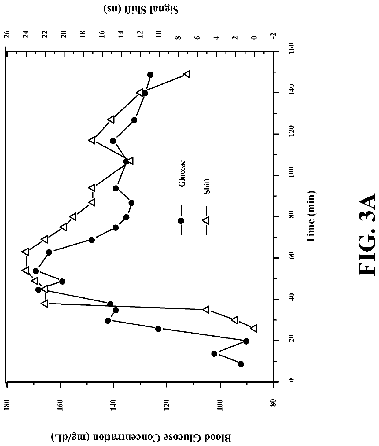
[0014]The measurement of dimensions or time of flight can be performed in a variety of tissues including, but not limited to: skin tissues (dermis, epidermis, subcutaneous fat), eye tissues (lens, anterior chamber, vitreous cavity, eye ball, sclera), mucosal tissues, nailbed, lunula, connective tissue, muscle tissue, blood vessels, cartilage tissue, tendon tissue. The dimension(s) of these tissues can change with blood glucose concentration. For instance, our studies demonstrated that increase of blood glucose concentration may decrease the thickness (and optical thickness) of and time of flight of ultrasound pulses in the skin tissues (namely, dermis). Measurements of dimensions of specific tissue layers (within one of these tissues) can be used for glucose monitoring. Measurement of one, two or more dimensions can be performed for more accurate, specific, and sensitive glucose monitoring. Ratios of dimensions of two or more tissues can be used for more robust, accurate, specific, and sensitive glucose monitoring. For instance, increasing blood glucose concentration may increase lens thickness and decrease anterior chamber thickness. The ratio of these changes may provide robust, accurate, and sensitive blood glucose monitoring. One can use measurement of total dimensions of complex tissues consisting of two or more different tissues. Measurement of time of flight of ultrasound or optical waves in these tissues, or optical thickness of these tissues can also be used for non-invasive glucose monitoring without calculating or determining geometrical thickness or other dimensions of these tissues.Wearable Devices
[0015]The present invention provides a wearable, noninvasive, and continuous glucose monitoring technique that is critically important for diabetic patients. Tight glucose control decreases dramatically complications and mortality associated with diabetes. Blood glucose monitoring is an important part of blood glucose control. At present, standard techniques for blood glucose monitoring are invasive and require a drop of blood or interstitial fluid for measurement. Continuous glucose monitoring (CGM) systems developed for diabetics require insertion of a sensor in skin and are not free of limitations associated with tissue trauma and inflammation, immune response, and encapsulation of the sensing area by proteins. A wearable, noninvasive, continuous glucose monitor would considerably improve the quality of life for diabetic patients, improve their compliance with glucose monitoring, and reduce complications associated with the disease.
[0017]The measurement of dimensions or time of flight can be performed using a wearable device in a variety of tissues including, but not limited to: skin tissues (dermis, epidermis, subcutaneous fat), eye tissues (lens, anterior chamber, vitreous cavity, eye ball, sclera), mucosal tissues, nailbed, lunula, connective tissue, muscle tissue, blood vessels, cartilage tissue, tendon tissue. The dimension(s) of these tissues can change with blood glucose concentration. For instance, our studies demonstrated that increase of blood glucose concentration may decrease the thickness (and optical thickness) of and time of flight of ultrasound pulses in the skin tissues (namely, dermis). Measurements of dimensions of specific tissue layers (within one of these tissues) can be used for glucose monitoring. Measurement of one, two or more dimensions can be performed for more accurate, specific, and sensitive glucose monitoring. Ratios of dimensions of two or more tissues can be used for more robust, accurate, specific, and sensitive glucose monitoring. For instance, increasing blood glucose concentration may increase lens thickness and decrease anterior chamber thickness. The ratio of these changes may provide robust, accurate, and sensitive blood glucose monitoring. One may use measurement of total dimensions of complex tissues consisting of two or more different tissues. Measurement of time of flight of ultrasound or optical waves in these tissues, or optical thickness of these tissues can also be used for non-invasive glucose monitoring without calculating or determining geometrical thickness or other dimensions of these tissues.

Problems solved by technology

Tight glucose control decreases dramatically complications and mortality associated with diabetes.
At present, standard techniques for blood glucose monitoring are invasive and require a drop of blood or interstitial fluid for measurement.
Continuous glucose monitoring (CGM) systems developed for diabetics require insertion of a sensor in skin and are not free of limitations.
However, conventional techniques for tightly controlling blood glucose have several limitations, including the need for frequent blood sampling and the risk that insulin administration will induce hypoglycemia (blood glucose <60 mg / dL) between sampling intervals and that hypoglycemia therefore will not be promptly diagnosed and treated.
Tight glucose control decreases dramatically complications and mortality associated with diabetes.
At present, standard techniques for blood glucose monitoring are invasive and require a drop of blood or interstitial fluid for measurement.
Continuous glucose monitoring (CGM) systems developed for diabetics require insertion of a sensor in skin and are not free of limitations associated with tissue trauma and inflammation, immune response, and encapsulation of the sensing area by proteins.

Method used


Image

  • Wearable, noninvasive glucose sensing methods and systems
  • Wearable, noninvasive glucose sensing methods and systems
  • Wearable, noninvasive glucose sensing methods and systems

Examples


example 1

[0087]Glucose-induced changes in skin thickness (and / or optical thickness) or time of flight measured with electromagnetic techniques.

[0088]Glucose-induced changes in skin tissue thickness (and / or optical thickness) can be measured by using electromagnetic waves including, but not limited to: optical radiation, terahertz radiation, microwaves, radiofrequency waves. Optical techniques include but not limited to reflection, focused reflection, refraction, scattering, polarization, transmission, confocal, interferometric, low-coherence, low-coherence interferometry techniques.

[0089]A wearable, like a wrist watch, optically-based glucose sensor can be developed.

example 2

[0090]Glucose-induced changes in time of flight in and thickness of skin measured with ultrasound techniques.

[0091]Glucose-induced changes in skin tissue thickness and time of flight can be measured by using ultrasound waves in the frequency range from 20 kHz to 10 Gigahertz. These techniques include but not limited to reflection, focused reflection, refraction, scattering, transmission, confocal techniques. It is well known that by using high frequency ultrasound can provide high-resolution images of tissues. One can use ultrasound frequencies higher than 10 MHz for measurement of skin thickness and time of flight.

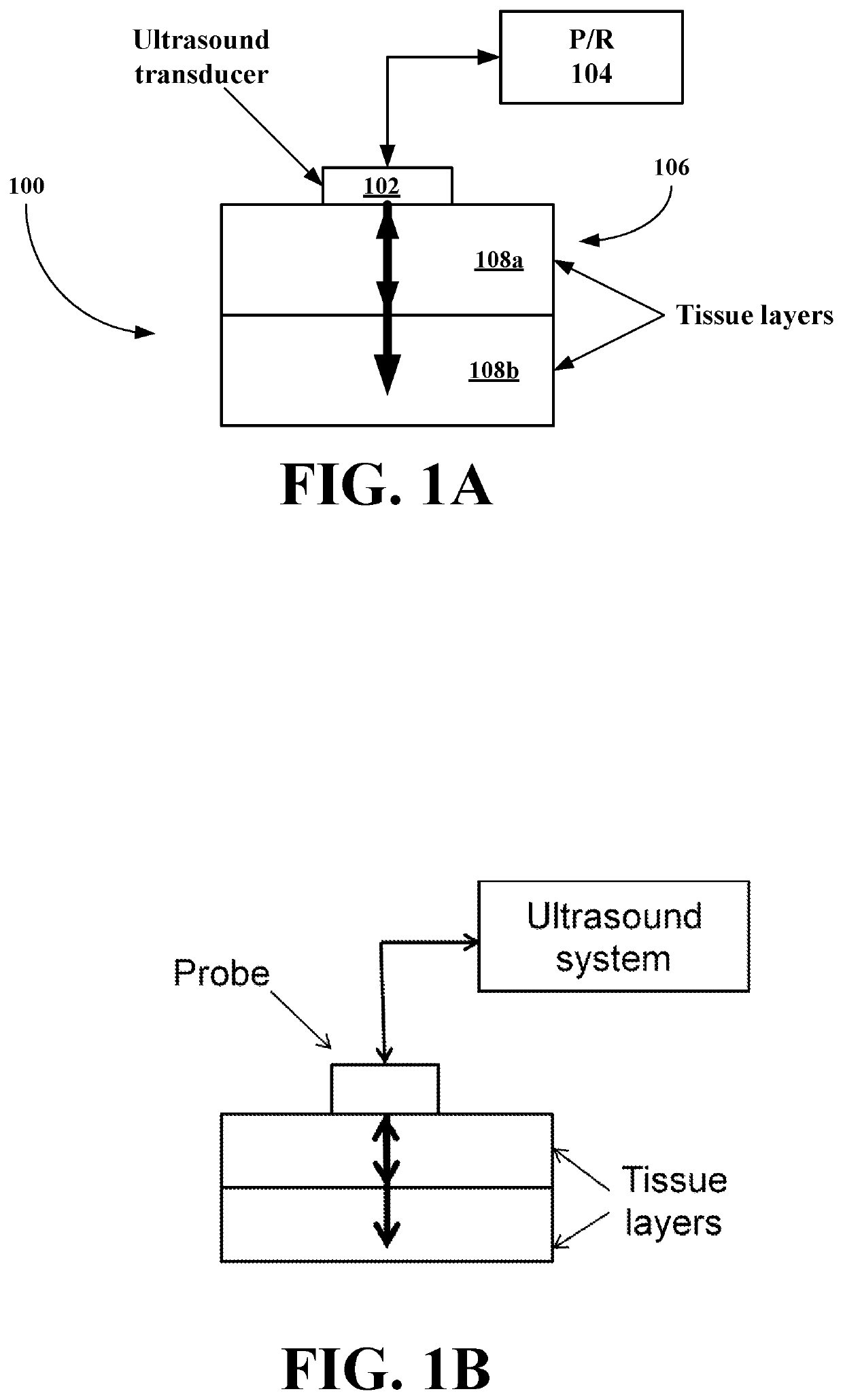
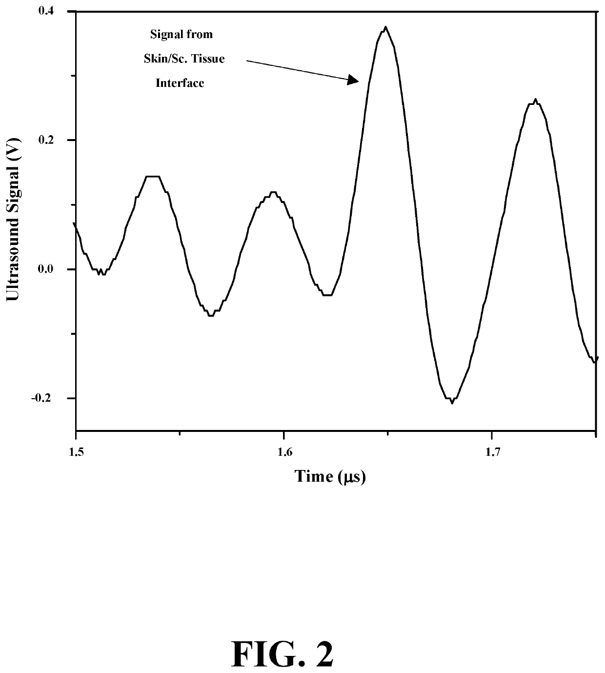
[0092]FIGS. 1 to 4D show different embodiments of the systems of this invention. In certain embodiments, the typical signal from skin / subcutaneous tissue interface, and glucose-induced signal shift (changes in time of flight) measured by the system. In other embodiments, compact ultrasound generators and sensors may be associated with wearable devices such as wrist watch,...

example 3

[0093]Glucose-induced changes in skin thickness and time of flight measured with optoacoustic or thermoacoustic techniques.

[0094]Glucose-induced changes in skin tissue thickness and time of flight can be measured by using optoacoustic or thermoacoustic techniques which may provide accurate tissue dimension measurement when short electromagnetic (optical or microwave) pulses are used in combination with wide-band ultrasound detection. FIG. 10 shows such a system. Optical detection of the ultrasound waves can be used instead of the ultrasound transducer.


PUM


Abstract

New wearable and non-wearable systems for noninvasive glucose sensing include an ultrasound generator, an ultrasound detector and a feedback unit. Methods for noninvasive glucose sensing using a wearable or nonwearable device include measuring a thickness (geometrical and / or optical) of a target tissue or a time of flight of ultrasound or optical pulses in the target tissue and determining a glucose value from the thickness of the target tissue or the time of flight in the target tissue in accordance with a target tissue thickness (geometrical and / or optical) or time of flight versus glucose calibration curve using new methodology for computing glucose concentrations with or without invasive measurements and simultaneously monitoring and generating a fitness index (FI), a body weight index (BWI), and / or a hydration index (HI).

Description

RELATED APPLICATION[0001]This application is a non-provisional of U.S. Provisional Patent Application Ser. No. 62 / 343,125 filed 30 May 2016 and a continuation-in-part of U.S. patent application Ser. No. 14 / 953,275 filed Nov. 27, 2015, which is a continuation-in-part of U.S. patent application Ser. No. 13 / 416,963 filed Mar. 9, 2012, now U.S. Pat. No. 9,167,993 issued 27 Oct. 2015.BACKGROUND OF THE INVENTION[0002]1. Field of the Invention[0003]The present invention relates to a method for noninvasive glucose sensing and a system for implementing the method and to methods for noninvasive glucose sensing with wearable devices and systems for implementing the methods.[0004]More particularly, the present invention relates to a method for noninvasive glucose sensing including the step of measuring a thickness of a target tissue or a time of flight of ultrasound or optical pulses in the target tissue and determining a glucose value from the thickness of the target tissue or the time of flig...

Claims


Application Information

Patent Timeline
Patent Type & Authority Patents(United States)
IPC IPC(8): A61B5/145A61B5/00A61B8/10A61B8/00A61B5/107G16H40/63A61B8/08G16H20/30A61B5/1455G16H40/67G16H50/30
CPCG16H40/63A61B5/1075A61B8/463A61B8/5223A61B8/4227A61B8/0891A61B8/5269A61B8/0858A61B5/14532A61B5/4839A61B5/4875A61B5/7225G16H20/30A61B8/56A61B8/10G16H50/30G16H40/67A61B5/1455A61B5/681A61B5/6803
Inventor ENSENALIEV, RINAT O.
Owner ENSENALIEV RINAT O
Why Eureka
Eureka Blog
Learn More