
Glycan arrays for high throughput screening of viruses

a high-throughput, virus-based technology, applied in the field of influenza virus subtype classification, can solve the problems of affecting the detection efficiency of influenza viruses, etc., to achieve the effect of high signal-to-noise ratio, rapid and easy control, and high signal-to-noise ratio

Active Publication Date: 2019-07-02
ACAD SINIC
View PDF423 Cites 3 Cited by
  • Summary
  • Abstract
  • Description
  • Claims
  • Application Information

AI Technical Summary

Benefits of technology

This approach provides a rapid, sensitive, and cost-effective diagnostic tool capable of distinguishing between influenza virus subtypes and detecting vaccination responses, suitable for use in low-resource areas, with the potential to identify new variants and drug resistance, and is adaptable for large-scale detection during outbreaks.

Problems solved by technology

Avian influenza, including the H5N1 strain, is a highly contagious and potentially fatal pathogen, but it currently has only a limited ability to infect humans.
The more complicated, and perhaps dangerous, scenario is the reassortment of Swine-Origin Influenza A with highly pathogenic avian H5N1 or with other serotypes.
This procedure might take a week and considerable effort.
Although virus isolation has long served as the standard for the diagnosis of influenza virus, the approach alone is inefficient when worldwide outbreaks occur as it is less sensitive, time-consuming, and requires considerable technical expertise.
Limitations of most of the conventional diagnostic methods are lack of accuracy, sensitivity and delay in getting results.
The current crop of quick test products on the market is only capable, due to limitations in their technology platform, of providing a yes / no answer to the presence of influenza.
While swine-origin influenza virus (S-OIV; National Virus Reference Laboratory, NVRL, Dublin) real time PCR detection is available, it may not be applicable for more-complex reassortant strains.
Further, these methods require relatively sophisticated laboratories, which are sometimes unavailable or inconvenient for clinical application.
The miniaturized array methodology is particularly well suited for investigations in the field of glycomics, since biological amplification strategies, such as the Polymerase Chain Reaction (PCR) or cloning, do not exist to produce usable quantities of complex oligosaccharides.
The development of glycan microarrays has been very slow for a number of reasons.
First, it is difficult to immobilize a library of chemically and structurally diverse glycans on arrays, beads or the like.
Second, glycans are not readily amenable to analysis by many of the currently available molecular techniques (such as rapid sequencing and in vitro synthesis) that are routinely applied to nucleic acids and proteins. methods of preparing glycan arrays have been described in PCT / US2005 / 007370 filed Mar. 7, 2005 titled “High Throughput Glycan Microarrays” (Blixt), and U.S. Pat. App. Pub. No. 20080220988 (Zhou).
Sensitive fluorescence detection commonly uses a laser and a confocal microscope, e.g., DNA microarray detector made by Affymetrix Inc., which are typically very expensive and need a trained technician to operate.
However, the reproducible preparation of highly purified antibody reagents is both challenging and time consuming (Jayasena, S. D. Clin. Chem.

Method used


Image

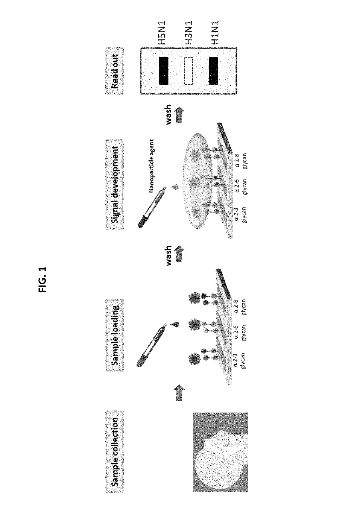
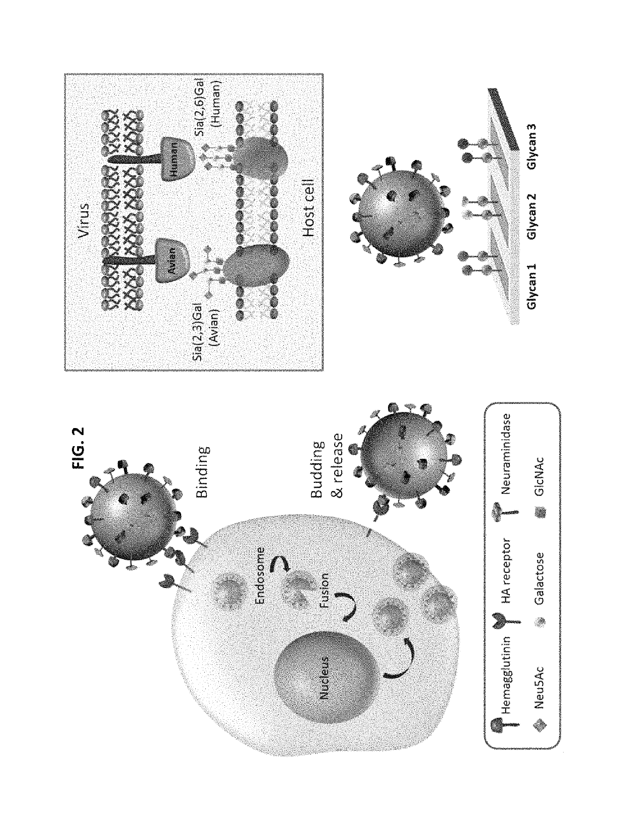
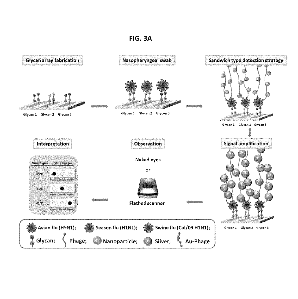
  • Glycan arrays for high throughput screening of viruses
  • Glycan arrays for high throughput screening of viruses
  • Glycan arrays for high throughput screening of viruses

Examples


example 1

nthesis

[0132]All oligosaccharides were prepared using synthetic procedures as described (Hsu, C.-H. et al. Chem. Eur. J. 2010, 16, 1754-1760; Hasegawa A, et al. Carbohydr. Res. 1995, 274, 165-181; Komba S, et al. Bioorg. Med. Chem. 1996, 4, 1833-1847; Otsubo N, et al. Carbohydr. Res. 1998, 306, 517-530; Yoshida M, et al. Glycoconjugate J. 1993, 10, 324). The synthetic oligosaccharides were further modified to cross-link to glass surface for the preparation of “glycan arrays” as described below.

example 2

paration

[0133]Samples of various viruses are collected from the Center for Disease Control and Prevention in TAIWAN, and provided by Dr. Jia-Tsong Jan (Genomic Research Center, Academia Sinica, Taiwan). All viruses were propagated in 10-day-old embryonated specific-pathogen free chicken eggs. Purified viruses were done by centrifugation at 100,000 g for 30 min and resuspended in PBS. Differences in the degree of virus purity did not influence significantly the pattern of receptor binding. Virus were inactivated by treatment with β-propiolactone (BPL; 0.05% v / v) for 60 minutes at 33° C., and resuspended in 0.01 M phosphate buffered saline pH 7.4 (PBS) and stored at −80° C. Comparison of samples of live and inactivated virus showed that BPL inactivation did not alter receptor binding specificity (Matrosovich M, et al. J. Virol. 1999, 73, 1146-1155.)

example 3

ray Fabrication

[0134]Amino-reactive N-hydroxysuccinimide (NHS)-activated glass microscope slides were used to fabricate glycan arrays by the standard protocol of Protein Application Nexterion Slide H. The NHS groups on the glass surface react readily with the primary amines of the amino modified glycans to form an amide linkage. Amine-modified glycans with desired concentration were dissolved in printing buffer (pH 8.5, 300 mM phosphate buffer with 0.005% (v / v) Tween 20) and were spotted on the glass microscope slides by robotic pin (BioDot, Cartesian Technologies; SMP3, Telechem International) deposition. Print slides were incubated under 80% humidity at 37° C. for 2 h followed by desiccation overnight. Before the binding assay, print slides were immersed with blocking buffer (50 mM ethanolamine in 50 mM borate buffer, pH 9.2) at room temperature for 1 h to remove remaining free NHS groups and then washed with washing buffer (PBST; 50 mM PBS buffer with 0.05% (v / v) Tween 20, pH 7.4...


PUM

PropertyMeasurementUnit
diameteraaaaaaaaaa
diameteraaaaaaaaaa
humidityaaaaaaaaaa

Abstract

Glycan arrays that can detect and distinguish between various sub-types and strains of influenza virus are provided. Methods for using the glycan arrays with assays using nanoparticle amplification technique are disclosed. Sandwich assays using gold nanoparticles conjugated to phage particles comprising influenza virus-specific antibodies for detecting multiple serotypes using a single reaction are provided. Plurality of glycans directed to specific target HA of influenza virus comprises the array. Detector molecules comprising noble metals conjugated to (a) phage display particles expressing antibodies against hemagglutinin and (b) neuraminidase binding agents are disclosed.

Description

[0001]This application is a U.S. National Stage entry of International Application No. PCT / US2011 / 032192 filed Apr. 12, 2011 which application claims priority under the Paris Convention to U.S. Provisional Patent Application Ser. No. 61 / 323,300, filed Apr. 12, 2010, the contents of which are incorporated herein by reference.TECHNICAL FIELD OF THE INVENTION[0002]The present invention relates to classification of influenza virus subtypes. Specifically, the invention relates to glycan arrays which can detect and distinguish between influenza virus subtypes. More specifically, the invention relates to methods for detecting influenza virus subtypes using nanoparticle-based detection methods. The invention relates to detector molecules comprising noble metals conjugated to (a) phage display particles expressing antibodies against hemagglutinin and (b) neuraminidase binding agents.BACKGROUND OF THE INVENTION[0003]Influenza has a long history of pandemics, epidemics, resurgences and outbrea...

Claims


Application Information

Patent Timeline
Patent Type & Authority Patents(United States)
IPC IPC(8): G01N33/569G01N33/53G01N33/543
CPCG01N33/56983G01N33/53G01N33/54346G01N2458/00G01N2400/00G01N2400/38G01N2333/11
Inventor WONG, CHI-HUEYWU, CHUNG-YILIANG, CHI-HUIYANG, AN-SUEI
Owner ACAD SINIC
Learn More