Vesicular stomatitis virus glycoprotein variant and construction method and application thereof
A technology of protein variants and membrane proteins, applied in the direction of viruses/bacteriophages, applications, viruses, etc., can solve the problems of virulence recovery and inapplicability, and achieve the effects of reducing infectivity, good safety, and reducing fusion ability
- Summary
- Abstract
- Description
- Claims
- Application Information
AI Technical Summary
Problems solved by technology
Method used
Image
Examples
Embodiment 1
[0069] Embodiment 1 Experiment related methods and processes
[0070] 1.1. Cell culture
[0071] HEK293T( CRL-11268) and Hela ( CRM-CCL-2) cells were cultured at 37°C, saturated humidity, 5% CO 2 in the cell culture incubator.
[0072] 1.2. Western blot
[0073] 1) Discard the culture medium and wash twice with PBS buffer;
[0074] 2) Add pre-cooled whole-cell protein lysate (50mM Tris (pH 7.4), 150mM NaCl, 1% NP-40, 0.5% sodium deoxycholate), about 30μL / mL cells, pipette to mix, place on ice for 10min ;
[0075] 3) Centrifuge the cell lysate suspension at 13300 rpm at 4°C for 10 min, transfer the supernatant to a new EP tube, and obtain the cell protein extract;
[0076] 4) Add 6x SDS loading buffer (6x loading buffer: 3.75mL 1M Tris pH 6.8, 1.2g SDS, 6mL glycerol, 0.006g bromophenol blue, 0.462g DTT) in proportion, and bathe in boiling water at 100°C for 5-10min;
[0077] 5) Load the sample on the SDS-PAGE gel. When the sample is in the stacking gel, electrophore...
Embodiment 2V
[0121] Cell fusion mediated by embodiment 2VSV-G
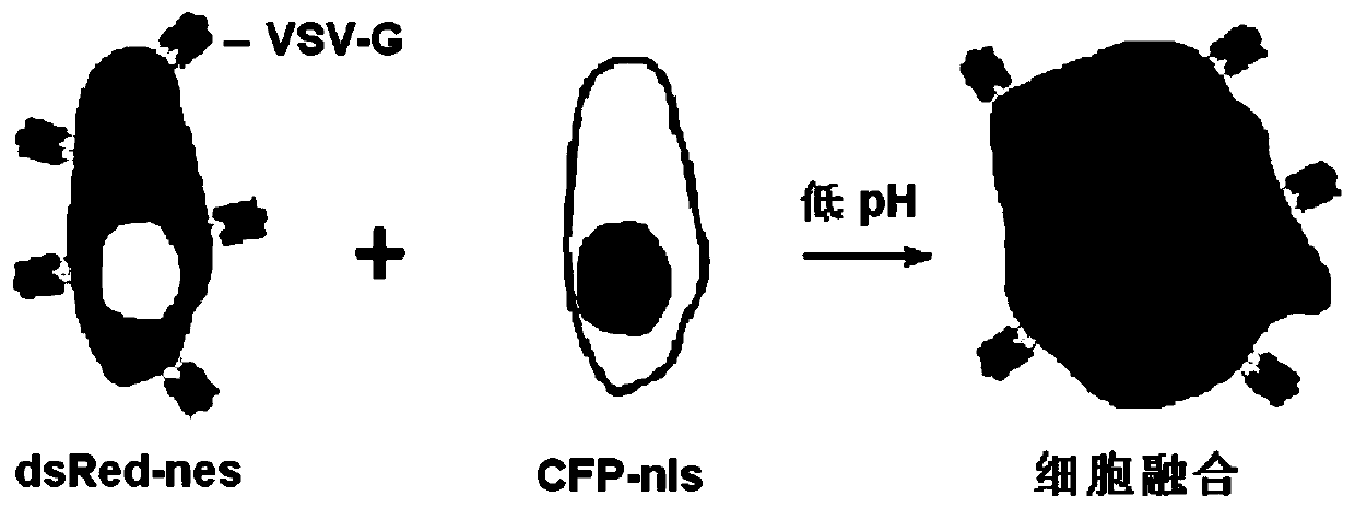
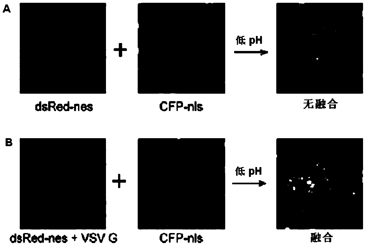
[0122] As a viral fusion protein, VSV-G's fusion ability has been confirmed. In order to quantitatively analyze its fusion function and monitor the kinetic process of fusion in real time, the inventors constructed a cell-cell fusion system. The red fluorescent protein (dsRED-nes) with nuclear export signal (dsRED-nes) and VSV-G were co-transfected into cells, the cytoplasm of the transfected cells was red under the fluorescence microscope, and VSV-G was expressed on the surface of the cell membrane; in addition, the cells with Cells were transfected with cyan fluorescent protein (CFP-nls) with nuclear localization signal, and the nuclei of the transfected cells were cyan under the fluorescence microscope. These two kinds of cells were co-cultured, and low pH was used to induce VSV-G-mediated fusion. If the cells with red cytoplasm and more than one cyan nucleus were fused cells, such as figure 1 and figure 2 shown.
[0...
Embodiment 3
[0148] Example 3 Preparation of Lentivirus Expressing VSV-G Variants and Cell Infection Experiment
[0149] As a viral fusion protein, VSV-G mediates the fusion of the viral envelope with the endocytic membrane of the target cell, and then invades the cell. The host range of VSV-G-mediated fusion is very wide. VSV-G can be expressed on the surface of genetically engineered lentivirus as a fusion agent of lentiviral transfection vector. Since the genetically engineered lentivirus has no self-replication ability, it is safe High reliability, the system has been widely used in research. In the lentiviral packaging system consisting of three plasmids, pLVTHM, pMD 2.G and psPAX2 (all purchased from Addgene), pMD 2.G carries the VSV-G gene, so that the obtained virus surface expresses VSV-G, which can mediate Fusion of virus and cells. The inventors used conventional molecular cloning techniques to modify VSV-G with reference to Example 2, replacing its transmembrane region or ins...
PUM
 Abstract
Description
Claims
Application Information
 - Generate Ideas
- Intellectual Property
- Life Sciences
- Materials
- Tech Scout
- Unparalleled Data Quality
- Higher Quality Content
- 60% Fewer Hallucinations
Browse by: Latest US Patents, China's latest patents, Technical Efficacy Thesaurus, Application Domain, Technology Topic, Popular Technical Reports.
© 2025 PatSnap. All rights reserved.Legal|Privacy policy|Modern Slavery Act Transparency Statement|Sitemap|About US| Contact US: help@patsnap.com


