


Quick Research
Generate reliable direction feasibility study reports for your R&D in just a few steps.

Technical Q&A
Discover and master advanced knowledge NOW. Basics, ideas, possibilities, all at once.

Find Solutions
As an expert in R&D theories, this can generate solutions to your technical problems instantly.

Evaluate Feasibility
Analyze your overall solution with one click, know your potential R&D risks in advance.

Monitor Landscape
Get weekly tech updates, stay abreast of the latest tech innovations and key insights.
MDCK-KOmavs cell line suitable for proliferating canine distemper virus and avian influenza virus, and application thereof
A technology of avian influenza virus and canine distemper virus, which is applied in the field of molecular biology, can solve the problems of insufficient source and quantity of chicken embryos, foreign virus contamination, gene variation, etc.
- Summary
- Abstract
- Description
- Claims
- Application Information
AI Technical Summary
Problems solved by technology
Method used
Image

Examples
Embodiment 1
[0099] Example 1 Construction of MDCK-KOmavs cell line suitable for canine distemper virus isolation and propagation
[0100] 1. Construction of replication-deficient lentivirus used to express Cas9
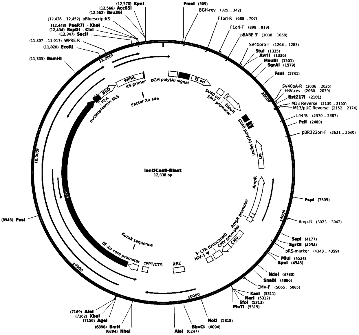
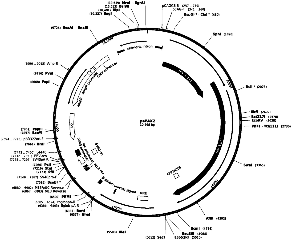
[0101] The 293FT cells were plated into a 6-well plate, and the lentivirus-packaged plasmids pMD2.G, psPAX2, lentiCas9-Blast were transfected into the cells according to 2:3:5, and the total amount of plasmids was 3μg. Among them, pMD2.G can form a viral envelope , PsPAX2 can express gag and pol for virus packaging. The lentiCas9-Blast plasmid not only contains packaging signals and long terminal repeats that are packaged into pseudoviral particles, but also contains Cas9 protein and blasticidin resistance screening genes; the solution is changed 8 hours after transfection, Collect the cell culture supernatant after 48h of transfection and 72h of transfection. After centrifugation for 10 minutes at 4°C, 12,000 rpm, the supernatant is the solution containing lentiviral particles.
[010...
Embodiment 2
[0159] Example 2 Isolation of CDV using the above MDCK-KOmavs cell line
[0160] Collect the eye and nose swabs and blood of the canine distemper cases that are positive by the colloidal gold test strip, centrifuge at 4℃, 3000rpm, 10min to take the supernatant, 4℃, 8000rpm, 5min, centrifuge again, and take the supernatant in the ultraclean table After filtering and sterilizing through a 0.22um filter, the supernatant samples were aliquoted into sterilized 1.5ml EP tubes and placed at -80°C for later use.
[0161] After the MDCK-KOmavs and MDCK are cultured to cover the monolayer, discard the old medium, wash it with pancreatin, add appropriate amount of pancreatin and put it in a 37℃ incubator for digestion for about 5 minutes, discard the pancreatin, and add an appropriate amount Resuspend in the serum-free DMEM medium, and connect the prepared cell resuspension and the processed CDV positive sample at a ratio of 1000:1, each of which is connected to a 12-well cell culture plate a...
Embodiment 3
[0163] Example 3 Isolation of AIV using the above MDCK-KOmavs cell line
[0164] Collect the eye and nose swabs and blood of avian influenza cases that are positive by the colloidal gold test strip, centrifuge at 4℃, 3000rpm, 10min to take the supernatant, 4℃, 8000rpm, 5min again, take the supernatant in the ultra-clean table After filtering and sterilizing with a 0.22um filter, the supernatant sample is aliquoted into a sterilized 1.5ml EP tube and placed at -80°C for use.
[0165] After culturing MDCK-KOmavs and MDCK in a 12-well cell culture plate to cover the monolayer, discard the old medium, wash twice with Hank's solution to remove residual serum, and add serum-free DMEM medium to each well 1mL, with medium and processed AIV positive samples in the ratio of 100:1, 1000:1, and 10000:1, each is connected to a 12-well cell culture plate and placed in a cell incubator for 2 hours. The infection solution was removed, washed twice with Hank's solution, and DMEM maintenance medium...
PUM
 Abstract
Description
Claims
Application Information
 - R&D Engineer
- R&D Manager
- IP Professional
- Industry Leading Data Capabilities
- Powerful AI technology
- Patent DNA Extraction
Browse by: Latest US Patents, China's latest patents, Technical Efficacy Thesaurus, Application Domain, Technology Topic, Popular Technical Reports.
© 2024 PatSnap. All rights reserved.Legal|Privacy policy|Modern Slavery Act Transparency Statement|Sitemap|About US| Contact US: help@patsnap.com