Fluorescent immunoblotting detection method of quantum dot nanosphere marker

A technique of fluorescent immunology and immunoblotting, applied in the field of immunoassay, to achieve the effects of controllable conditions, uniform particle size, and easy control of concentration

Inactive Publication Date: 2018-10-16
上海昆道生物技术有限公司
View PDF3 Cites 4 Cited by
  • Summary
  • Abstract
  • Description
  • Claims
  • Application Information

AI Technical Summary

Problems solved by technology

And at present, the application of quantum dots in Western Blot detection is seldom compared with the chemiluminescence color method commonly used in laboratories.

Method used


Image

  • Fluorescent immunoblotting detection method of quantum dot nanosphere marker

Examples


Embodiment 1

[0040] Example 1: Preparation of Quantum Dot Nanosphere Fluorescent Secondary Antibody

[0041] The preparation of CdSe / ZnS quantum dot-polymer composite nanosphere comprises the following steps:

[0042] A. Oil phase components: Disperse CdSe / ZnS quantum dots with an emission wavelength of 610nm and polystyrene-maleic anhydride copolymer in chloroform solution, the concentration of quantum dots is 0.1μmol / L, and the concentration of polymer is 0.05mg / mL;

[0043] B, water phase component: configure polyvinyl alcohol and sodium dodecylsulfonate into an aqueous solution, wherein the concentration of polyvinyl alcohol is 2.5%, and the concentration of sodium dodecylsulfonate is 0.5%;

[0044] C. In a water bath at 4°C, under magnetic stirring, add the oil phase solution to the water phase solution, and keep stirring for 60 minutes, and then use a sonicator to sonicate the solution for 10 minutes to obtain a uniform and stable microemulsion.

[0045] D, at room temperature, th...

Embodiment 2

[0050] Example 2: Western blot analysis of target protein

[0051] (1) SDS-PAGE electrophoresis

[0052] Prepare 10% separating gel and 5% stacking gel, load the treated Hela cell lysate protein, perform constant voltage electrophoresis, first wait for the protein to run to the interface of the stacking gel and separating gel at 80V voltage, and then electrophoresis to the target at 120V voltage protein separation.

[0053] (2) Protein electrotransfer from the gel to the membrane

[0054] The PVDF membrane was soaked in methanol and then washed 3 times with the membrane transfer buffer, and then the separated protein electrophoresis gel was removed from the stacking gel, stacked according to 3 layers of filter paper, gel, PVDF membrane, and 3 layers of filter paper, and placed in a semi-dry Transfer the protein to the membrane with 2mA / cm2 for 40 minutes on the electrotransfer instrument.

[0055] (3) Incubate the primary antibody

[0056] The transferred membrane was wash...

Embodiment 3

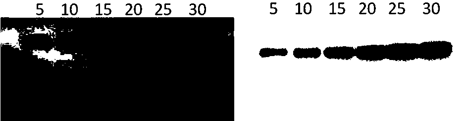
[0058] Example 3: Analysis of target protein by chemiluminescence

[0059] The membrane obtained in Example 2 after incubation with the primary antibody was further incubated with HRP enzyme-labeled goat anti-mouse secondary antibody (5000-fold dilution), incubated at room temperature for 1 h, and then washed 3 times with TBST buffer;

[0060] Chemiluminescence substrate ECL kit, color development, exposure with a chemiluminescence imager, and save the image obtained under a better exposure time.


PUM

PropertyMeasurementUnit
Particle sizeaaaaaaaaaa

Abstract

The invention belongs to the technical field of immunoassay and in particular relates to a method for detecting trace protein by fluorescent immunoblotting of quantum dot nanospheres. The method comprises the following steps: firstly, carrying out gel electrophoresis separation on a protein mixture to be detected; transferring gel subjected to the electrophoresis separation into a solid-phase carrier film; then sealing a protein-loaded solid-phase film; after adding a specific primary antibody of protein to be detected, washing; then reacting with a fluorescent secondary antibody of the quantum dot nanosphere marker and washing; finally, developing and photographing through a protein blot film under the illumination of exciting light. Compared with chemiluminescence, the method for detecting the protein through the fluorescent immunoblotting, provided by the invention, has the characteristics that luminescent substrates are not needed, a detection signal is more stable and can be stored for a long period, results have consistency and repeatability and the like; compared with near-infrared fluorescent immunoblotting, the method has the characteristics that an additional signal amplification step is not needed, a fluorescent signal is more stable, light-shielding operation is not needed, an expensive near-infrared laser scanner does not need to be used for detecting and the like.

Description

technical field [0001] The invention belongs to the technical field of immunodetection, and in particular relates to a method for detecting trace protein by fluorescent immunoblotting marked with quantum dot nanospheres. Background technique [0002] Western Blot is to transfer the cell or tissue lysate separated by electrophoresis from the gel to a solid-phase membrane (usually nitrocellulose membrane or polyvinylidene fluoride membrane), and then use a specific antibody to detect a specific antigen. A protein detection technology, which is a commonly used protein analysis and detection method in biological laboratories. [0003] At present, the commonly used immunoblotting color development methods in the laboratory include: enzymatic substrate chemiluminescence and near-infrared fluorescence. Chemiluminescence generally uses horseradish peroxidase (HRP)-labeled secondary antibody plus chemiluminescent substrate (ECL) for development. , the chemiluminescent signal needs c...

Claims


Application Information

Patent Timeline
IPC IPC(8): G01N21/64G01N33/532G01N33/68
CPCG01N21/64G01N33/532G01N33/6803
Inventor 不公告发明人
Owner 上海昆道生物技术有限公司
Why Eureka
Try Eureka