Cervical cancer cell detection kit
A detection kit, cervical cancer cell technology, applied in the direction of measuring devices, microbiological determination/inspection, instruments, etc., can solve the problems of prone to photobleaching and photodegradation, poor photochemical stability, wide emission spectrum, etc., and achieve optical resistance Strong interference ability, improved detection sensitivity, and large amount of information
- Summary
- Abstract
- Description
- Claims
- Application Information
AI Technical Summary
Problems solved by technology
Method used
Image

Examples
Embodiment 1
[0035] The preparation of embodiment 1 kit
[0036] (1) Preparation of B-GQDs
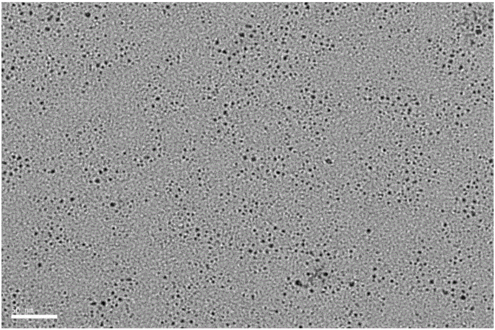
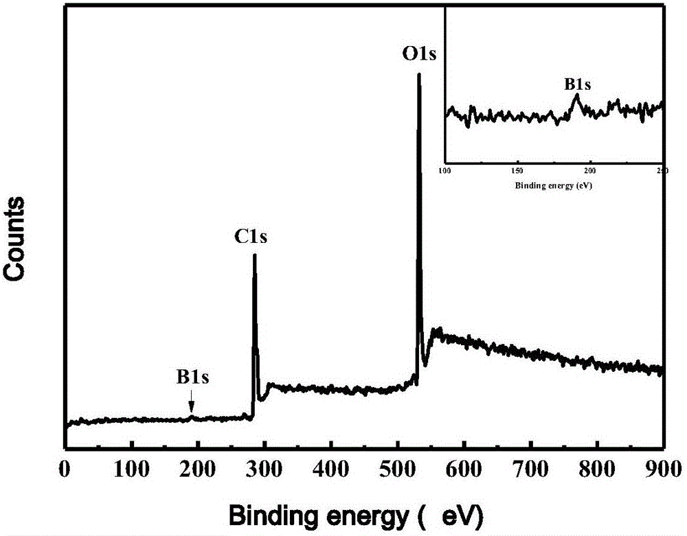
[0037] Boron-doped graphene quantum dots (B-GQDs) were synthesized by potentiostatic chronoamperometry via an electrochemical workstation (CHI 660B). The specific method is: prepare 0.1mol / L borax aqueous solution, and take 20mL as electrolyte. A high-purity graphite rod was used as the working electrode, a platinum wire electrode was used as the counter electrode, and a calomel electrode was used as the reference electrode, and the working potential was 3V. After reacting for 2 hours, the solution was filtered through a 0.22 μm filter membrane to remove graphene sheets. Dialyze the above solution through a 3500Da dialysis bag for 24 hours to remove Na in the solution + and B 4 o 7 2- , to get B-GQDs. Gained B-GQDS is evenly distributed, and its particle size is between 3-8nm ( figure 1 ); Its elemental analysis found that it contains B element, the content is 3.2% ( figure 2 ).
[0038]...
Embodiment 3
[0045] Embodiment 3 establishes the working curve that detects HeLa cell concentration
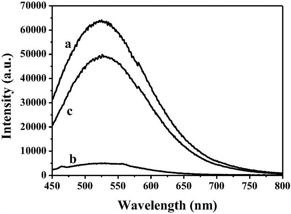
[0046] Take 1.0 mL of reagent A and 20.0 μL of reagent B, mix well, add 10.0 μL of reagent C to make a mixed reagent, mix the mixed reagent with different concentrations of cervical cancer cell HeLa (10-10 6 cell / mL) at 37°C, 5% CO 2 Incubate for 2 h in the incubator; record the fluorescence signal F of B-GQDs by fluorescence spectrometer i ( Figure 4 ), Figure 4 The middle a-g respectively represent the cell concentration of 0, 10, 10 2 、10 3 、10 4 、10 5 、10 6 cell / mL. Calculate the difference ΔF of the response signal (ΔF=F i -F 0 ); The concentration of described ΔF and HeLa cells is drawn into ΔF-c working curve ( Figure 5 ); thereby established the HeLa cell concentration detection working curve based on the fluorescence response of B-GQDs; its linear range is 10-10 6 cell / mL.
Embodiment 4
[0047] Detection of HeLa cells in the sample to be tested in embodiment 4
[0048] Get the HeLa cell to be tested, act on the reagent in the kit according to the same method as in Example 3 and detect its fluorescence signal, the experimental results show that when the HeLa cell exists, the fluorescence signal of the quantum dots obviously recovers; according to the fluorescence signal of the HeLa cell The degree of change, from the standard curve obtained in Example 3, can further obtain the concentration of HeLa cells to be tested. Human embryonic kidney epithelial HEK 293T cells were used as the detection object. When it interacted with the kit, there was no significant change in the fluorescence signal of the quantum dots. It shows that the kit can be used for the specific detection of cells (such as HeLa cells) positively expressing alkaline phosphatase.
PUM
| Property | Measurement | Unit |
|---|---|---|
| particle size | aaaaa | aaaaa |
| particle diameter | aaaaa | aaaaa |
Abstract
Description
Claims
Application Information
 - R&D
- Intellectual Property
- Life Sciences
- Materials
- Tech Scout
- Unparalleled Data Quality
- Higher Quality Content
- 60% Fewer Hallucinations
Browse by: Latest US Patents, China's latest patents, Technical Efficacy Thesaurus, Application Domain, Technology Topic, Popular Technical Reports.
© 2025 PatSnap. All rights reserved.Legal|Privacy policy|Modern Slavery Act Transparency Statement|Sitemap|About US| Contact US: help@patsnap.com