Visualization and quantitation of cellular cytotoxicity using cell-permeable fluorogenic protease substrates and caspase activity indicator markers

a fluorogenic protease substrate and cytotoxicity technology, applied in the field of immunology, can solve the problems of large background signal, inability to study direct ctl killing of primary host target cells, and little information about the kinetic interaction of effectors and targets

Active Publication Date: 2011-04-19
ONCOIMMUNIN
View PDF15 Cites 14 Cited by
  • Summary
  • Abstract
  • Description
  • Claims
  • Application Information

AI Technical Summary

Problems solved by technology

Second, CTL killing of primary host target cells often cannot be studied directly as only certain types of cells, primarily immortalized cell lines, can be efficiently labeled with 51Cr (Nociari et al.
Third, target-cell death is measured at the end point of the entire process and thus provides little information about the kinetic interaction of effectors and targets at the molecular and cellular levels.
Fourth, the radio active conventional assay using chromium 51 isotope (51Cr) results in a very large background (noise) signal due to a large amount of spontaneous nonspecific release of the isotope from the target cells and often very heterogeneous loading of the isotope in the selected target cells.
Consequently, the conventional chromium release assay has difficulty in detecting definite but less potent cytotoxic effects, i.e., it is difficult to distinguish a signal caused by cell-mediated cytotoxic activity from the assay's background radioactivity.
Measurement of 51Cr release does not permit monitoring the physiology or fate of effector cells as they initiate and execute the killing process.
Finally, radioactive materials require special licensing and handling, which substantially increases cost and complexity of the assay.
More recently developed immunologic methods, including major histocompatibility complex (MHC)-tetramers, intracellular cytokine detection and ELISPOT assays, have greatly improved sensitivity to enumerate antigen-specific T cells; however, these newer methods do not assess the cytolytic function of antigen-specific cell-mediated cytotoxicity (Altman et al.
However, none of these assays reveal the fundamental processes responsible for the initiation and execution of target-cell killing, and none have yet been applied to analyses of primary cell-mediated cytotoxicity generated in vivo following antigenic exposure.

Method used


Image

  • Visualization and quantitation of cellular cytotoxicity using cell-permeable fluorogenic protease substrates and caspase activity indicator markers
  • Visualization and quantitation of cellular cytotoxicity using cell-permeable fluorogenic protease substrates and caspase activity indicator markers
  • Visualization and quantitation of cellular cytotoxicity using cell-permeable fluorogenic protease substrates and caspase activity indicator markers

Examples


example 1

Visualization and Quantification of T Cell-Mediated Cytotoxicity Using Cell-Permeable Fluorogenic Caspase Substrates

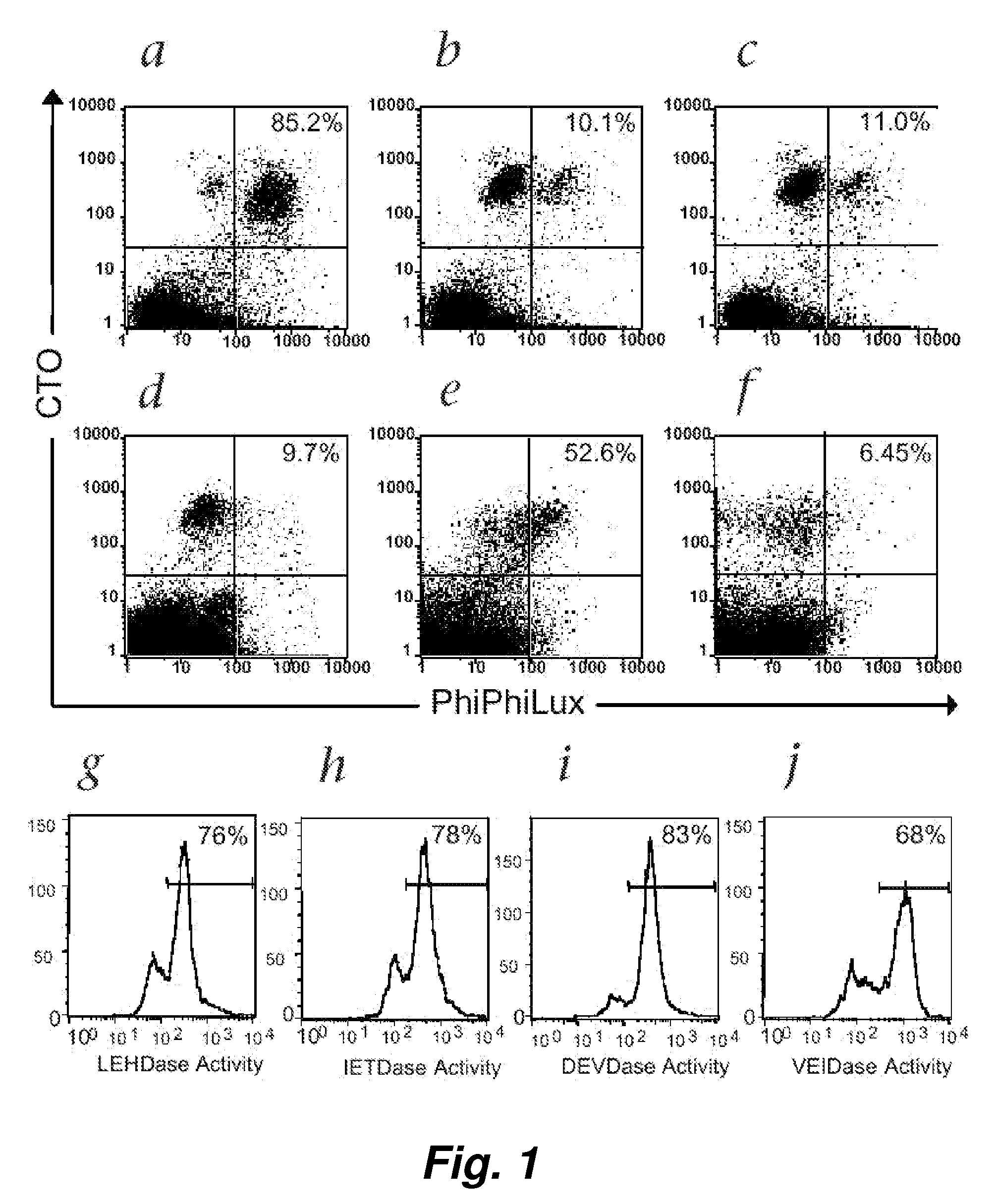
[0100]Following T-cell receptor recognition of antigenic peptide-MHC class I complexes on the surface of target cells, cytotoxic effector cells (e.g., CTLs) induce target-cell apoptosis either through directed exocytosis of perforin and granzymes or through ligation of “death receptors” in the Fas / Fas ligand (FasL) pathway. An immediate event following both types of cytotoxic signaling is the activation of the caspase cascade within the target cells (Atkinson et al. (1998) J. Biol. Chem. 273: 21261-21266). We have used a novel class of cell-permeable fluorogenic caspase substrates to develop a fluorescence-based cellular cytotoxicity (FCC) assay that detects CTL-induced caspase activation within individual target cells (Packard et al. (1996) Proc. Natl. Acad. Sci. USA 93: 11640-11645; Komoriya et al. (2000) J. Exp. Med. 191: 1819-1828). These reagents are composed of t...

example 2

Cell Permeable Fluorogenic Caspase Substrates Show the Presence of Memory Cells where as the Tradition Chromium Release Assay Did not

[0122]Detection of memory CTL responses using chromium release assay generally requires a 5 to 6-day in vitro restimulation and the expansion of CTL precursors in culture. With the improved sensitivity of fluorescence cellular cytotoxicity (FCC) assays described herein, we believed that we would be able to detect a memory CTL response with limited or no in vitro restimulation.

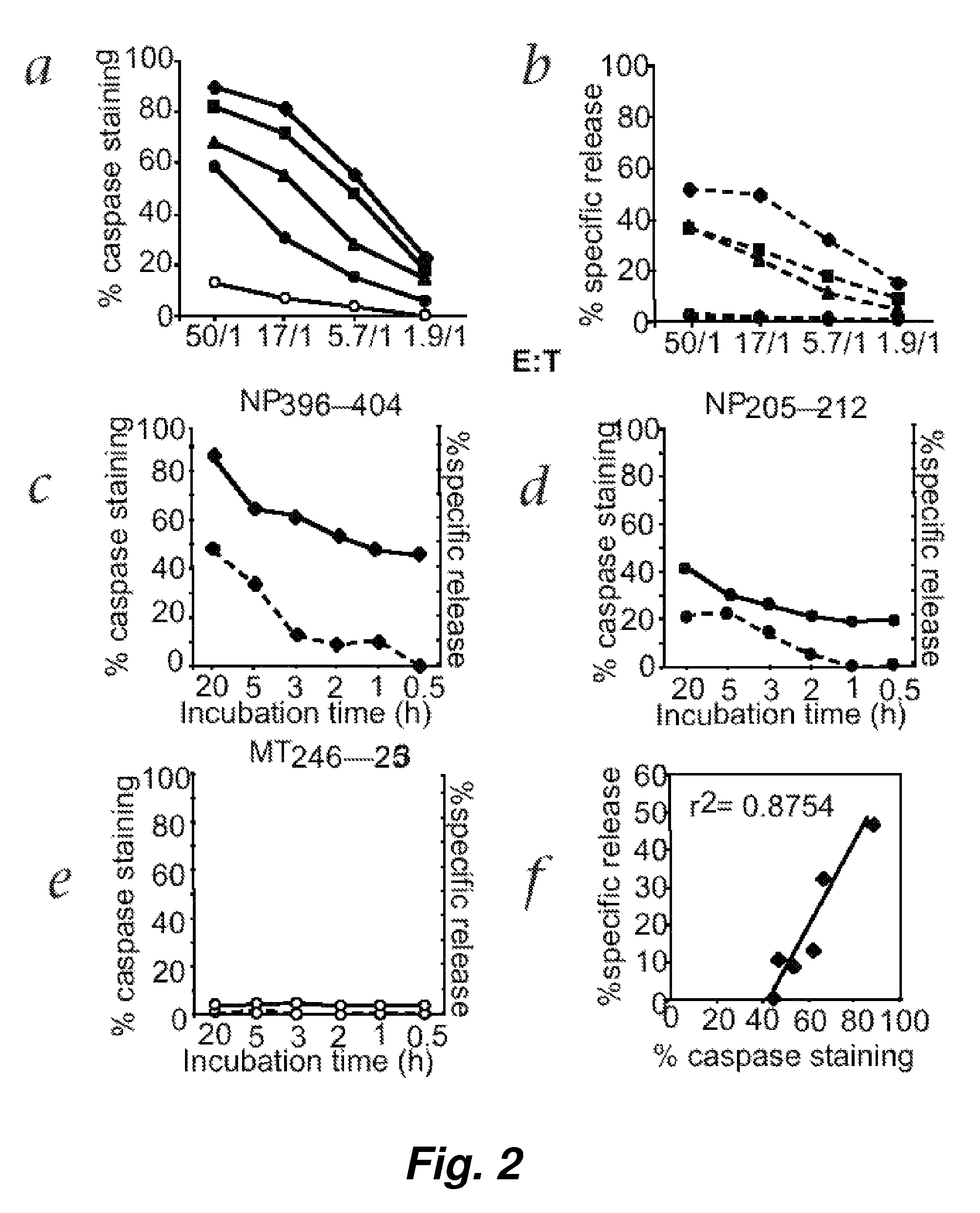
[0123]To test this hypothesis, direct ex vivo memory CTL activity specific for NP396-404 was measured using both the fluorescence cellular cytotoxicity (FCC) and 51chromium release assays. Freshly prepared spleen cells obtained from LCMV-infected C57BL / 6 mice 32 days after initial LCMV infection were incubated at various E / T ratios with target EL4 cells for 5 hours. FIG. 5 shows that, surprisingly, the fluorescence cellular cytotoxicity (FCC) assay but not 51chromium release assay...

example 3

Cellular Cytotoxicity Assay Using Various Adherent and Suspension Cells as the Target Cells

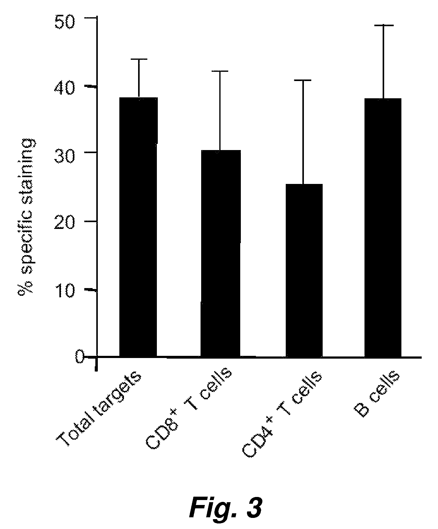
[0124]In order to evaluate the broader applicability of the present invention, we have tested assay performance using both an adherent cells as well as additional suspension cells as the target cells. The effector cells in this experiment were human NK cells. The percentage killing (see Table 2) observed for just 1 hour of co-incubation of the effector cells with an effector to target cell ratio of 5 to 1 shows clearly that the cell-mediated cytotoxicity described herein works very well with these widely different cell types, i.e. the adherent human breast adenocarcinoma cell and human suspension cells (Jurkat and K562).

[0125]An alternative assay was also performed where the VEIDase substrate (VEID, SEQ ID NO:23) rather than DEVDase substrate (DEVD, SEQ ID NO:22) was used. It has been reported that this caspase 6 substrate with the VEID tetrapeptide amino acid sequence is also recognized by cy...


PUM

PropertyMeasurementUnit
molecular weightaaaaaaaaaa

Abstract

This invention provides a non-radioactive assay to monitor and quantify the target-cell killing activities mediated by cytotoxic T lymphocytes (CTLs). This assay is predicated on the discovery that apoptosis pathway activation and, in particular, granzyme B activity, provides a measure of cytotoxic effector cell activity. In one embodiment, measurement of CTL-induced granzyme B activation in target cells is achieved through detection of the specific cleavage of fluorogenic granzyme B substrates. This assay reliably detects antigen-specific CTL killing of target cells, and provides a more sensitive, more informative and safer alternative to the standard 51Cr-release assay most often used to quantify CTL responses. The assay can be used to study CTL-mediated killing of primary host target cells of different cell lineages, and enables the study of antigen-specific cellular immune responses in real time at the single-cell level. As such, the assay can provide a valuable tool for studies of infectious disease pathogenesis and development of new vaccines and immunotherapies.

Description

CROSS-REFERENCE TO RELATED APPLICATIONS[0001]This application claims benefit of and priority to U.S. Ser. No. 60 / 763,804, filed on Jan. 30, 2006, which is incorporated herein by reference in its entirety for all purposes. This application is also a continuation-in-part of U.S. Ser. No. 10 / 353,791, filed Jan. 28, 2003, which claims benefit of and priority to U.S. Ser. No. 60 / 353,712, filed Jan. 29, 2002, all of which are herein incorporated by reference in their entirety for all purposes.STATEMENT AS TO RIGHTS TO INVENTIONS MADE UNDER FEDERALLY SPONSORED RESEARCH AND DEVELOPMENT[0002][Not Applicable]FIELD OF THE INVENTION[0003]This invention pertains to the field of immunology. In particular, this invention provides improved cellular assays for determining the presence or activity of cytotoxic effectors cells that will mount a cytotoxic response against a particular cell or antigen including pathogen.BACKGROUND OF THE INVENTION[0004]Cytotoxic T lymphocytes (CTLs) have crucial roles i...

Claims


Application Information

Patent Timeline
Patent Type & Authority Patents(United States)
IPC IPC(8): C12N5/00C12Q1/37G01N33/53
CPCA61K38/07A61K38/08G01N33/5047G01N33/582G01N33/5055G01N33/533G01N33/505
Inventor PACKARD, BEVERLYKOMORIYA, AKIRA
Owner ONCOIMMUNIN
Why Eureka
Try Eureka