Virosome-like-particles

Active Publication Date: 2011-03-08
BESTEWIL HOLDING BV
View PDF6 Cites 11 Cited by
  • Summary
  • Abstract
  • Description
  • Claims
  • Application Information

AI Technical Summary

Benefits of technology

[0017]Here we demonstrate that the spikes of influenza virus and SFV can be efficiently solubilised and reconstituted with a short-chain phospholipid, such as DHPC or DCPC, by dialysis. Our results show that we found a new method for the reconstitution of viral envelope proteins, such as HA and E1 / E2, and that the membrane fusion activities of the reconstituted viral envelopes corresponded well to the fusion characteristics of the intact viruses.

Problems solved by technology

However, while the injection of live viruses generally induces a strong immune response, protecting against future infections by the virus, protein preparations may fail to do so.
These phagocytic cells patrol the body, ingesting particles of the size of viruses all the time, but they do not efficiently take up the purified proteins of split virus or subunit vaccines [1-2].
However, although ISCOMs have been used in a number of veterinary vaccines, and strongly enhance the immunogenicity of the viral membrane proteins, the development of such vaccines for humans has been inhibited by concerns about their toxicity and the complexity of the mixture [3].
While these multiple bacterial proteins may act as adjuvants, the complex nature of such mixtures, consisting of multiple proteins, lipids and other substances, will present a regulatory issue.
This step is thought to be necessary for a functional reconstitution of the native viral envelope, but quite difficult to control.
Current detergent removal protocols that result in reconstitution are mostly based on detergents with a low critical micelle concentration (cmc), and such detergents are particularly difficult to remove, in contrast to detergents with a comparatively high cmc, which can be removed by dialysis or ultrafiltration.
However, it was found that the latter type of detergent does not generally properly reconstitute viral membrane proteins, including the influenza virus hemagglutinin, leading predominantly to the formation of empty membranes on the one hand, and protein aggregates on the other.
However, removal of detergent from the supernatant by a hydrophobic resin can hardly be scaled up sufficiently.
However, the use of low-cmc detergents has a disadvantage in that they cannot be readily removed by dialysis.

Method used


Image

  • Virosome-like-particles
  • Virosome-like-particles
  • Virosome-like-particles

Examples


example 1

Preparation of a Virosome-Like-Particle Containing the Influenza Hemagglutinin Glycoprotein

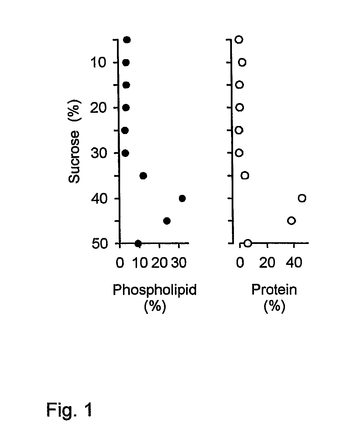
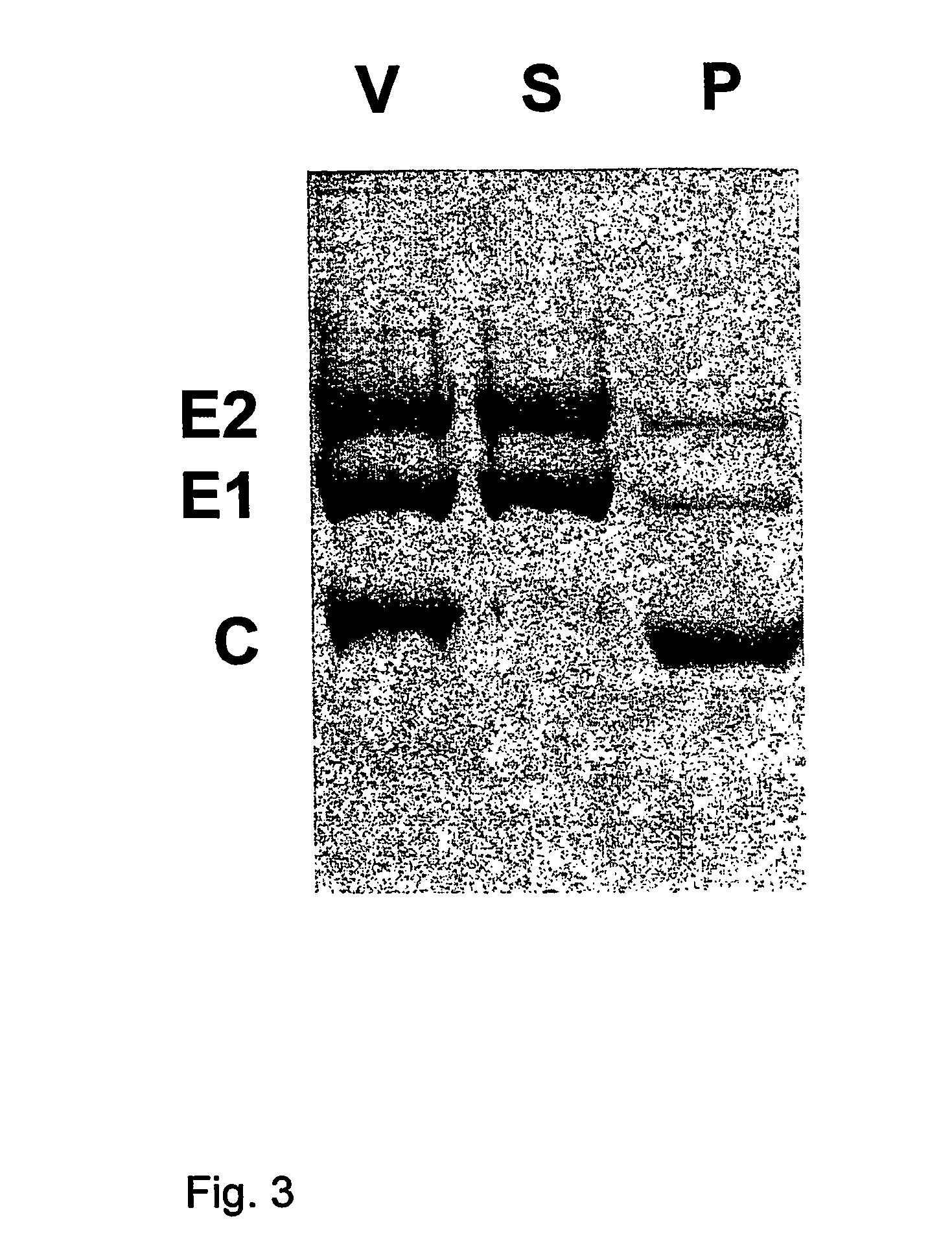
[0037]Influenza virus was produced by growing virus acquired from the World Influenza Center or the American Type Tissue Culture Collection (ATCC, Rockville, Md.), using methods known to persons skilled in the art, for instance by growing the virus on embryonated eggs or in cultured cells such as PER.C6™ or MDCK cells. The virus was then purified, by density gradient ultracentrifugation, and may subsequently be inactivated by treatment with beta-propiolactone or formaldehyde according to established procedures. Inactivation is not required for this procedure, and may affect the immune response. The virus may be further purified by the resuspension of sedimented virus in 5.0 mM Hepes (pH 7.4), containing 0.15 mM NaCl and 0.1 mM EDTA (HNE-buffer), and loading this preparation atop linear 10-60% (w / v) sucrose gradients in HNE, centrifuging at 90,000×g for 36 hr at 4° C. Virus can be pooled and al...

example 2

Preparation of a Virosome-Like-Particle Containing the Semliki Forest Virus Spike Glycoproteins

[0043]Semliki Forest virus, obtained from sources such as the ATCC, was produced on cell lines known in the art such as baby hamster kidney cells (BHK-21, from the ATCC) according to methods known in the art [23]. These cells were grown in Glasgow minimum essential medium, supplemented with 10% tryptose phosphate broth, 2.0 mM L-glutamine and 10% fetal bovine serum (all from GibcoBRL Life Technologies Inc., Paisley, UK). The medium was clarified by low-speed centrifugation (1,000×g) at 4° C. for 10 min, and the virus was pelleted from clarified cell-culture medium by ultracentrifugation at 100,000×g. The virus was further purified by the resuspension of sedimented virus in 6.0 mM Hepes (pH 7.4), containing 0.15 mM NaCl and 0.1 mM EDTA (HNE-buffer), and loading this preparation atop linear 10-60% (w / v) sucrose gradients in HNE, centrifuging at 90,000×g for 36 hr at 4° C. Virus was pooled an...

example 3

Membrane Fusion Activity of Influenza Virosomes

[0046]For analysis of the membrane fusion activity of the a virosome preparation, the X-47 strain of influenza virus (0.25 μmol phospholipid) was first sedimented by ultracentrifugation, and subsequently resuspended with 0.55 ml HNE-buffer, containing 25 mM DCPC. After a 30 min incubation at 0° C., the viral membrane was solubilized, and the nucleocapsids were removed by centrifugation for 20 min at 100,000×g at 4° C. The supernatant was then added to a dried thin film of 10 nmol of (1-pyrenedecanoyl)-sn-phosphatidylcholine (pyrPC) (Molecular Probes, Leiden, The Netherlands). After incubation for 20 min at 4° C., virosome-like-particles were then formed from this mixture by dialysis as described above. For the measurement of membrane fusion, the resulting virosome-like-particles were mixed with erythrocyte ghosts, prepared as described previously [25] from human red blood cells, at neutral pH (pH 7.4) at 37° C. At time 0, the medium was...


PUM

PropertyMeasurementUnit
Amphiphilicaaaaaaaaaa
Pharmaceutically acceptableaaaaaaaaaa
Solutionaaaaaaaaaa

Abstract

The invention relates to the production of virosome-like-particles. The invention provides a method for producing a virosome-like-particle comprising contacting an enveloped virus with a solution containing a short-chain phospholipid allowing solubilisation of the viral envelope of said virus further comprising removing short-chain phospholipid from said solution allowing formation of a functionally reconstituted viral envelope.

Description

[0001]This application is the U.S. National Phase of International Application No. PCT / NL2004 / 000101 filed on 11 Feb. 2004, which is incorporated herein by reference.BACKGROUND OF THE INVENTION[0002]The invention relates to the production of virosome-like-particles.[0003]Vaccines against membrane-containing (enveloped) viruses mostly consist of killed or live attenuated viruses, or a preparation of their proteins (e.g. split virus vaccines or subunit preparations). Vaccination with killed viruses and protein preparations is safer than vaccination with live attenuated viruses, because the latter may mutate or revert back to wild-type virus. Subunit vaccines have the clear advantage that they can be prepared from viral proteins expressed by cells rather than from virus, making production safer and eliminating the risk of contaminating vaccine preparations with live viruses. However, while the injection of live viruses generally induces a strong immune response, protecting against futu...

Claims


Application Information

Patent Timeline
IPC IPC(8): C12N7/04A61K9/127A61K39/12A61K39/145A61K39/295A61K39/39
CPCA61K9/1271A61K39/145A61K39/39A61K9/127A61K2039/5258A61K2039/55516C12N2760/16134C12N2760/16122C12N2760/16123C12N2770/36122C12N2770/36123A61K2039/55555A61K39/12A61P31/04A61P31/12A61P33/00A61P35/00
Inventor HUCKRIEDE, ANKE LUISE WILHELMINEDE JONGE, JORGEN MARTINSTEGMANN, ANTONIUS JOHANNES HENDRIKUSSCHOEN, PIETER JOSEPHWILSCHUT, JAN CHRISTIAAN
Owner BESTEWIL HOLDING BV
Why Eureka
Try Eureka