Imaging method, controller and imaging system, for monitoring a patient post evar

Active Publication Date: 2020-05-21
KONINKLJIJKE PHILIPS NV
View PDF0 Cites 0 Cited by
  • Summary
  • Abstract
  • Description
  • Claims
  • Application Information

AI Technical Summary

Benefits of technology

The invention provides a method to monitor an aneurysm after a stent graft procedure using two scans taken at different times. The scans can be compared by identifying the same recognizable features in the stent in the two images and aligning them based on the recognizable features. This approach avoids bias between different measurements in time, avoids an invasive procedure and applies to different imaging modes. The method can also include a user-guided registration and an imaged-based refinement of the registration to improve the alignment of the images.

Problems solved by technology

And as an aneurysm increases in size, the risk of rupture increases.
A ruptured aneurysm can lead to bleeding.
A ruptured AAA typically leads to hypovolemic shock and, if left untreated, death.
A type I endoleak is more serious, and results from the stent not sealing properly at the proximal or distal ends of the stent.
Less common but serious complications include type III and type IV endoleaks, which involve mechanical failure of the stent itself, which may result in surgical intervention.
Life-long surveillance after EVAR is troublesome for patients and physicians alike.
The surveillance requires significant resources, and may be associated with harm owing to the risk of potentially unnecessary interventions, repeated exposure to radiation, and contrast nephrotoxicity from the contrast agent.

Method used


Image

  • Imaging method, controller and imaging system, for monitoring a patient post evar
  • Imaging method, controller and imaging system, for monitoring a patient post evar
  • Imaging method, controller and imaging system, for monitoring a patient post evar

Examples


Embodiment Construction

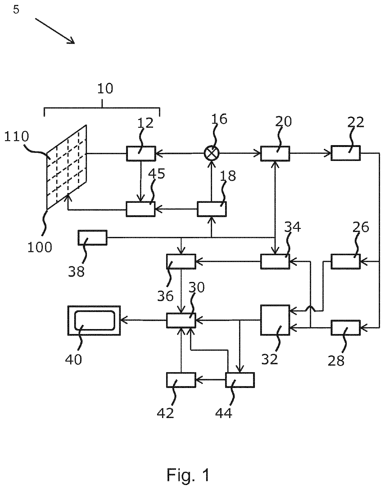
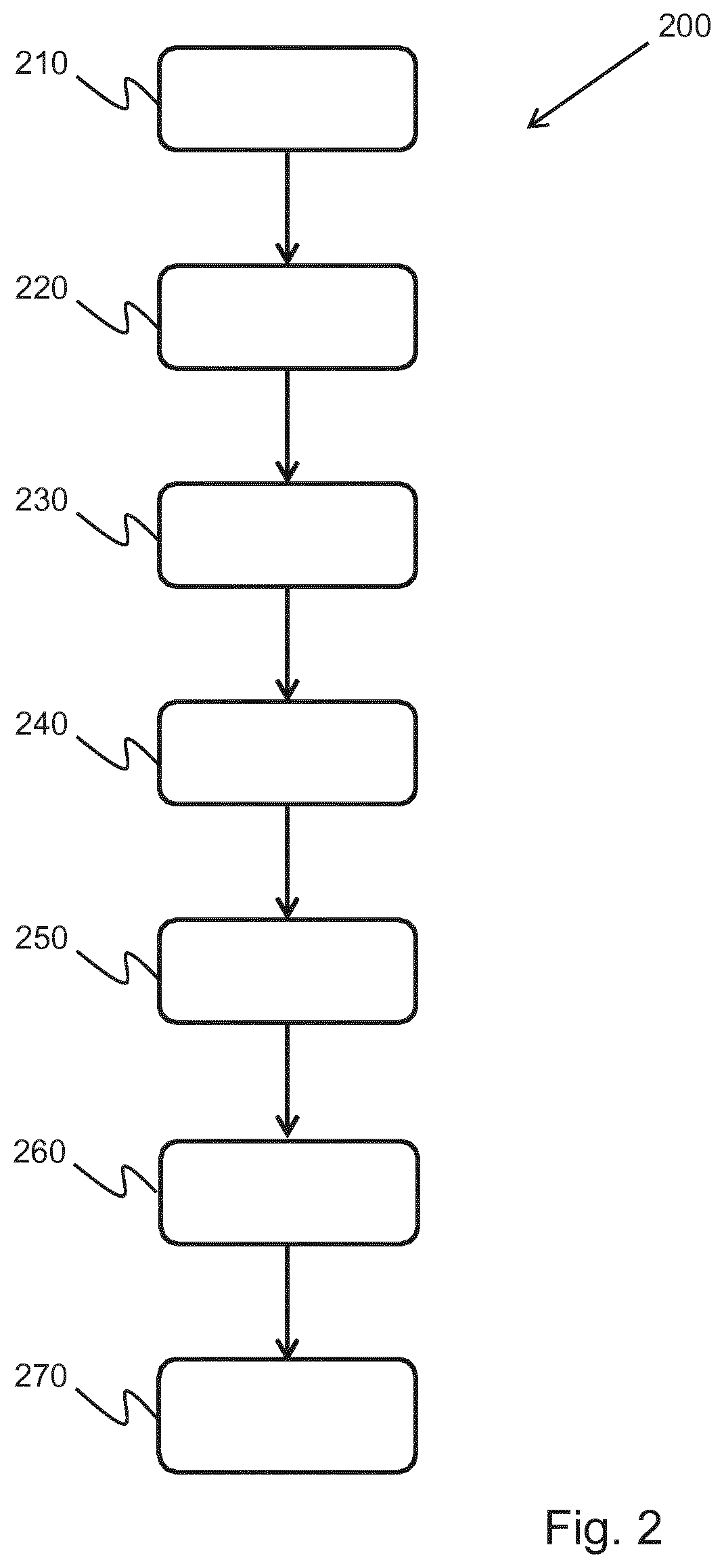
[0044]The invention provides a method of patient monitoring following EVAR using 3D-imaging to show any changes in size of the stent over time.

[0045]The invention provides a method of patient monitoring following endovascular aneurysm repair (EVAR) with a stent graft. It particularly applies to abdominal aortic aneurysm repair.

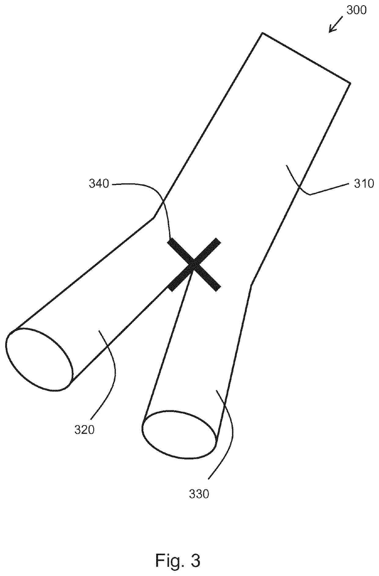
[0046]A first 3D volume scan of the stent in situ is provided and a second, subsequent 3D volume scan of the stent is also provided. One or more fiducial markers are generated in a first image derived from the first scan and in a second image derived from the second scan. The fiducial markers identify recognizable features of the stent, such as the bifurcation point in the stent graft. A rigid 3D transform mapping based on the one or more fiducial markers is extracted and then a registration is applied to the first and second scans based on the 3D rigid transform between the first and second images. A size value is derived at the same location in the aneurysm ...


PUM


Abstract

The invention provides a method of patient monitoring following endovascular aneurysm repair (EVAR) with a stent graft. It particularly applies to abdominal aortic aneurysm repair. A first 3D volume scan of the stent in situ is provided and a second, subsequent 3D volume scan of the stent is also provided. One or more fiducial markers are generated in a first image derived from the first scan and in a second image derived from the second scan. The fiducial markers identify recognizable features of the stent, such as the bifurcation point in the stent graft. A rigid 3D transform mapping based on the one or more fiducial markers is extracted and then a registration is applied to the first and second scans based on the 3D rigid transform between the first and second images. A size value is derived at the same location in the aneurysm from the first and second scans. The same location is determined with reference to the registered first and second images of the stent. A comparison of the size value from the first and second scans is used to assess a change in size of the aneurysm between the first and second scans. This change in size allows the clinician to evaluate disease progression.

Description

FIELD OF THE INVENTION[0001]This invention relates to an imaging method and apparatus for monitoring a patient post-endovascular aneurysm repair (EVAR). It also provides a computer program and associated hardware to implement the imaging method.BACKGROUND OF THE INVENTION[0002]An aneurysm is a localized dilation of a blood vessel and is characterized by a bulge in the wall of the blood vessel at the point of the aneurysm. Aneurysms may occur in any blood vessel. The natural evolution of an aneurysm is a diameter increase. And as an aneurysm increases in size, the risk of rupture increases. A ruptured aneurysm can lead to bleeding. A common example is an abdominal aortic aneurysm (AAA or “triple A”). A ruptured AAA typically leads to hypovolemic shock and, if left untreated, death.[0003]Treatment of an aneurysm is often by way of an endovascular aneurysm repair (EVAR). EVAR is a type of endovascular surgery which involves the placement of an expandable stent graft within the vessel t...

Claims


Application Information

Patent Timeline
IPC IPC(8): A61B6/00G16H30/40G16H50/50A61B6/03A61B5/00A61B8/00
CPCG16H50/50G16H30/40A61B6/5247A61B6/463A61B6/032A61B5/4842A61B6/504A61B8/54
Inventor ROUET, LAURENCECOLLET-BILLON, ANTOINEDUFOUR, CECILEENTREKIN, ROBERT RANDALL
Owner KONINKLJIJKE PHILIPS NV
Why Eureka
Try Eureka