
Regenerative tissue matrix

a tissue matrix and tissue technology, applied in the field of regenerative tissue matrix, can solve the problems of insufficient decellularization of tissue, insufficient recellularization of tissue, and insufficient current technology to deal with the presence of agents in the damaged myocardium, etc., and achieve the effect of reducing phospholipids and lipids

Inactive Publication Date: 2014-11-20
VIVEX BIOLOGICS GRP INC
View PDF17 Cites 18 Cited by
  • Summary
  • Abstract
  • Description
  • Claims
  • Application Information

AI Technical Summary

Benefits of technology

The present invention is about a process for preparing a tissue matrix that can be used for regenerative repair of soft tissues. This process aims to remove or modify certain elements present in native tissues that hinder cell-based regenerative repair, such as phospholipids, lipids, nucleic acids, major histocompatibility antigens, and endotoxins. By doing so, the tissue matrix becomes more conducive to cell-based regenerative repair, allowing cells to infiltrate, propagate, and differentiate. The process also includes making certain changes to the collagenous structure of the tissue matrix to facilitate cell infiltration. Overall, this process produces a tissue matrix that can promote recellularization and regenerative repair in both in vitro and in vivo applications.

Problems solved by technology

As another example, many heart attacks are the result of occluded vasculature and the resultant ischemic damage to the cardiomyocytes comprising the contractile component of the heart.
The currently available technologies fail to deal with the presence of agents in the damaged myocardium that will act in a negative manner towards the cells being added such that the cells become directed along a repair pathway leading to scar tissue formation rather than a regenerative pathway leading to restoration of functional cardiac muscle tissue.
Such tissues were described as having recellularized following implantation into a patient; however, initial outcomes indicated considerable calcification of the tissues without indicating the cause or corrective actions needed with this method for decellularizing tissues.
However, to date such tissues have not been produced which will contribute to a regeneration of soft tissues to produce tissue materials that are functionally equivalent to the native tissue into which they are implanted.
However, the method used to produce this devitalized tissue is only generally described as involving the use of hypotonic saline and peroxides.
Moreover, it is not sufficient to merely decellularize a tissue, nor is sufficient to simply add agents to a processed tissue that act in a positive manner to stimulate cellular proliferation and differentiation.
It is not sufficient to just decellularize a tissue, nor is sufficient to just add agents to a processed tissue that act in a positive manner to stimulate cellular proliferation and differentiation.

Method used


Image

  • Regenerative tissue matrix
  • Regenerative tissue matrix
  • Regenerative tissue matrix

Examples


example 1

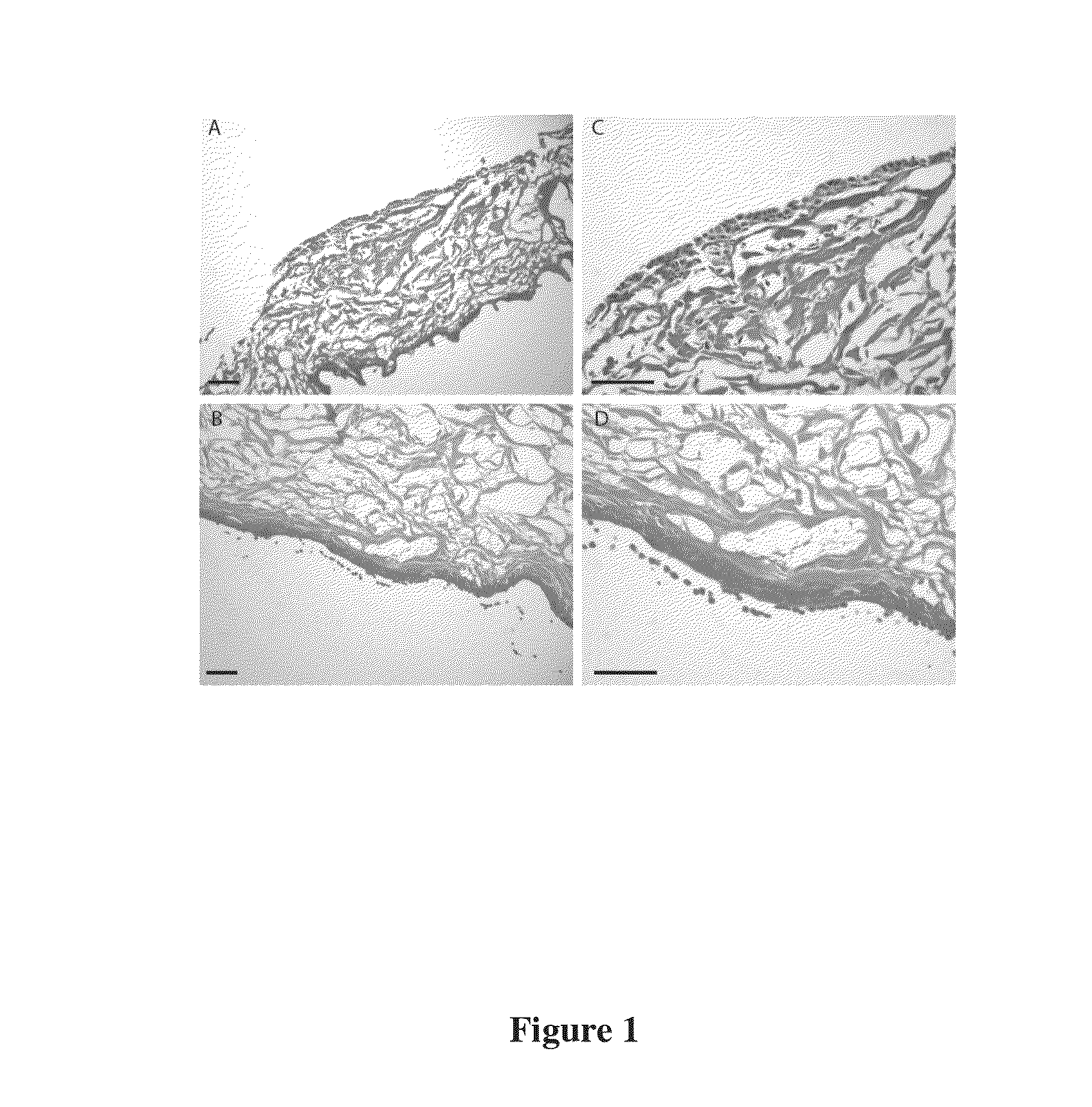
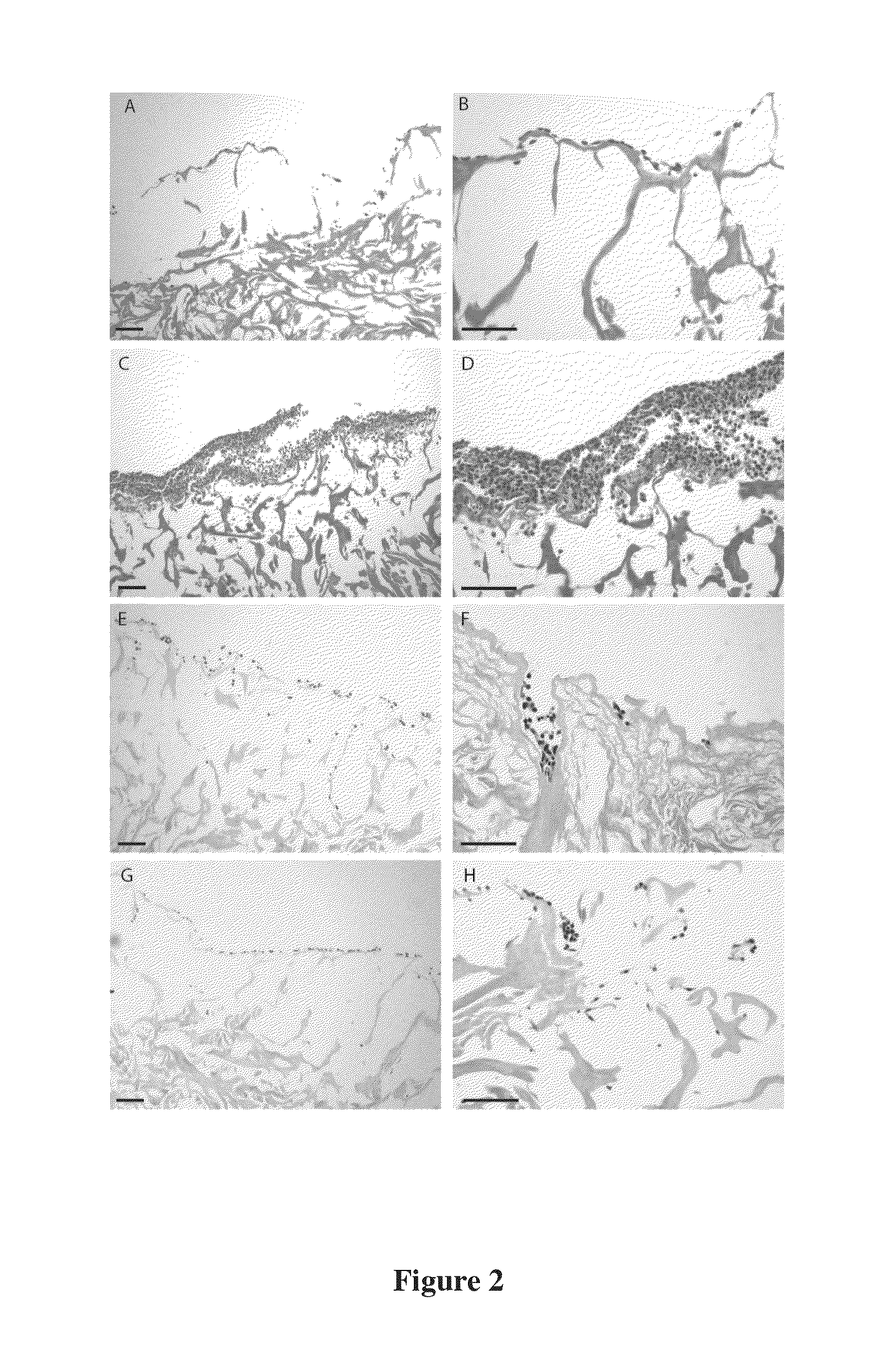
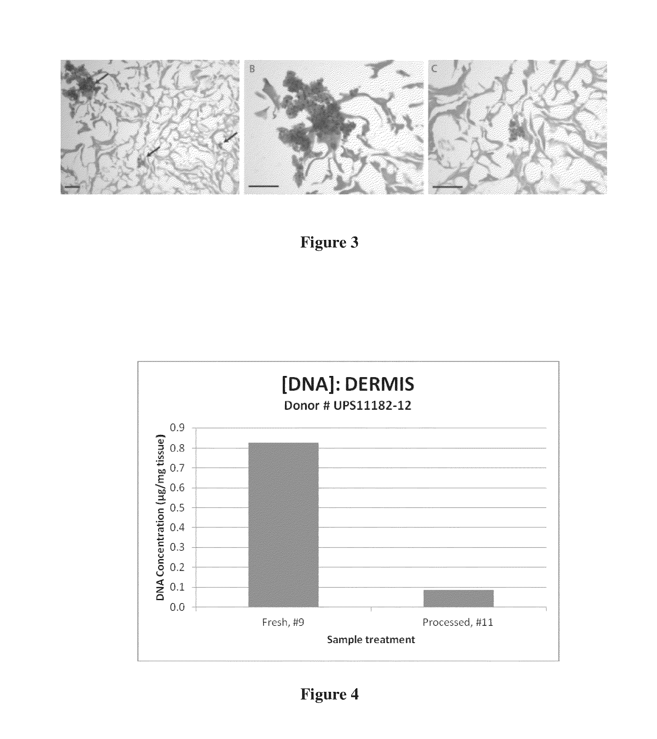
Processing (A) of Skin Tissue in the Production of a Dermal Matrix Tissue

1. Procedure

[0086]1.1 Preparation of Skin Connective Tissues[0087]1.1.1 If previously frozen in 1.5 M Sodium Chloride solution, remove wrapped skin package from freezer, place on orbital shaker at 60 RPM, and allow skin to thaw completely for 24 hours at 36° C.±1° C. If freshly recovered, proceed to processing as detailed below. If converting cryo-preserved skin to de-cellularized dermis, remove skin package from freezer and place in an incubator at 36° C.±1° C. After thawing, remove skin from sterile packaging and continue to section 1.2.[0088]1.2 Inspection, Cleaning and Weighing of Tissues[0089]1.2.1 Using aseptic techniques, present skin tissues to the sterile field.[0090]1.2.2 Place each piece of skin with the epidermal side up on a cutting board.[0091]1.2.3 Inspect the skin tissues for holes, moles, warts, and tattoos and cut out these areas using a scalpel.[0092]1.2.4 Examine the tissues for hair and rem...

example 2

Processing (B) of Skin for the Production of Dermal Matrix

2 Procedure

[0156]2.1 Preparation of Skin Connective Tissues[0157]2.1.1 If previously frozen in 1.5M Sodium Chloride solution, remove wrapped skin package from freezer, place on orbital shaker at 60 RPM, and allow skin to thaw completely for 24 hours at 36° C.±1° C. DO NOT UNSEAL THE PACKAGING. Skip to section 2.2[0158]2.1.2 If freshly recovered, proceed to processing as detailed below. Continue to section 2.2[0159]2.1.3 If converting cryo-preserved skin to de-cellularized dermis, remove skin package from freezer and place in an incubator at 36° C.±1° C. After thawing, remove skin from sterile packaging and continue to section 2.2.[0160]2.2 Inspection, Cleaning and Weighing of Tissues[0161]2.2.1 Using aseptic technique; present skin tissues to the sterile field.[0162]2.2.2 Place each piece of skin with the epidermal side up on a cutting board.[0163]2.2.3 Inspect the skin tissues for holes, moles, warts, and tattoos and cut out...

example 3

Preparation (A) of Fascia for Connective Tissue Matrix

Preparation of Fascia Connective Tissues

[0233]2.1 Rinsing of Tissues[0234]Based on the weight of the tissue determined in section 2.2, determine the volume of antibiotic-free normal saline solution to be utilized for one rinsing step.[0235]2.1.1 Add fresh, antibiotic-free normal saline solution (5 ml of saline per 1 gram of fascia) into the sterile plastic jar, secure jar with its lid, and agitate aggressively for 15 seconds.[0236]2.1.2 Remove the lid and decant the solution.[0237]2.1.3 Repeat this rinsing step three (3) times.[0238]NOTE: USE THE PRESCRIBED VOLUME OF NEW SOLUTION FOR EACH RINSE.[0239]2.1.4 After the last rinse, decant solution.[0240]2.2 Rinse[0241]2.2.1 Transfer the fascia tissues into a sterile plastic jar.[0242]2.2.2 Add fresh, sterile water (5 ml of saline per 1 gram of fascia) into the sterile plastic jar, secure jar with its lid, and agitate aggressively for 15 seconds.[0243]2.2.3 Remove the lid and decant t...


PUM

PropertyMeasurementUnit
diameteraaaaaaaaaa
sizeaaaaaaaaaa
diameteraaaaaaaaaa

Abstract

A process for the preparation of a tissue matrix suitable for regenerative repair of tissues, including contacting an isolated connective tissue with an amount of detergent and an amount of disinfectant to significantly reduce at least one of lipids, phospholipids, nucleic acids, major histocompatibility (MHC) antigens, contaminating microorganisms, and endotoxins. The process further provides for less significant reduction in proteoglycan content while retaining the overall structure of the tissue matrix produced. Processing may further include micronizing the tissue matrix. Also, a tissue matrix having a scaffold portion and non-structural portion, collectively structured to promote cellular infiltration, attachment, and proliferation. The tissue matrix may be in the form of a sheet, thick sheet, or micronized. Also, kits including a prepared tissue matrix for use in regenerative repair, and kits for preparing a tissue matrix.

Description

CLAIM OF PRIORITY[0001]This application claims the benefit of co-pending U.S. Provisional Patent Application having Ser. No. 61 / 581,803 filed on Dec. 30, 2011, the contents of which are incorporated by reference herein in its entirety.STATEMENT REGARDING FEDERALLY-SPONSORED RESEARCH AND DEVELOPMENT[0002]Not applicable.BACKGROUND OF THE INVENTION[0003]1. Field of the Invention[0004]This invention relates generally to the field of tissue matrices for regenerative repair. More particularly, this invention relates to methods for processing tissues for use in regenerative repair of soft tissue defects and the resulting tissue matrices.[0005]2. Description of Related Art[0006]When damage to soft tissue occurs, an inflammatory response is initiated and cells and molecular factors are recruited to the site of injury to begin the healing process. In order for healing to occur, these cells must be able to infiltrate and proliferate into the wounded area.[0007]Cellular proliferation in tissues...

Claims


Application Information

Patent Timeline
Patent Type & Authority Applications(United States)
IPC IPC(8): A61K35/12
CPCA61K35/35A61L27/362A61L27/3687A61K35/36A61L2300/414A61L2400/06
Inventor MORRIS, PAUL R.KAST, NATHANTEMPLE, THOMAS H.WOLFINBARGER, LLOYD
Owner VIVEX BIOLOGICS GRP INC
Learn More