Methods and compositions for targeted imaging
- Summary
- Abstract
- Description
- Claims
- Application Information
AI Technical Summary
Benefits of technology
Problems solved by technology
Method used
Image
Examples
example 1
Synthesis and Modification of Peptides
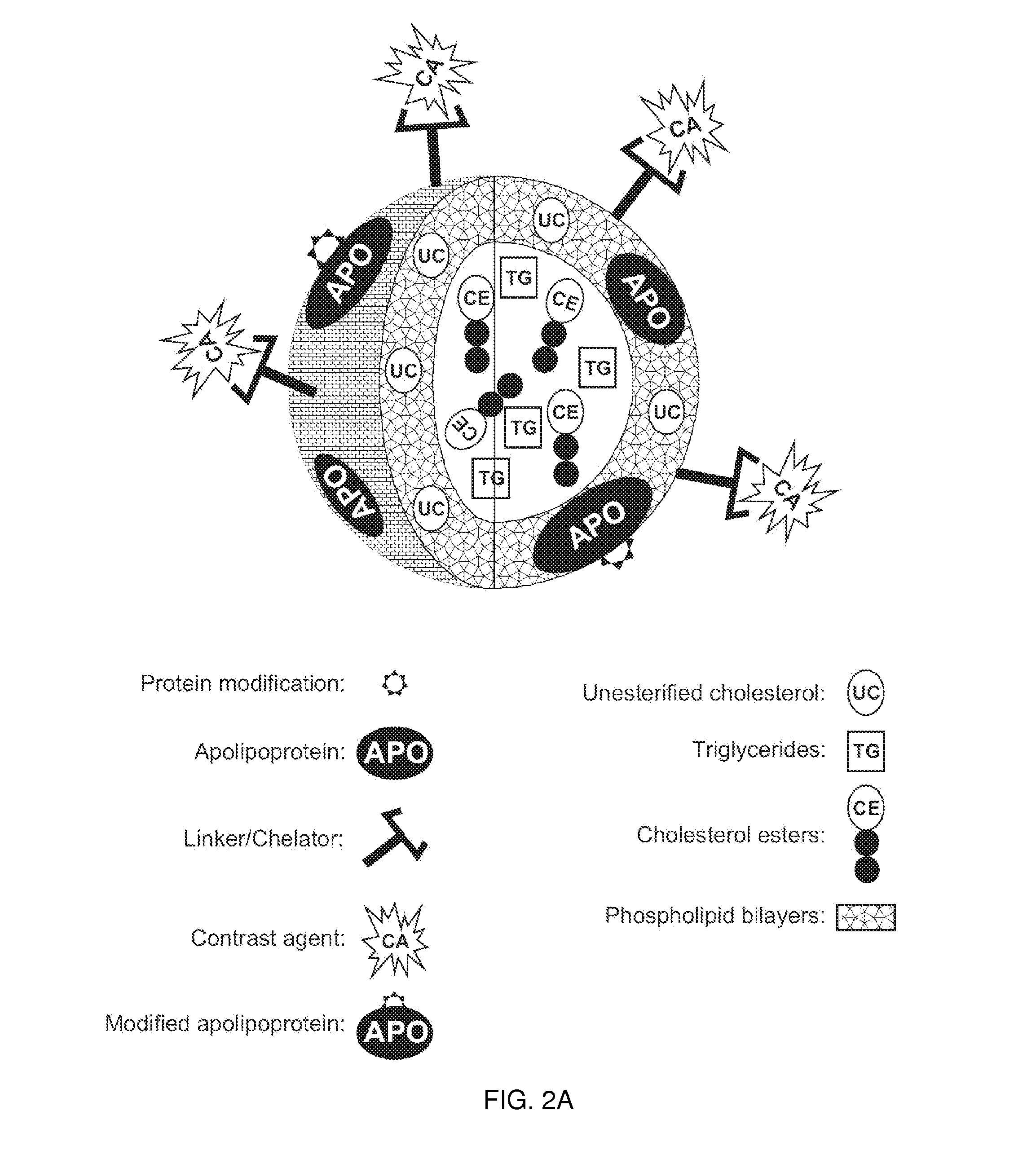
[0239]This example demonstrates one embodiment of a synthesized apo A-I peptide containing methionine residues at position 148 as referred to the full-length apo A-I primary sequence. It also illustrates one embodiment of a modified synthetic apo A-I peptide containing methionine sulfoxide at positions 148 as referred to the full-length apo A-I primary sequence. Although it is not necessary to understand the mechanism of an invention, it is believed that being incorporated into the rHDL compositions of the present invention, this modified peptide targets the rHDL of the invention to sites of interest such, for example, as macrophages in atherosclerotic plague.
[0240]It is well known to those of ordinary skill in the art that other apo A-I peptide fragments such as those containing methionine residues at positions 86 or 112 can be easily synthesized using standard procedures described below or manufactured by any technique for peptide synthesis kn...
example 2
Isolation and Purification of Apolipoproteins A-I and A-II
[0246]As discussed herein throughout, the present invention is related to imaging compositions that comprise rHDL as a backbone structure. The apolipoproteins for the production of the rHDL composition may be derived from an animal as a source of the apolipoproteins for the production of the rHDLs. In a preferred method for the producing rHDLs the apolipoproteins are from human HDL.
[0247]To isolate and purify human apolipoproteins A-I and A-II the standard procedure known in the art can be used (Sigalov et al. J Chromatogr 1991; 537:464-8). Briefly, HDL of density 1.063-1.210 g / ml were isolated from fasting serum of normo-lipidaemic donors by sequential ultracentrifugation in a Beckman (Berkeley, Calif., U.S.A.) Model L8-7Q ultracentrifuge using a 45.Ti rotor. The isolated HDL fraction was extensively dialysed against 50 mM ammonium hydrogencarbonate buffer (pH 8.2), lyophilized and delipidated by an original procedure using ...
example 3
Characterization of Apolipoproteins A-I and A-II
[0248]Apolipoproteins A-I and A-II isolated and purified as described in Example 2 were quantified according to Lowry et al. (Lowry et al. J Biol Chem 1951; 193:265-75) and spectrophotometrically at 280 nm using extinction coefficients of 1.22 and 1.82 AU / mg protein / ml for apo A-I and apo A-II, respectively (Edelstein et al. J Biol Chem 1972; 247:5842-9). Lipid phosphorus analysis was performed utilizing the method of Bartlett (Bartlett G. R. J Biol Chem 1959; 234:466-8). No phospholipid was detected in either the isolated apo A-I or apo A-II. Homogeneity was confirmed using the standard procedures well known in the art such as SDS-PAGE on 15% polyacrylamide gels under both reducing (FIG. 7) and non-reducing conditions and by urea-PAGE. Identification of apo A-I and apo A-II was confirmed by electrophoretic mobility and immunoelectrophoresis with monospecific antibodies to human apo A-I and apo A-II (Clarke H. G. & Freeman T. Clin Sci ...
PUM
| Property | Measurement | Unit |
|---|---|---|
| molar concentrations | aaaaa | aaaaa |
| molar concentrations | aaaaa | aaaaa |
| molar concentrations | aaaaa | aaaaa |
Abstract
Description
Claims
Application Information
 - R&D
- Intellectual Property
- Life Sciences
- Materials
- Tech Scout
- Unparalleled Data Quality
- Higher Quality Content
- 60% Fewer Hallucinations
Browse by: Latest US Patents, China's latest patents, Technical Efficacy Thesaurus, Application Domain, Technology Topic, Popular Technical Reports.
© 2025 PatSnap. All rights reserved.Legal|Privacy policy|Modern Slavery Act Transparency Statement|Sitemap|About US| Contact US: help@patsnap.com


