
Renal Function Analysis Method and Apparatus

a function analysis and renal function technology, applied in the field of medical methods and devices, can solve the problems of increased mortality rate, inability to reliably apply two approaches over the full range of gfr, and independent association of aki, so as to accurately quantify and track the degree of renal function, the effect of convenient operation and near real-time efficiency

Active Publication Date: 2011-08-18
PHARMACOPHOTONICS INC
View PDF2 Cites 25 Cited by
  • Summary
  • Abstract
  • Description
  • Claims
  • Application Information

AI Technical Summary

Benefits of technology

[0074]A rapid and accurate measurement of GFR in an early stage of acute kidney injury is important for diagnosis, stratification of extent of injury and therapeutic purposes. An advantage of the present invention is that it will rapidly identify and determine the extent of injury allowing for early treatment, including dialysis initiation, as well as enrollment and stratification for clinical studies. It could also be used to determine the effect of a clinical maneuver on GFR, such as volume resuscitation. Therefore, this technical advance is of major clinical importance, especially in high risk patients where intense surveillance is necessary for early diagnosis, injury stratification and determination of therapeutic potential.
[0075]The inadequacies of methods currently clinically used for estimating GFR are established both in literature and in practice. While progress is being made to identify biomarkers for detecting presence of injury, little progress has been made in finding a functional marker that is practical enough for broad acceptance. The inventors' method represents a true advancement in the ability to accurately quantify and track the degree of kidney function with near real-time efficiency. The inventors have also developed a device that is easy to operate in a busy medical environment—a critical adoption barrier in medical technology.
[0076]The optical technique developed by the inventors is based on plasma clearance measurements of a fluorescent bioreporter molecule and allows for the rapid, frequent, and safe evaluation of GFR. To further validate the values, other standard GFR tests, including but not limited to inulin clearance, may be performed. Upon comparison of these values, a correction factor may be applied to the data obtained using this novel method if needed.
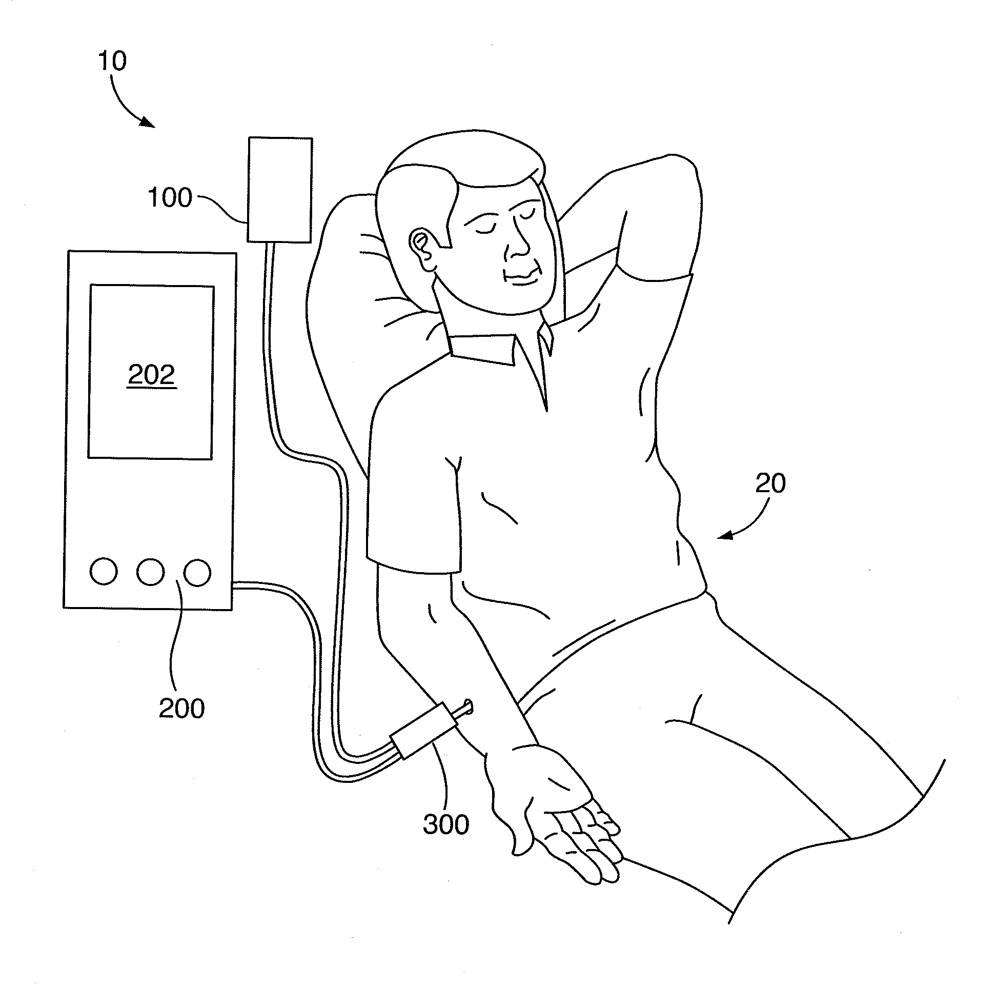
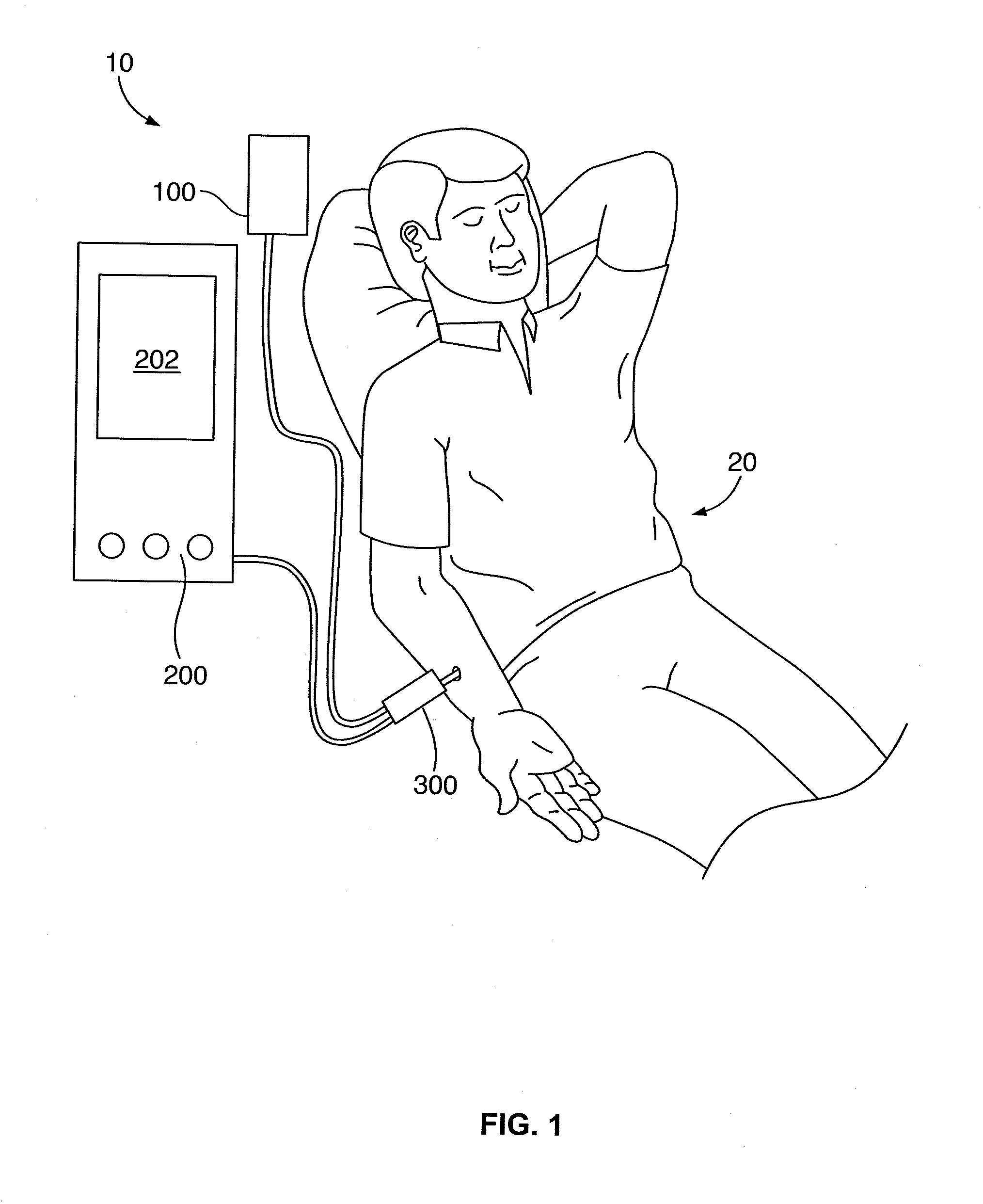
[0077]Referring to FIG. 1, a system 10 which incorporates a method of the present invention is illustrated. The system 10 comprises a source of a GFR measurement composition 100, a kidney fluorescent detector 200, and a light transfer probe 300. The GFR measurement composition 100, which comprises a plurality of reporter molecules and a plurality of marker molecules, is introduced into the blood stream of a human subject 20 via the light transfer probe 300. The fluorescent detector 200 includes a light source to provide one or more exciting lights which travel from the detector 200 to the blood stream via the light transfer probe 300 to excite the GFR measurement composition 100 in the blood stream of the subject. The fluorescent detector 200 monitors the level of the GFR measurement composition within the blood stream and reports an operating condition of the human subject's kidney in at least substantially real time. This apparatus measures volume of plasma distribution based on a fluorescence of a marker molecule relative to the fluorescence of a reporter molecule. “Substantially real time” is intended to encompass the duration elapsed between measurement of the levels of the reporter and the marker within the blood stream, calculation of the operating condition of the kidney, and reporting of that condition. It is contemplated by the inventors that this elapsed time will be very near real time as to be negligible in relation to the prior techniques discussed above.The GFR Measurement Composition
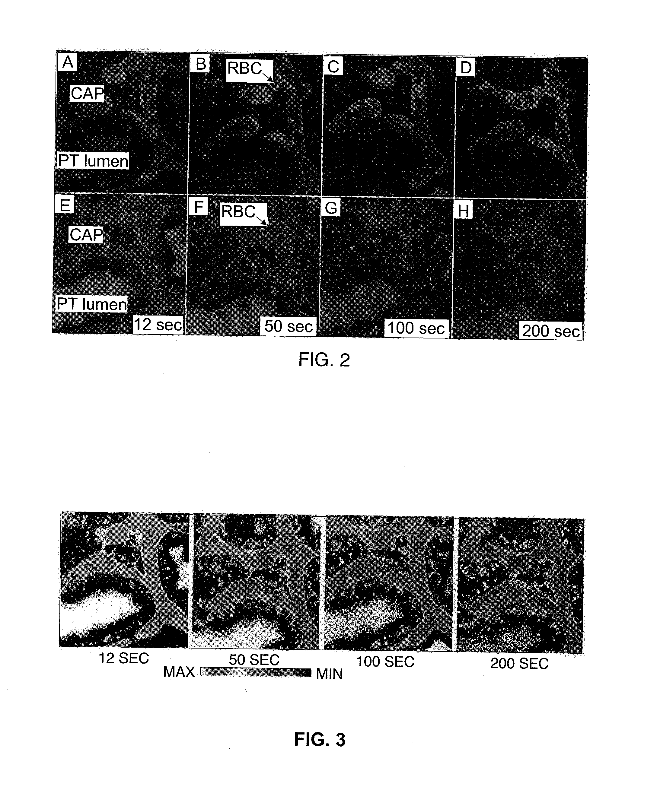
[0078]By utilizing intravital multi-photon microscopic imaging of the kidney, the inventors have quantified glomerular filtration and tubular reabsorption processes independent of each other. The inventors have developed ratiometric imaging techniques permitting quantitative analysis of fluorescence signals within local regions of the kidney using multi-fluorescent probe experiments. To measure GFR by plasma clearance, the inventors use a fluorescent GFR reporter molecule, e.g. fluorescein isothiocyanate (FITC) dextran, together with a large different fluorescent marker molecule that does not pass through the glomerular filtration barrier. This large fluorescent marker serves to quantify the plasma volume of distribution in the vascular space and allow for the ratiometric technique.
[0079]The inventors have been able to quantify plasma clearance of the fluorescent GFR marker by examining the ratio of fluorescence intensities of the two molecules from within the blood vessel regions of the image. GFR can be rapidly determined using this ratio technique. This method has been tested in a number of animal models. Since the fluorescent signals are being measured from within the blood vessels to quantify the kinetics of plasma clearance, the ratio signal of the two fluorescent molecules is independent of the body location where the measurement is performed.

Problems solved by technology

AKI is independently associated with increased mortality rates in several clinical situations, including subsequent to administration of radio contrast dye and cardiovascular surgery.
Unfortunately, these two approaches are not reliable over the full range of GFR, and neither can be used in AKI, since both muscle mass (creatinine is a breakdown product of creatine, which is an important part of muscle) and GFR determine a patient's serum creatinine level.
However, even these formulas are inaccurate and often misleading in estimating GFR below 20 or above 60 ml / min.
Therefore, this is another reason they cannot be used in the setting of AKI.
100121 Serum creatinine determinations as a measure of GFR may also be severely limiting because of the time it takes to reach equilibrium values required for an accurate conversion.
This slow rise in serum creatinine limits the physician's ability to diagnose the injury for 12-24 hours after the event, and it is also not possible to determine the extent of injury for days.
This has markedly limited the ability to conduct a therapeutic trial in AKI.
It is widely held that beginning therapy after 12-24 hours of AKI may limit the success rate of any potential therapeutic agent.
However, improvements sought utilizing these structural biomarkers may not be significant because they were developed using population results that may not apply to an individual.
Collection of a 24 hour urine and invasive techniques exist to accurately determine a patient's GFR, but these are cumbersome, error prone, expensive, time consuming, or expose the patient to radiation or radio contrast media.
Also, there is no rapid and accurate measurement technique that can determine GFR reliably in patients with acute kidney injury when the serum creatinine is rising.

Method used


Image

  • Renal Function Analysis Method and Apparatus
  • Renal Function Analysis Method and Apparatus
  • Renal Function Analysis Method and Apparatus

Examples


example 1

Measurement of GFR in Swine Using Fluorescence Ratiometric Analysis Method

[0142]The data obtained using the described optical device (also known as the fluorescence ratiometric analysis method herein) from a female Osawbaw swine weighing 29.5 kg are shown in FIG. 18. The anesthetized pig was administered 75 mg 5 kDa FITC-inulin and 75 mg 150 kDa Texas Red-dextran. The fluorescent data after the completion of the mixing in the vascular space following the bolus infusion are shown. FIG. 18A shows the normalized fluorescence time course for both small and large conjugates. While the signal from the large non-filterable dextran (red channel) remained stable over time, the signal of the filterable 5 kDa FITC-inulin (green channel) decreased rapidly initially indicating a combined inter-compartment movement (from the vascular space to the interstitial space) and kidney clearance, which was followed by a slower decay caused only by kidney clearance (from the vascular space). As a result, t...

example 2

Measurement of GFR in Dog Using the Fluorescence Ratiometric Analysis Method

[0143]Data from the dog study are shown in FIG. 19. The dog was administered 175 mg 5 kDa aminofluorescein-dextran and 75 mg 150 kDa 2-sulfohexamine rhodamine-dextran (2SHR-dextran). The higher noise in the data was caused by the movement during the data collection as the dog was not anesthetized. The plasma volume was determined to be 1720 ml. The GFR of the 33.0 kg dog was determined using three different methods. The value determined from the optical device was 4.49 ml / min / kg and the value obtained from iohexol analysis reported from Dr. Schwartz's laboratory was 4.50 ml / min / kg.

example 3

Corrleation of GFR Determined by Fluorescence Ratiometric Analysis Method and by the Iohexol Plasma Clearance Method in Gentamycin-Induced Acute Kidney Injury (AKI) Dogs

[0144]Five spayed female young adult hound-type mogrel dogs were confirmed to be in good health based on physical examination, complete blood count, blood chemistry analysis, urinalysis, urine protein-to-creatine ratio (UPC) and aerobic urine culture. Acute tubular necrosis was induced in the 5 dogs with IV gentamycin (10 mg / Kg IV q8 hours for 10 days; first day of injection was Day 0. Serum creatinine, blood urea nitrogen (BUN), urine specific gravity (USG), UPC and GFR via iohexal plasma clearance method and ratiometric fluorescence analysis method were determined on days 3, 6 and 9. All dogs were euthanized on day 9. Table 1 shows the serum creatinine and BUN concentrations, USG and UPC in the five dogs with gentamycin-induced AKI. Greater than 25% increases over baseline serum creatinine, reduced urine concentrat...


PUM


Abstract

A method for measuring a glomerular filtration rate of a mammalian subject comprises a source of reporter and marker fluorescent molecules. The fluorescent molecules are introduced into the vascular system of the mammalian subject. Over a period of time, a measurement of the intensities of the reporter and marker fluorescent molecules is taken. A ratio is calculated to determine the glomerular filtration rate.

Description

CROSS REFERENCE TO RELATED APPLICATIONS[0001]The present application is a continuation-in-part of co-pending U.S. patent application Ser. No. 12 / 425,827 filed on Apr. 17, 2009, which claims the benefit of U.S. Provisional Patent Application No. 61 / 046,273, filed on Apr. 18, 2008, the disclosures of which are incorporated by reference as if fully set forth herein.TECHNICAL FIELD[0002]The invention relates to medical methods and devices used in conjunction with analyzing organ functions of a mammalian subject. More particularly, the present invention is directed to an apparatus and method used for real time measurement of the glomerular filtration rate (GFR) of the kidney in a mammalian subject.BACKGROUND OF THE INVENTION[0003]Acute kidney injury (AKI) is a serious and deadly disease process affecting 5-10% of all hospitalized patients. The mortality rate in these cases often exceeds 50%. AKI is independently associated with increased mortality rates in several clinical situations, in...

Claims


Application Information

Patent Timeline
Patent Type & Authority Applications(United States)
IPC IPC(8): A61B6/00A61K49/00A61P43/00
CPCA61B5/0071A61B5/0075A61B5/0084A61K49/0054A61B5/412A61K49/0017A61K49/0043A61B5/201A61P43/00
Inventor WANG, EXINGMEIER, DANIELBUNCH, ROBERTMOLITORIS, BRUCESANDOVAL, RUBENRUBIN, MATTHEWSHERIDAN, ERINN
Owner PHARMACOPHOTONICS INC
Learn More