
Compositions and uses thereof for the treatment of wounds

a technology of topical formulations and wounds, applied in the field of topical formulations, can solve the problems of complex wounds that are often chronic, damage to the skin layer of a subject, and complication of dermal ulcers, and achieve the effects of enhancing the proliferation of cells, reducing or preventing apoptosis and/or necrosis

Inactive Publication Date: 2010-07-01
PHYLOGICA +1
View PDF42 Cites 0 Cited by
  • Summary
  • Abstract
  • Description
  • Claims
  • Application Information

AI Technical Summary

Benefits of technology

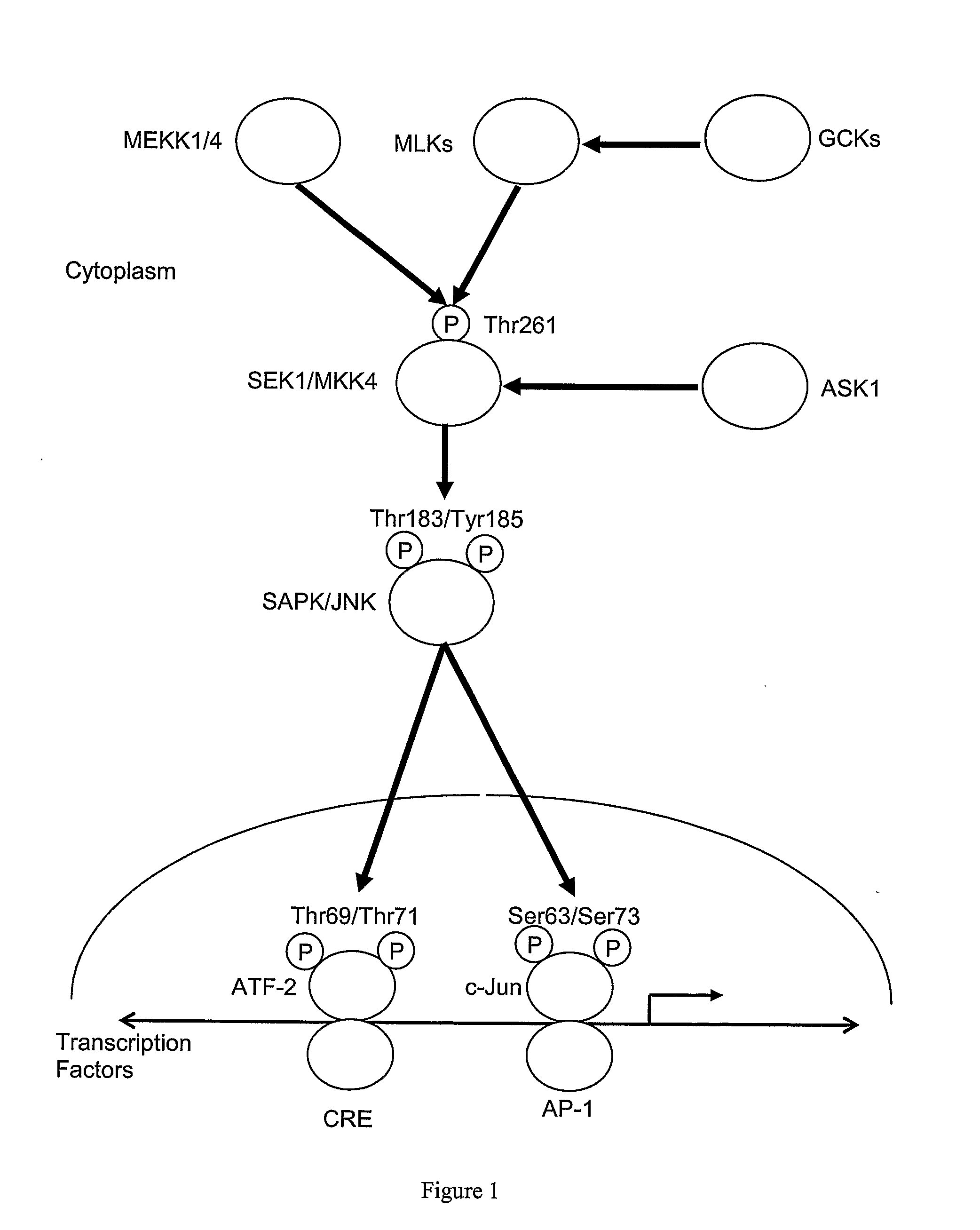
[0057]The present invention is based in part on the production by the inventors of topical formulations of peptides that inhibit AP-1 signaling, e.g., by inhibiting c-Jun homodimerization and / or heterodimerization, and in part, on the finding that such topical formulations inhibit or delay or reduce apoptosis and / or necrosis following dermal wounding in vivo and promote wound healing in vivo.
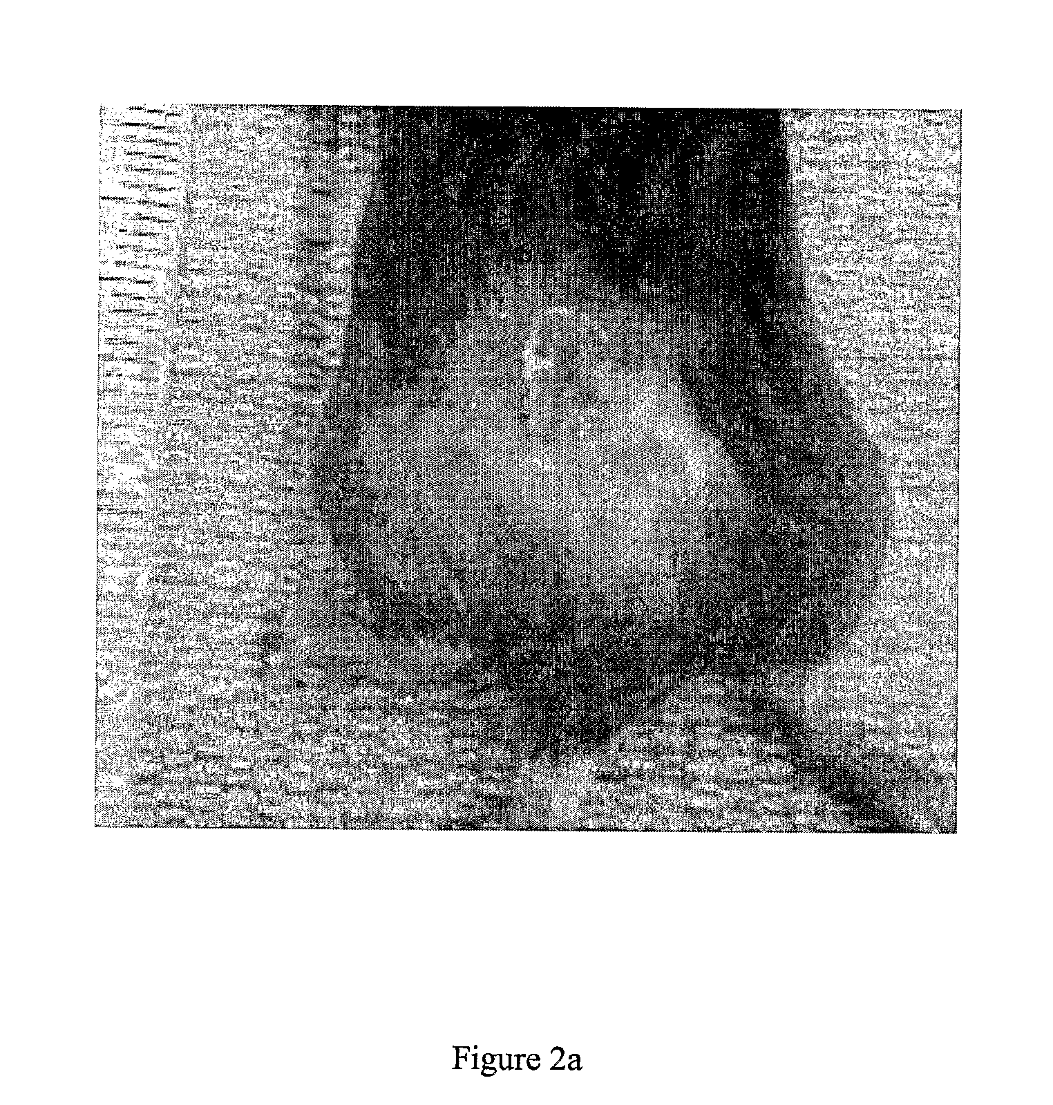
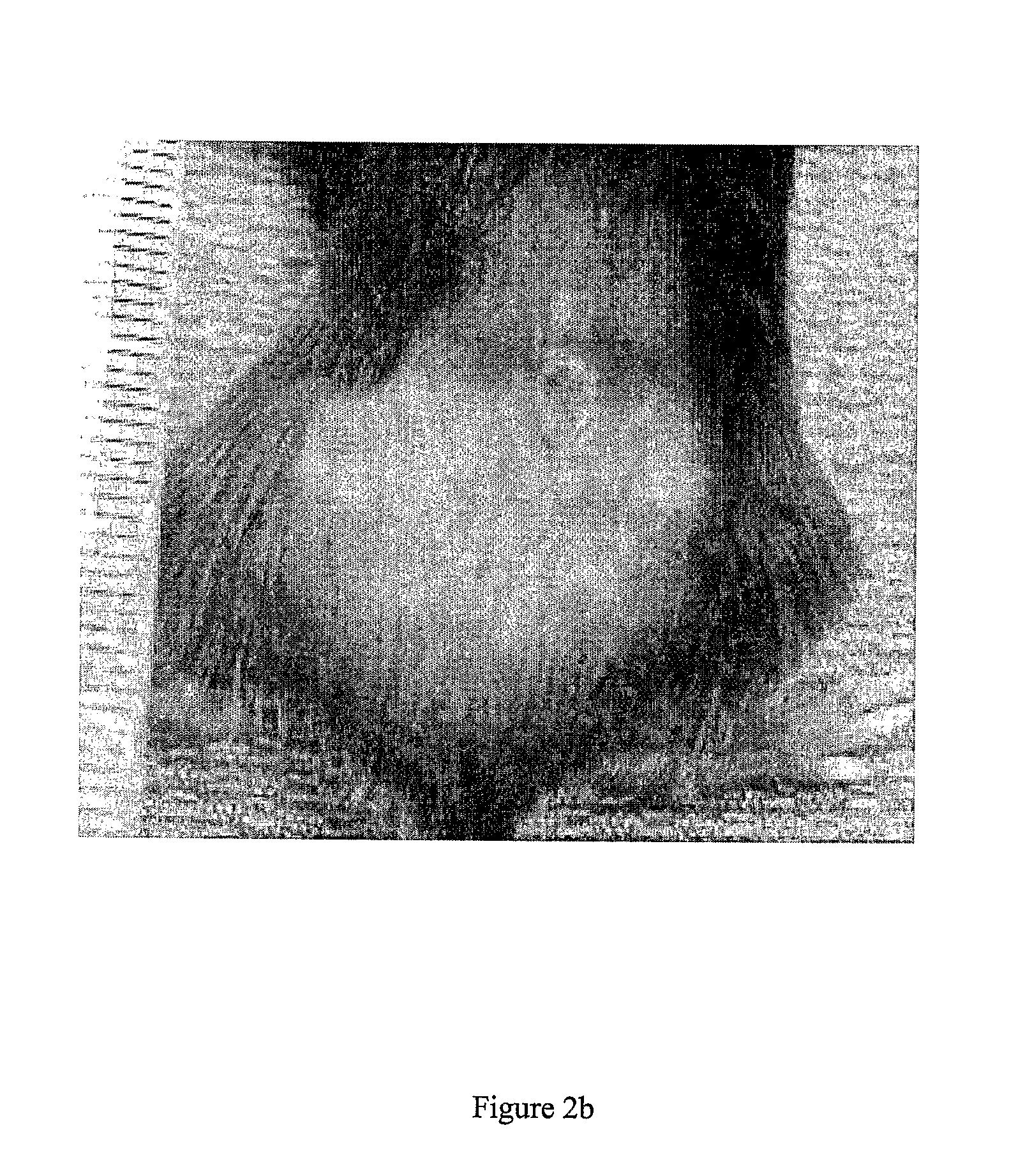
[0059]For example, in a rodent model of full-thickness burn injury, the inventor found that treatment with a topical formulation of one or more peptide inhibitors of AP-1 signaling (e.g., by inhibiting c-Jun dimerization) inhibits, reduces or delays apoptosis and / or necrosis in the epidermis e.g., in the zone of stasis. Wound healing is also promoted or enhanced, as determined, for example, by early and / or more rapid re-epithelialization of the wound and separation of the eschar. Without being limited to any theory or mode of action, the inhibited, delayed or reduced apoptosis and / or necrosis may promote healing of a burn wound.

Problems solved by technology

The dermal layers of a subject may be damaged or wounded by any of a variety of insults, such as, for example, a burn and / or development of an ulcer and / or an infection.
Complex wounds, such as, burns and ulcers are of particular concern, because these wounds may damage all layers of the skin, i.e., is a full-thickness wound, often requiring a skin graft for treatment.
Moreover, such complex wounds are often chronic, because the body is unable to successfully heal the wound by the normal healing process.
Dermal ulcers are also a common complication and frequent cause of hospitalization for subjects suffering from diabetes.
However, in the absence of suitable treatment, cells in the zone of stasis die by necrosis and / or apoptosis resulting in tissue loss; and(iii) Zone of hyperanemia—is the area of skin surrounding the zone of stasis in which tissue perfusion is increased relative to the level of perfusion in the zone of stasis.
Such a hypermetabolic response may cause death in some subjects.
Of these, approximately 70,000 burn injuries require intensive care treatment and approximately 10,000-12,000 burn injuries result in death.
However, in many cases, these forms of treatment are ineffective in the treatment of chronic wounds or severe wounds.
However, skin grafts are only useful in the case of well-vascularized wounds (Braddock et al., Int. J. Dermatol., 38: 808-817, 1999).
Accordingly, skin grafts may not be useful for the treatment of severe burns, i.e., in which vascular tissue in the hypodermis has been damaged or destroyed, or in subjects suffering from reduced circulation, e.g., diabetic subjects.
Moreover, notwithstanding that artificial skin demonstrates good biocompatibility and healing characteristics for the treatment of wounds, these materials are very expensive, which limits their widespread use.
Whilst, cadaver skin is a relatively efficient and cheap approach for wound coverage, the risk of transmission of infections, e.g., human immunodeficiency virus (HIV), cytomegalovirus (CMV), herpes simplex virus (HSV) and / or hepatitis is a significant concern, thereby limiting application of cadaver skin.
One disadvantage of such keratinocyte-based therapies is the time required to produce an autologous keratinocyte suspension for application to a subject.
Clearly this form of treatment is not amenable to the rapid treatment of a dermal wound.
Moreover, application of cultured cells to a subject increases the risk of infection as a result of contamination during the culturing process.
Laser Surg. Med. 18: 397-405, 1996 have shown that photodynamic therapy actually results in delayed wound healing
However, there are adverse side effects of treatment with IGF-I, such as hypoglycemia, mental status changes, edema, fatigue and headache, which limit the therapeutic utility of IGF-I in the treatment of burns (Jabri, et al.
Similarly, despite positive in vitro results, in vivo studies have shown that administration of interleukin-1 are ineffective in the treatment of pressure ulcers (Robson et al., Wound Repair Regen., 2: 177-185, 1994).

Method used


Image

  • Compositions and uses thereof for the treatment of wounds
  • Compositions and uses thereof for the treatment of wounds
  • Compositions and uses thereof for the treatment of wounds

Examples


example 1

Isolation of an Inhibitor of AP-1 Signaling

[0322]Nucleic acid was isolated from the following bacterial species:

1Archaeoglobus fulgidis2Aquifex aeliticus3Aeropyrum pernix4Bacillus subtilis5Bordetella pertussis TOX66Borrelia burgdorferi7Chlamydia trachomatis8Escherichia coli Kl29Haemophilus influenzae (rd)10Helicobacter pylori11Methanobacterium thermoautotrophicum12Methanococcus jannaschii13Mycoplasma pneumoniae14Neisseria meningitidis15Pseudomonas aeruginosa16Pyrococcus horikoshii17S nechosistis PCC 680318Thermoplasma volcanium19Thermotoga maritima

[0323]Nucleic acid fragments were generated from the genomic DNA of each genome using 2 consecutive rounds of primer extension amplification using tagged random oligonucleotides with the sequence:

5′-GACTACAAGGACGACGACGACAAGGCTTATCAATCAATCAN6-3′.

The PCR amplification was performed using the Klenow fragment of E. coli DNA polymerase I in the following primer extension reaction:

ReagentDNA (100-200 ng)VolumeOligonucleotide comprising SEQ ID NO...

example 2

Treatment of Burns Using an Inhibitor of c-Jun Dimerization

1.1 Materials and Methods

Animals

[0352]8 week old C57 / B16 mice were purchased from the Animal Resource Center, Murdoch University, Western Australia and used for all experiments. All experiments were approved by the necessary institutional animal ethics committees and were performed in accordance with the National Health and Medical Research Council (NHMRC) Australian Code of Practice for the Care and Use of Animals for Scientific Purposes.

Peptide

[0353]A peptide used was synthesised by Mimotopes Pty Ltd (Melbourne, Australia) and supplied as a lyophilised powder (purity>90%). The peptide (H-rerrsssqiGGsrisqyaGrrrqrrkkrG-NH2) (SEQ ID NO: 104) is a D-form retro-inverted mimic of a peptide originally identified in a screen for inhibitors of AP-1 signaling e.g., by virtue of inhibiting c-Jun dimerization (as described in Example 1, i.e, a peptide comprising an amino acid sequence set forth in SEQ ID NO: 57), and is fused to the T...

example 4

An Inhibitor of AP-1 Signaling Enhances and / or Induces Cellular Proliferation

[0364]Cultures of keratinocytes (HaCaT cells) or fibroblasts were cultured in a microwell plate the presence of a peptide analog comprising the amino acid sequence set forth in SEQ ID NO: 104 at a concentration of 100 nm, 1 μM or 10 μM. Following an incubation period 24 hours, 48 hours, or 72 hours, cells were incubated in the presence of MTT for a sufficient time for a mitochondrial dehydrogenase enzyme from viable cells to cleave the tetrazolium rings of the pale yellow MTT and form dark blue formazan crystals. Cells were then solubilized and the level of formalin crystals determined using a microtitre plate reader at 492 nm.

[0365]As shown in FIG. 8a, the peptide analog enhanced and / or induced proliferation of HaCat keratinocytes, which are associated with wound healing. The peptide analog did not stimulate proliferation of fibroblast cells, which are associated with scar formation.


PUM

PropertyMeasurementUnit
Volumeaaaaaaaaaa
Volumeaaaaaaaaaa
Volumeaaaaaaaaaa

Abstract

The present invention provides a topical composition comprising (i) an amount of an AP-1 signaling inhibitor sufficient to reduce, delay or prevent apoptosis and / or necrosis induced by dermal wounding and / or to induce and / or enhance proliferation of a cell; and (ii) a suitable carrier or excipient e.g., a topical carrier or excipient or other carrier or excipient for dermal application. For example, the AP-1 signaling inhibitor is a peptide analog comprising the sequence set forth in SEQ ID NO: 104. The present invention also provides a method of treating a dermal wound comprising topically administering said topical composition to a subject suffering from a dermal wound.

Description

RELATED APPLICATION DATA[0001]The present invention claims priority from U.S. Patent Application No. 60 / 826,208 filed Sep. 19, 2006 which is herein incorporated by reference in its entirety.FIELD OF THE INVENTION[0002]The present invention relates to topical formulations for the treatment of dermal wounds e.g., involving apoptosis and / or necrosis.BACKGROUND OF THE INVENTIONGeneral[0003]This specification contains nucleotide and amino acid sequence information prepared using PatentIn Version 3.3, presented herein after the claims. Each nucleotide sequence is identified in the sequence listing by the numeric indicator <210> followed by the sequence identifier (e.g., <210>1, <210>2, <210>3, etc). The length and type of sequence (DNA, protein (PRT), etc), and source organism for each nucleotide sequence, are indicated by information provided in the numeric indicator fields <211>, <212> and <213>, respectively. Nucleotide sequences referred to in...

Claims


Application Information

Patent Timeline
IPC IPC(8): A61L15/16A61K38/00A61K35/12
CPCA61K38/00C07K14/4703C07K2319/00A61P9/10A61P17/02A61P25/06A61P25/08A61P25/16A61P25/28
Inventor FEAR, MARKWATT, PAULHOPKINS, RICHARDMILECH, NADIA
Owner PHYLOGICA
Learn More