
Method of luminescent solid state dosimetry of mixed radiations

a solid state dosimetry and mixed radiation technology, applied in the field of radiation dosimetry techniques, can solve the problems of difficult discrimination between radiations, low saturation fluence, and difficulty in neutrons and heavy charged particles (hcp) to achieve the effect of low saturation fluence, low saturation fluence, and laborious wet-chemistry processing

Active Publication Date: 2010-04-29
LANDAUER INC
View PDF19 Cites 54 Cited by
  • Summary
  • Abstract
  • Description
  • Claims
  • Application Information

AI Technical Summary

Benefits of technology

[0005]According to a first broad aspect of the present invention, there is provided a method comprising the following steps: (a) providing one or more respective values of one or more respective dosimetric parameters for a luminescent material irradiated with one or more radiations; and (b) determining an absorbed dose for the one or more radiations based on the respective values of the one or more respective dosimetric parameters, wherein the one or more dosimetric parameters are each based on a spatial frequency domain analysis of one or more high spatial resolution fluorescence images of the luminescent material.
[0006]According to a second broad aspect of the present invention, there is provided a method comprising the following steps: (a) providing a depth profile of one or more dosimetric parameters for one or more radiations based on measurements of one or more high spatial resolution fluorescence images at different depths below the surface of a luminescent material covered by one or more converters; and (b) determining a median energy of one or more radiations based on the depth profile analysis.
[0007]According to a third broad aspect of the present invention, there is pr

Problems solved by technology

The measurement of neutrons and heavy charged particles (HCP) remains one of the most challenging tasks in radiation dosimetry.
TLDs have the strong neutron energy dependence and the difficulty in discriminating between radiations having low- and high linear energy transfer (LET).
PNTDs have good neutron / gamma discrimination but require laborious wet-chemistry processing and have low saturation fluence.
SBDs are very sensitive to neutrons but bulky and environmentally unstable.
All these difficulties have stimulated the search for a new approach.

Method used


Image

  • Method of luminescent solid state dosimetry of mixed radiations
  • Method of luminescent solid state dosimetry of mixed radiations
  • Method of luminescent solid state dosimetry of mixed radiations

Examples


example 1

[0124]Calibration of detector sensitivity to gamma radiation. Detectors made of a luminescent Al2O3:C,Mg crystal in the form of plates with dimensions 4 mm×6 mm×0.5 mm are cut along the optical c-axis and polished on the larger opposing sides to obtain optically transparent surfaces. The crystalline detectors have a green coloration due to the optical absorption band at 435 nm with an absorption coefficient of 13 cm−1. The dosimeter configuration is similar to the configuration of FIGS. 1, 2 and 3 provides the luminescent crystalline detector and two converters: a HDPE converter for gamma plus fast neutron measurements, and a PTFE converter for gamma dose measurements.

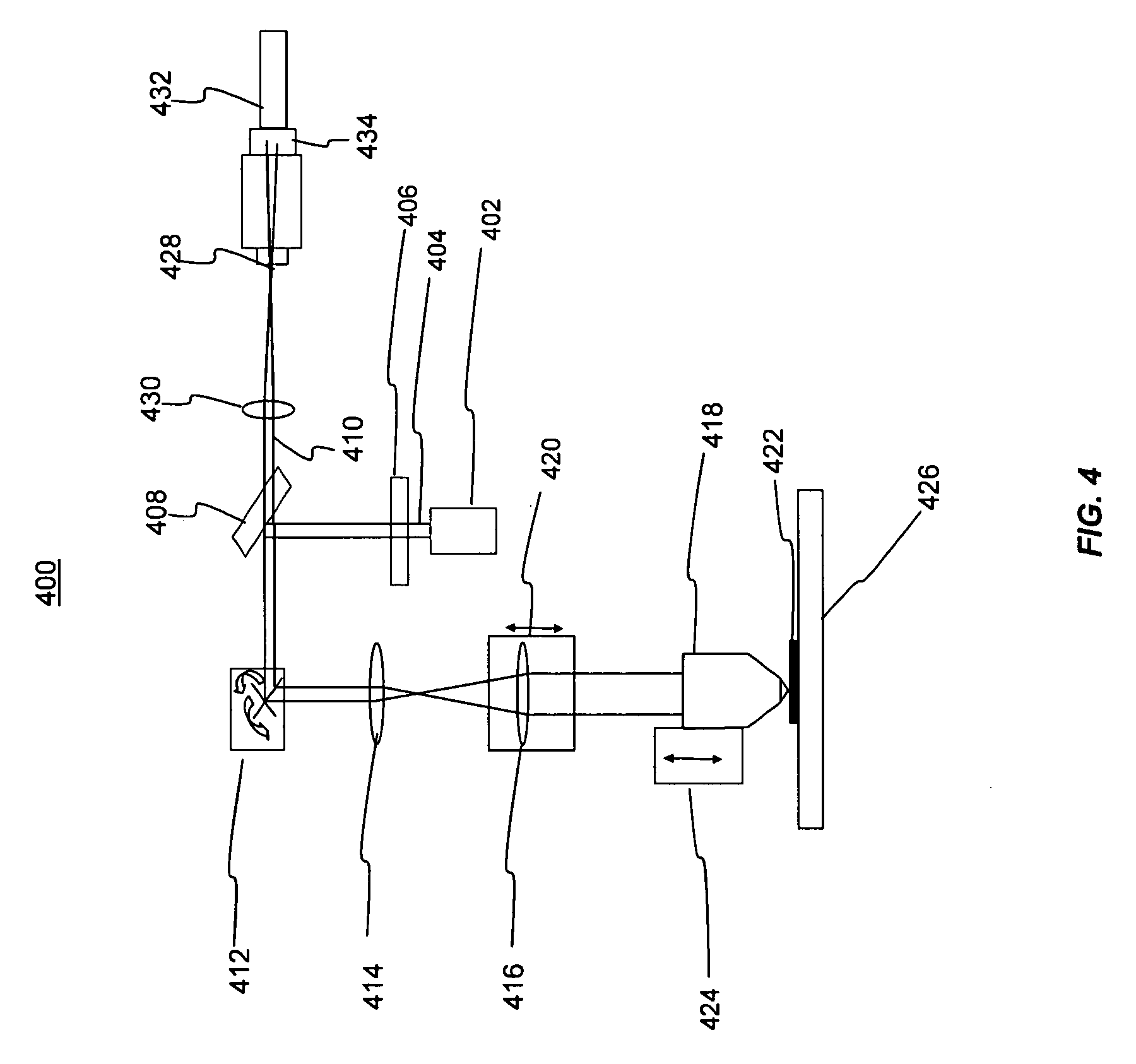
[0125]A set of Al2O3:C,Mg luminescent detectors are irradiated with gamma photons from a 137Cs source with known absorbed dose in tissue in the range from 3 mGy to 10 Gy. The detectors are then read using the laser scanning confocal system schematically shown in FIG. 4 starting with the side in contact with the HDPE co...

example 2

[0127]Calibration of the detector sensitivity to fast neutron radiation. Another set of Al2O3:C,Mg detectors having both PTFE and HDPE converters are irradiated with neutrons from a 241AmBe source having an activity of 185 MBq with a range of known doses, for example from 1 mSv to 1,000 mSv in tissue. The plates are then read using the laser scanning confocal system in FIG. 4 starting with the side in contact with the HDPE converter and then the side in contact with the PTFE converter. The resulting images are processed by performing the Fast Fourier Transform. The magnitude of the FFT is calculated and squared yielding the three-dimensional power spectrum of the image similar to FIG. 7. The power spectrum is then integrated in cylindrical coordinates sequentially from the lowest useful spatial frequency (0.01 μm−1) to the largest useful spatial frequency (1.5 μm−1) resulting in the amplitude of the power (one-dimensional power spectrum) at frequencies in the image in a range of 0.0...

example 3

[0129]Discriminating neutrons and gamma using PE and polytetrafluoroethylene converters. The dosimeter configuration similar to the configuration of FIGS. 1, 2 and 3 provides the luminescent crystalline detectors and two converters: a HDPE converter for gamma plus fast neutron measurements, and a PTFE converter for gamma dose measurements.

[0130]Irradiations of the single crystal detectors are performed with fast neutrons at a distance of 200 mm from a bare 241AmBe source having an activity of 185 MBq. Gamma irradiation of the same detector is performed from a 137Cs source in addition to neutron irradiation to imitate a mixed neutron-gamma field. Different ratios of neutron and gamma doses are delivered to the dosimeters and are listed in Table 1 of FIG. 19. Table 1 is a summary of processing mixed neutron-gamma irradiations. The total dose equivalent in tissue contains gamma component from the 241AmBe irradiations. The plates are processed with the same method as Examples 1 and 2 to...


PUM


Abstract

A method of determining the doses of neutrons, gamma and X-ray photons, beta, alpha and other ionizing radiations using a method of image processing in spatial and frequency domain that produces parameters that are related to the radiation dose absorbed in a luminescent material. Portions of the luminescent material may be covered by different converters to allow for doses of different radiations to be discriminated.

Description

BACKGROUND OF THE INVENTION[0001]1. Field of the Invention[0002]This invention relates generally to radiation dosimetry techniques.[0003]2. Background of the Technology[0004]The measurement of neutrons and heavy charged particles (HCP) remains one of the most challenging tasks in radiation dosimetry. Among the most widely investigated and used passive detector technologies are: plastic nuclear track detectors (PNTD) based on CR-39 plastic and its derivatives, pairs of 6LiF and 7LiF thermoluminescent detectors (TLD) in an albedo configuration and superheated bubble detectors (SBD). See Benton et al., “Proton Recoil Neutron Dosimeter for Personnel Monitoring. Health Phys., 40, pp. 801-809 (1981); Piesch et al., “Albedo Dosimetry System for Routine Personnel Monitoring,” Radiat. Prot. Dosim., 23(1 / 4), pp. 117-120 (1988); d'Errico, “Radiation Dosimetry and Spectrometry with Superheated Emulsions,” Nuclear Instr. Meth. B, 184, pp. 229-254 (2001). TLDs have the strong neutron energy depen...

Claims


Application Information

Patent Timeline
IPC IPC(8): G01J1/58
CPCG01T5/00G01T1/10
Inventor AKSELROD, MARK S.SYKORA, GARRETT J.
Owner LANDAUER INC
Learn More