Identification of ligands that enable endocytosis, using in vivo manipulation of neuronal fibers

Inactive Publication Date: 2006-12-14
FERGUSON IAN +1
View PDF10 Cites 3 Cited by
  • Summary
  • Abstract
  • Description
  • Claims
  • Application Information

AI Technical Summary

Benefits of technology

[0126] An in vivo screening process is disclosed, which can identify and isolate ligand molecules that can activate and drive ligand-mediated endocytosis (internalisation) of molecules into mammalian cells. These ligands can provide efficient transport of “passenger” or “payload” molecules (such as therapeutic drugs, diagnostic or analytical compounds, or non-viral genetic vectors) into specifically targeted classes of cells.
[0127] This in vivo screening process involves placement of an assortment of candidate ligands, in a suitable form (such as a bacteriophage display library) inside the body of a rat or other animal, in a location where the candidate ligands will contact nerve fibers (such as a sciatic nerve bundle, in a rat leg). In a preferred embodiment, a ligature loop is also tightened around the same nerve fibers at a different location, such as near the animal's hip. A period of time is allowed to pass, to enable ligands that bind to endocytotic receptors on the nerve fibers to be internalised. After entry into the nerve fibers, internalised ligands will be transported through the fibers in a retrograde direction (i.e., toward the spinal cord), by axonal transport. The animal is sacrificed, and a segment of nerve fibers is harvested (such as immediately adjacent to the hip ligature) that will contain the internalised ligands. The internalised ligands are collected from the harvested nerve fiber segments, and treated in any desired manner (such as by reproducing and analyzing the phage particles that carried internalised polypeptide sequences). Since nerve fiber segments are harvested from a site that is located a distance away from the ligand placement site, the ligands that actually undergo endocytotic uptake and axonal transport are separated from the other candidate ligands that did not enter the nerve fibers. In this way, false positives are avoided or minimized.
[0129] Similarly, this screening method can be adapted in various ways for use with combinatorial chemical synthesis. This can enable, for example, improved in vivo screening of candidate ligand compounds that are not polypeptides.

Problems solved by technology

However, those types of procedures are not enough to enable the identification and selection of particular phages that can trigger and then drive the process of endocytosis (i.e., active transport of selected phage particles into cells, via endocytotic receptors).
The challenges of selecting and identifying phages that can drive endocytosis, in living cells, are substantially more difficult than the problems of merely selecting phages that will be immobilized inside an affinity column.
These phagemids usually cannot not generate infective phage particles, because they are missing other essential parts of the phage.
As briefly mentioned above, some difficult problems arise, when researchers try to push the selection of phage display libraries beyond a level of simply binding to immobilized molecules in affinity columns, and into the realm of active endocytosis and uptake into cell interiors.
For the most part, these problems center on two factors: (i) multiple different phages will usually bind to multiple different proteins and other molecules, on the surfaces of cells, without being taken into the cells; and, (ii) it is very difficult to rinse off, wash off, or otherwise reliably remove any and all phages that are clinging to the surfaces of cells, and that have not been taken inside the cells, without killing and lysing the cells or otherwise creating severe problems that will interfere with other desired processing of the cells and / or internalized phages.
Because of these two factors, it is very difficult to prevent “false positives” from being selected, during efforts to identify endocytotic ligands by screening a phage display library.
In addition, because of the two problematic factors listed above, the screening of phage display libraries, in efforts to identify and select endocytotic ligands, is almost always limited to cell culture tests.
This type of endeavor is technically very challenging, difficult, tedious, and plagued with false positives.
Those problems apply to even the simplest tissue culture systems, where all of the cells can be clonal duplicates and have exactly the same receptor types.
By contrast, there have been few efforts and only very paltry and limited progress, in using phage display libraries to treat other diseases, or to create genetic vectors that can enable the transformation of neurons and other cells that are present in cohesive tissue, inside the body.
Under the prior art, the challenges and difficulties of eliminating false positives, when phage display libraries are screened for endocytotic uptake into neurons and other cohesive tissue cells in intact animals, in in vivo tests, have been so severe, and so formidable, that they have effectively blocked and prevented any substantial progress in that field of research.

Method used


Image

  • Identification of ligands that enable endocytosis, using in vivo manipulation of neuronal fibers
  • Identification of ligands that enable endocytosis, using in vivo manipulation of neuronal fibers
  • Identification of ligands that enable endocytosis, using in vivo manipulation of neuronal fibers

Examples


example 1

Phage Types and Libraries

[0213] M13KO7 helper phages can be purchased from various commercial suppliers, such as New England Biolabs (www.neb.com) and Amersham Biosciences (www4.amershambiosciences.com). This strain of helper phage contains fully functional genes that encode both the pIII and pVIII coat proteins. It also contains an origin of replication (from plasmid p15a) which is tightly controlled in a manner that results in low copy numbers in bacterial cells. It also contains a mutated (Met-40-Ile) copy of the phage M13 pII gene, which is essential for phage replication; this mutation causes it to be secreted by E. coli cells, as phage particles, in low copy numbers. It also carries a kanamycin resistance gene, inserted at the Ava I site within the M13 origin of replication. This kanamycin gene functions as a selectable marker in E. coli host cells that are not resistant to kanamycin. Additional information on using and culturing these helper phages is available from commerci...

example 2

Cell Types, Traits, and Methods

[0219] Except as otherwise noted, all phage amplification and titering used the TG1 strain of E. coli, from Cambridge Antibody Technology. This strain, which was specifically designed and developed for working with M13 phages, is also sold by companies such as Stratagene (La Jolla, Calif.; www.stratagene.com). Additional information describing culturing and transformation methods for this strain can be downloaded at no cost from the websites of commercial suppliers.

[0220] Among other features, the TG1 strain has a “lacIq” repressor gene which, together with catabolite repression by glucose, negatively regulates a “lac” promoter that has been placed in control of expression of the M13 gene that encodes the pIII phage coat protein. Most types of M13 phages that are used with TG1 cells contain an “amber” stop codon, inserted at the start of the pIII gene. As described below, this allows expression of pIII polypeptides (including chimeric pill polypeptid...

example 3

Cross-Linking of P75 Receptor-Binding Antibodies MC192) to M13KO7 Helper Phages

[0228] A monoclonal antibody preparation known as MC192 (and by similar terms, such as clone 192; originally described in Chandler et al 1984) is commercially available from various suppliers, such as Cell Sciences (www.cellsciences.com) and Chemicon (www.chemicon.com). These monoclonal antibodies bind to “low affinity” (p75) nerve growth factor receptors on rat neurons. Monoclonal antibodies that bind to human p75 receptors are also available, from companies such as United States Biological (www.usbio.net).

[0229] Unlike various other monoclonal antibodies that also bind to p75 receptors in rats, the MC192 antibody can trigger endocytosis of the antibody-receptor complex, leading to neuronal uptake of the MC192 antibody. This has been shown by studies using radiolabelled antibodies (Johnson et al 1987, Yan et al 1988).

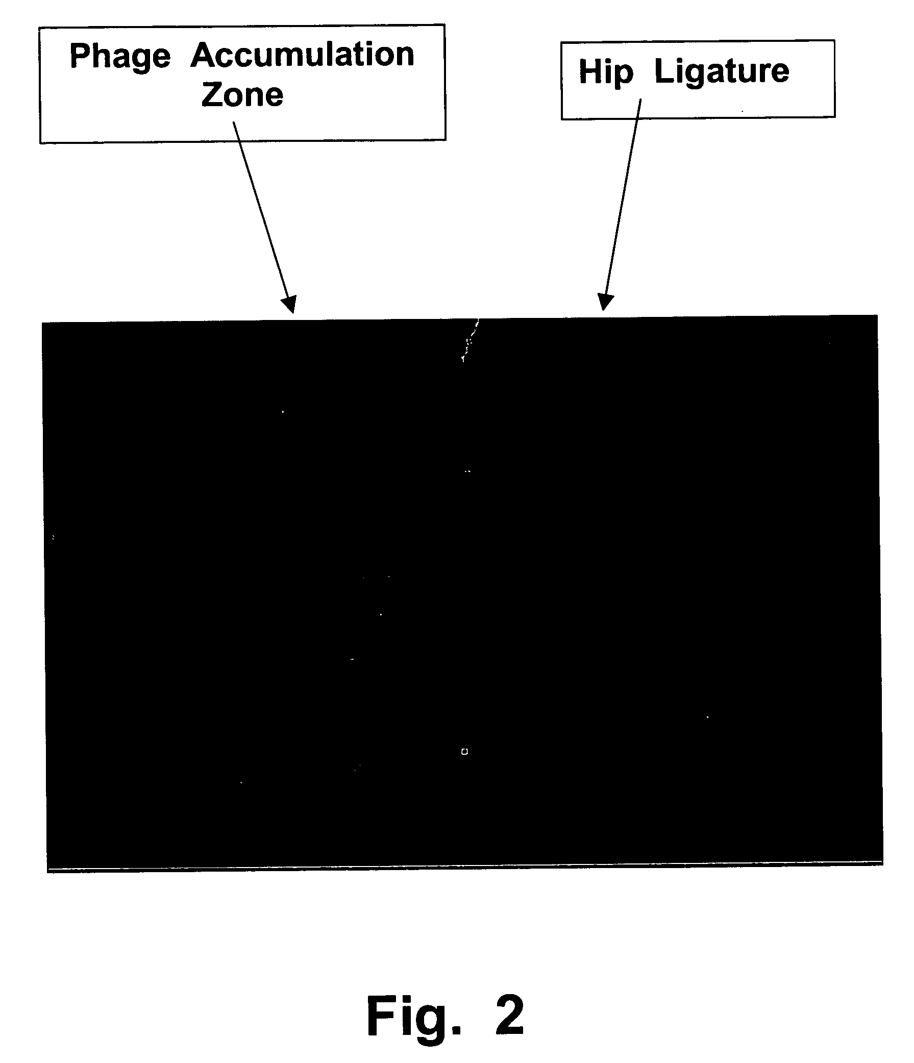
[0230] To evaluate the ability of the MC192 antibody to drive endocytosis of phages i...


PUM

PropertyMeasurementUnit
Transport propertiesaaaaaaaaaa
Molecular structureaaaaaaaaaa
Affinityaaaaaaaaaa

Abstract

In vivo screening is used to identify and isolate ligands that drive endocytosis (internalisation) of molecules into animal cells. These ligands can transport passenger molecules (drug or diagnostic compounds, genetic vectors, etc.) into targeted classes of cells. A population of candidate ligands, such as a phage display or combinatorial library, is placed in a rat leg, in contact with a sciatic nerve bundle, and a ligature is tightened around the same nerve bundle at the hip. After a delay, to enable ligands that bind to endocytotic receptors on the nerve fibers to be internalised and transported within the fibers, fiber segments are harvested from the ligature site in the hip. Ligands that entered the harvested nerve segments can be isolated, sequenced, reproduced, etc. If desired, rats can be transformed to express human endocytotic receptors, to allow selection of ligands that will be transported into targeted human cells.

Description

RELATED APPLICATIONS [0001] This application is a continuation-in-part of U.S. utility patent application Ser. No. 10 / 188,184, filed on Jul. 2, 2002, which in turn was a C-I-P of U.S. application Ser. No. 09 / 705,428, filed on Nov. 4, 2000, now abandoned. The '428 application also claimed the benefit of an Australian provisional patent application, number PL-8405, filed Nov. 4, 1999. [0002] This application also claims priority based on Australian provisional application PS-1935, filed on Apr. 26, 2002.FIELD OF THE INVENTION [0003] This invention relates to biochemistry, genetic engineering, and medicine. In particular, it relates to delivery of molecules into cells, by making use of specific binding molecules (called ligands) that will bind only to particular molecules on the surfaces of targeted cells. This allows a ligand to transport other attached molecules (such as therapeutic, diagnostic, or analytical compounds, or genetic engineering vectors) into targeted cells. BACKGROUND ...

Claims


Application Information

Patent Timeline
IPC IPC(8): C40B30/06C40B50/06
CPCC12N15/1037
Inventor FERGUSON, IANTANI, HIROAKI
Owner FERGUSON IAN
Why Eureka
Try Eureka