
Delivery of physiological agents with in-situ gels comprising anionic polysaccharides

Inactive Publication Date: 2005-04-21
NANOTHERAPEUTICS INC
View PDF99 Cites 44 Cited by
  • Summary
  • Abstract
  • Description
  • Claims
  • Application Information

AI Technical Summary

Benefits of technology

[0019] The compositions can be formulated to improve the stability and / or storage life of sensitive biopharmaceuticals including peptides, proteins, antigens, vaccines, nucleic acids, viruses, whole cells or fragments thereof. The compositions can be administered by injection into body tissues, organs, or cavities, so as to contact body fluids such as blood or serum, or the compositions can be administered to the various mucosal surfaces of the body, including those of the oral / digestive tract, or the nasal and lung cavities. In some embodiments, administration of biomolecules such as vaccines, antigens, peptides, and / or proteins via in-situ gels formed in the nasal cavity can be advantageous.
[0020] In some aspects of the invention, inclusion of or co-administration of solid or gel inducing agents and / or compositions comprising divalent or multivalent cations in the compositions can provide improved gel formations. The in-situ gels, once formed can slow and / or modulate the release, or improve the bio-availability of the physiologically active agents.

Problems solved by technology

The thermally gelling polymers (Poloxamer, Pluronics, PEO-PLLA diblock copolymer, PEG-PLGA-PEG triblock copolymer, and Matrigel) also have the disadvantage of gelling before administration due to temperature change during packaging or storage.
Unfortunately some of these polymers are not biodegradable such as Poloxamer or require manipulation of the temperature before administration (PEO-PLLA diblock copolymer) or during formulation (Pluronics and Gelrite).
Such polymers are therefore not well suited for medical applications in humans and animals.
Biotechnology and associated methods for delivering drugs and related biopharmaceutical agents has been a subject of intense studies over recent years, but only limited progress has been made in the area of delivery of these agents, especially biopharmaceutical agents.
Biopharmaceutical agents, such as peptides, proteins, nucleic acids, vaccines, antigens, and bioengineered cells, microorganisms, and viruses tend to be unstable, both in storage and after application.
Injection of such agents into the tissues of an animal or human is sometimes successful, but is often economically and aesthetically undesirable, especially if frequent administrations are required.
Therefore, many long felt but as yet unfulfilled needs exist in the area of administration of biopharmaceutical agents.

Method used


Image

  • Delivery of physiological agents with in-situ gels comprising anionic polysaccharides
  • Delivery of physiological agents with in-situ gels comprising anionic polysaccharides
  • Delivery of physiological agents with in-situ gels comprising anionic polysaccharides

Examples


example 1

In-Situ Gelation of Aloe Pectins

Extraction of Aloe Pectin

[0166]Aloe pectin was extracted from cell wall fibers prepared from either pulp or rind of Aloe vera leaves. The general methods of extracting pectins have been reported. See, Voragen et al, In Food polysaccharides and their applications. pp 287-339. Marcel Dekker, Inc. New York, 1995. See also, U.S. Pat. No. 5,929,051, the entire content of which is hereby specifically incorporated by reference. The extraction of Aloe pectin was achieved with a chelating agent such as EDTA or under other conditions including hot water, hot diluted acid (HCl, pH 1.5-3), and cold diluted base (NaOH and Na2CO3; pH 10).

[0167] Following the initial extraction, the remaining fibers were removed by coarse and fine filtrations. The pectin was precipitated with ethanol. The pectin precipitates were further rinsed with ethanol solutions before being dried.

[0168]Aloe pectins obtained in this manner from either pulp or rind cell wall fibers were cha...

example 2

In-Situ Gelation Following Topical Application to a Wound Surface

[0176]Aloe pectin preparation (0.5%, w / v) in physiological saline was directly applied to fresh full-thickness excisional skin wounds on mice or rats. A 0.5% (w / v) CMC preparation in physiological saline and a commercial hydrogel wound dressing were used as a control. The wounds were made with a biopsy punch in accordance with animal use protocols. After 4 hrs, rats were sacrificed and wounds surgically removed. Wounds were fixed in formalin, sectioned, and stained with H&E. A layer of gel was clearly visually observable on the surface of wounds with the Aloe pectin preparation but not with CMC or the commercial hydrogel wound dressing.

example 3

Pectin In-Situ Gelation Mediated by Calcium Ions in Body Fluids, as Measured by A Gel Frontal Migration Assay

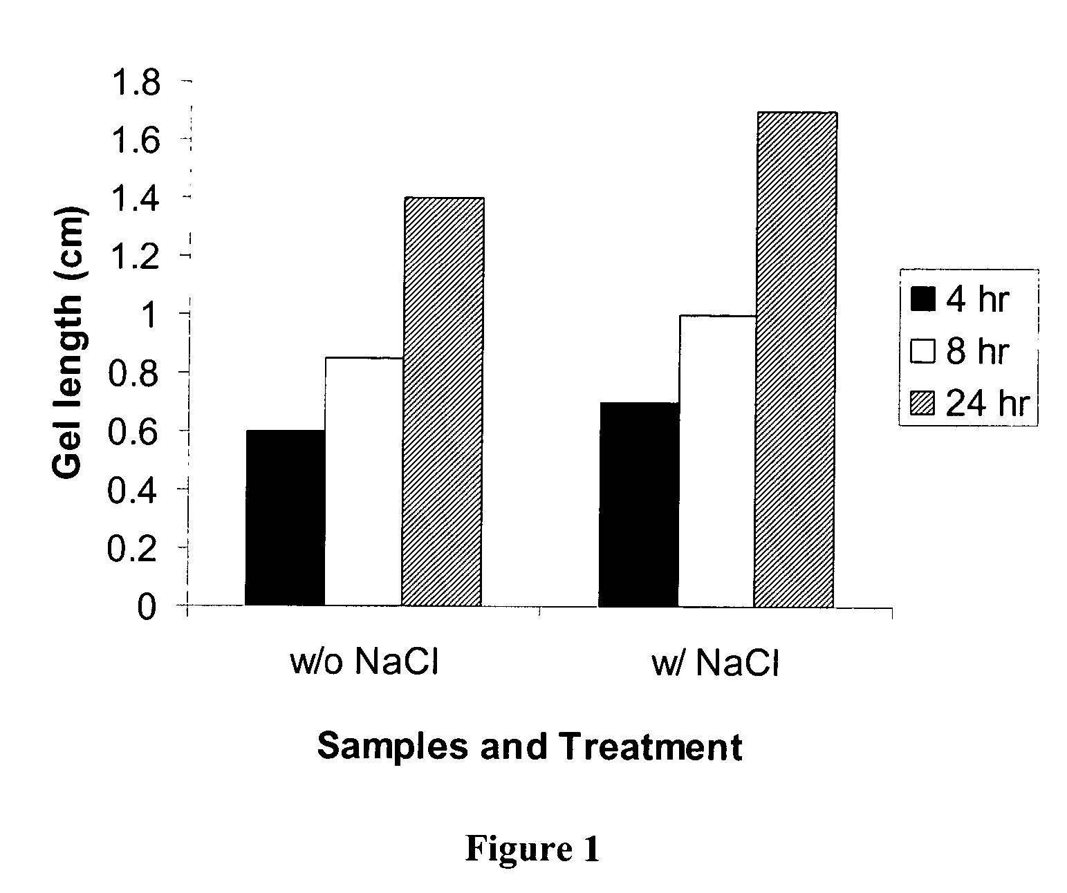
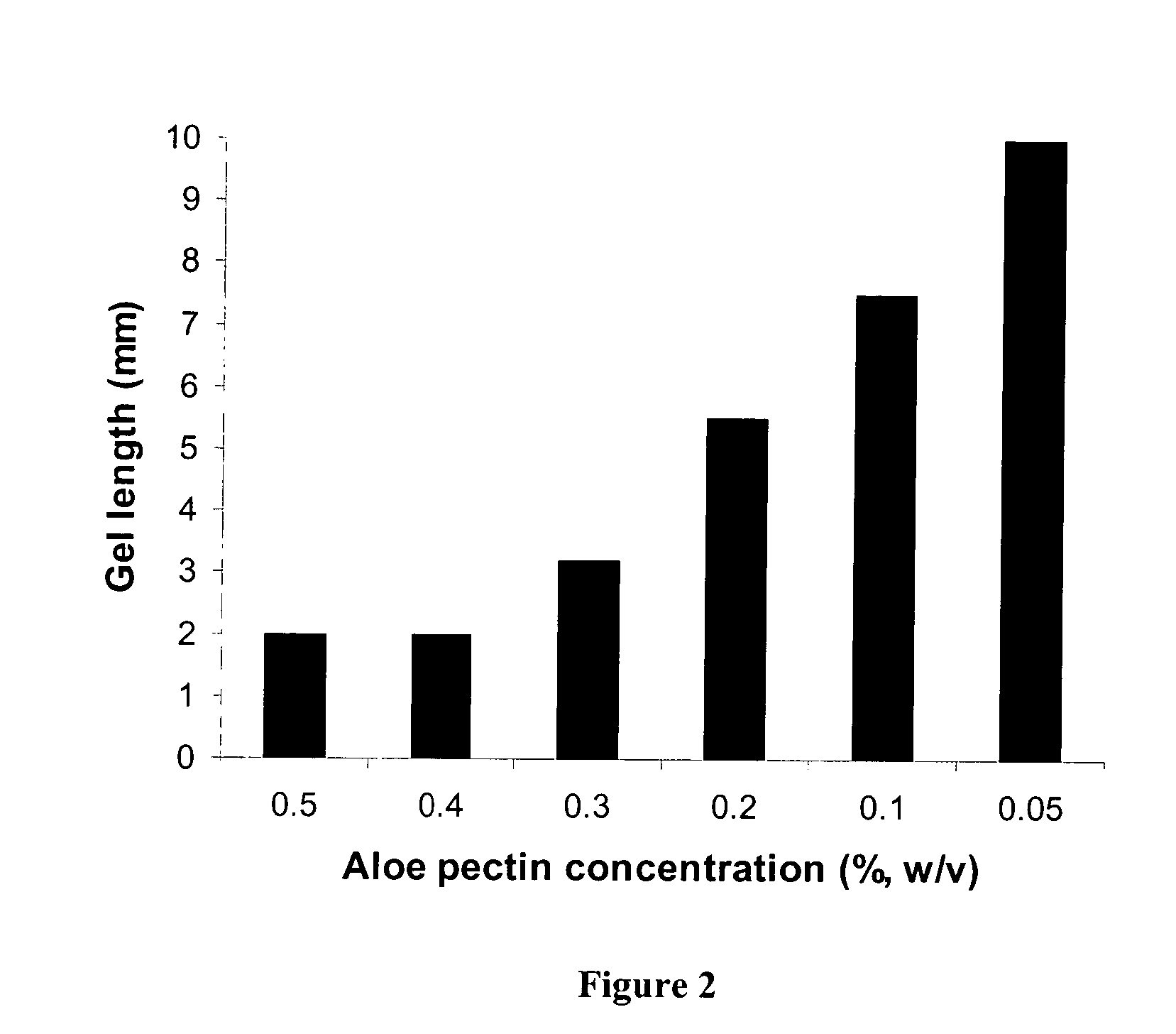
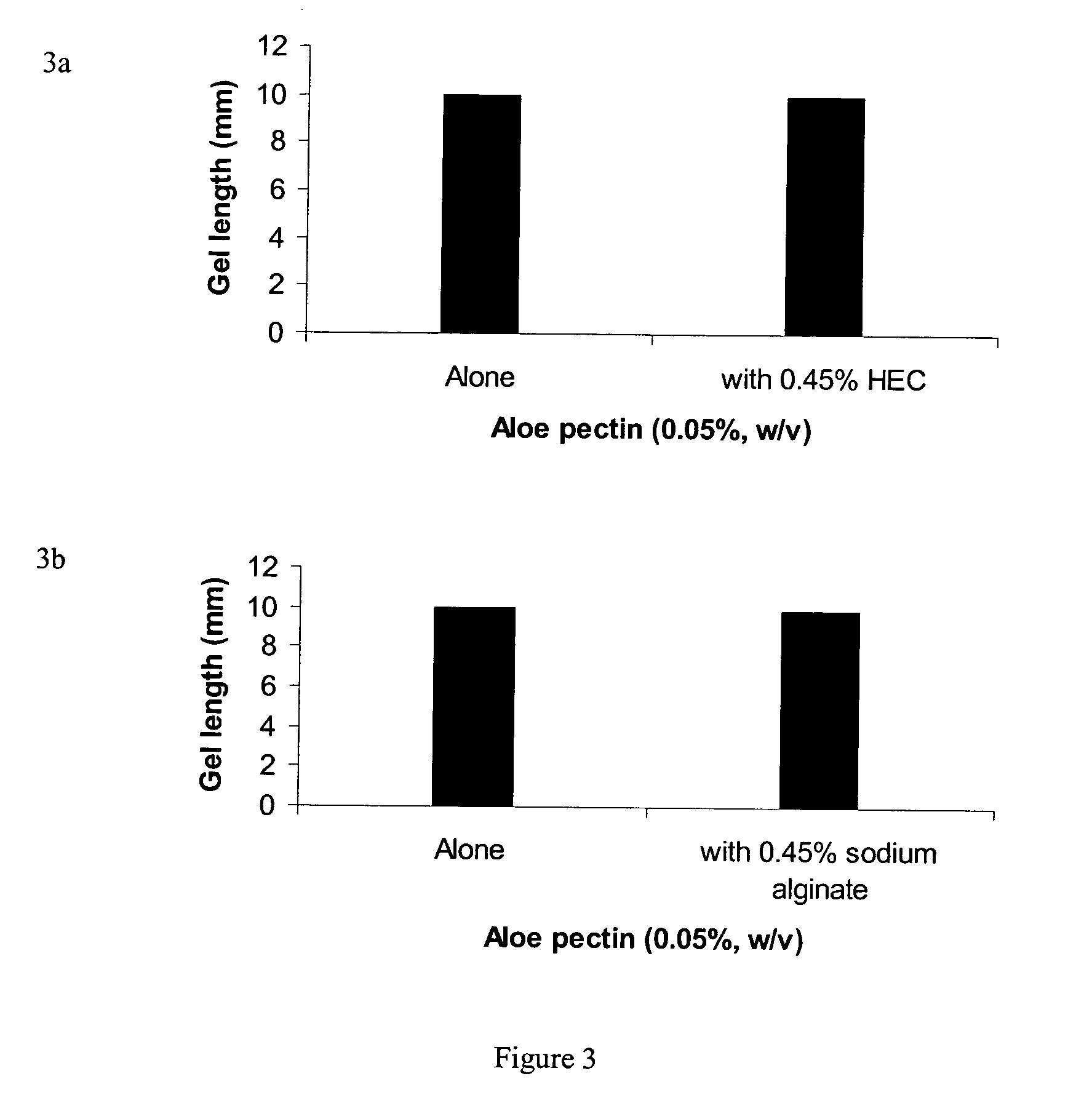
[0177] Body fluids such as blood, lacrimal fluid, lung fluid and nasal secretions contain calcium ions (8.5-10.3 mEq / dl in blood, for example). Since Aloe pectin forms calcium gel, the role of calcium in the in-situ gelation of Aloe pectin was examined using an in vitro gelling assay with animal serum that simulates in-situ gel formation. This in vitro assay is described as a gel frontal migration assay. Animal serum was placed at the bottom of a glass tube and the Aloe pectin solution was layered on top of the serum (the pectin solution may also be placed at the bottom of the tube dependent on the density of the test solution in relation to the pectin solution). Tissue culture grade normal calf serum was used. Two ml of serum was placed at the bottom of a glass tube (0.8×11 cm) and 1 ml pectin solution (0.5-0.75%, w / v) was placed on top of it.

[0178] Gel formation was immed...


PUM

PropertyMeasurementUnit
Fractionaaaaaaaaaa
Fractionaaaaaaaaaa
Fractionaaaaaaaaaa

Abstract

In-situ gelation of a pectic substance. Composition, method of preparation, and method of use of a pectin in-situ gelling formulation for the delivery and sustained release of a physiologically active agent to the body of an animal. The pectin can be isolated from Aloe vera.

Description

REFERENCE TO RELATED APPLICATIONS [0001] This application is a continuation-in-part of, and claims priority to co-pending U.S. Utility application Ser. No. 09 / 795,987, filed Feb. 28, 2001, the disclosure of which parent application is hereby incorporated herein in its entirety by this reference.BACKGROUND OF THE INVENTION [0002] A variety of polymer-based drug delivery systems for achieving sustained or controlled drug release have been described (See Langer, Nature, 392 (supplement), 5-10, 1998 and references therein). The goal of many of those systems was typically one or more of prolonging drug release, improving drug bioavailability, and / or providing non-injectable drug delivery systems that improve patient compliance and comfort. The polymers, either synthetic or natural, provide delivery of the various agents by various mechanisms, depending on the properties of the polymer. [0003] The polymer-based systems have been variously formulated as, for example, a liquid, a suspension...

Claims


Application Information

Patent Timeline
IPC IPC(8): A61K9/06A61KA61K9/00A61K38/04A61K39/00A61K47/04A61K47/36A61K47/38A61K47/40A61K47/42A61L15/44A61P17/02
CPCA61K9/0024A61K9/06A61K38/1825A61K39/39A61K47/36A61K2039/70A61K2039/55505A61K2039/55583C12N2760/16134A61K39/12A61K2039/543A61K39/05A61K2039/5252A61P17/02A61P31/16A61P37/04A61K9/20A61K9/48B82Y5/00
Inventor NI, YAWEIYATES, KENNETH M.
Owner NANOTHERAPEUTICS INC
Learn More