
Device and method for preventing magnetic resonance imaging induced damage

a magnetic resonance imaging and device technology, applied in the field of medical assist systems, can solve problems such as present problems, medical assist devices are vulnerable to external sources of severe electromagnetic noise, and impaired the desired functionality of pacemakers

Inactive Publication Date: 2004-10-07
MEDTRONIC INC
View PDF8 Cites 200 Cited by
  • Summary
  • Abstract
  • Description
  • Claims
  • Application Information

AI Technical Summary

Benefits of technology

0031] A first aspect of the present invention is directed to a medical assist system. The medical assist system includes a primary device housing, the primary device housing having a control circuit therein; a lead system to provide an electrical p

Problems solved by technology

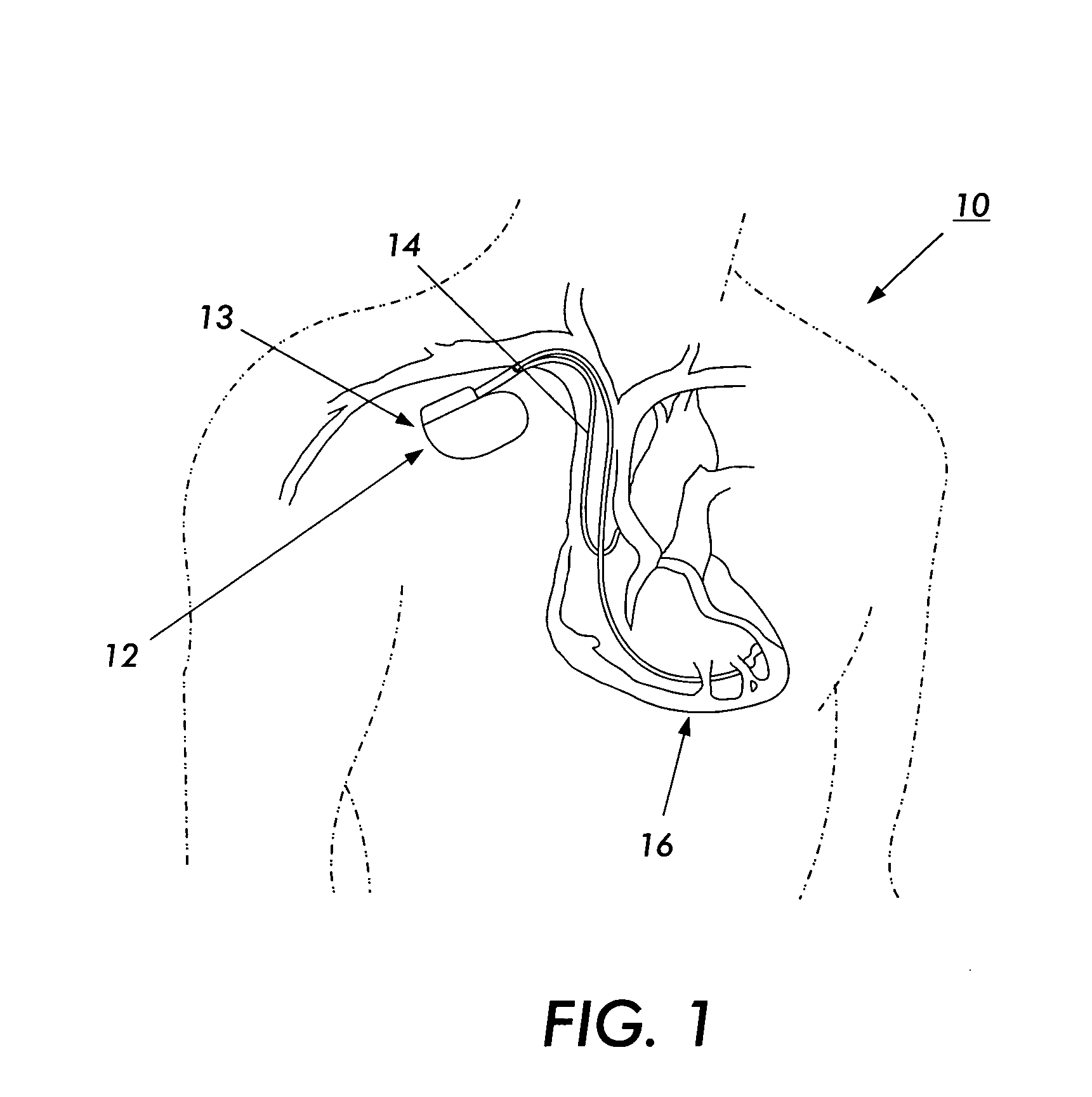
The use of the magnetic-resonance imaging process with patients who have implanted or non-implanted medical assist devices; such as, but not limited to, cardiac assist devices, implanted insulin pumps, catheter guide wires, leads for neurostimulation probes, intraluminal coils, guided catheters, temporary cardiac pacemakers, temporary esophageal pacemakers; often presents problems.
Since the sensing systems and conductive elements of these medical assist devices are responsive to changes in local electromagnetic fields, the medical assist devices are vulnerable to external sources of severe electromagnetic noise, and in particular, to electromagnetic fields emitted during the magnetic-resonance imaging (magnetic-resonance imaging) procedure.
A common implantable pacemaker can, under some circumstances, be susceptible to electrical interference such that the desired functionality of the pacemaker is impaired.
Such electrical interference can damage the circuitry of the cardiac assist systems or cause interference in the proper operation or functionality of the cardiac assist systems.
For example, damage may occur due to high voltages or excessive currents introduced into the cardiac assist system by voltages or currents induced in the cardiac assist system circuitry or on the wire leads leading to and from the cardiac assist system circuitry.
Therefore, it is required that such voltages and currents be limited at the input of such cardiac assist systems, e.g., at the interface.
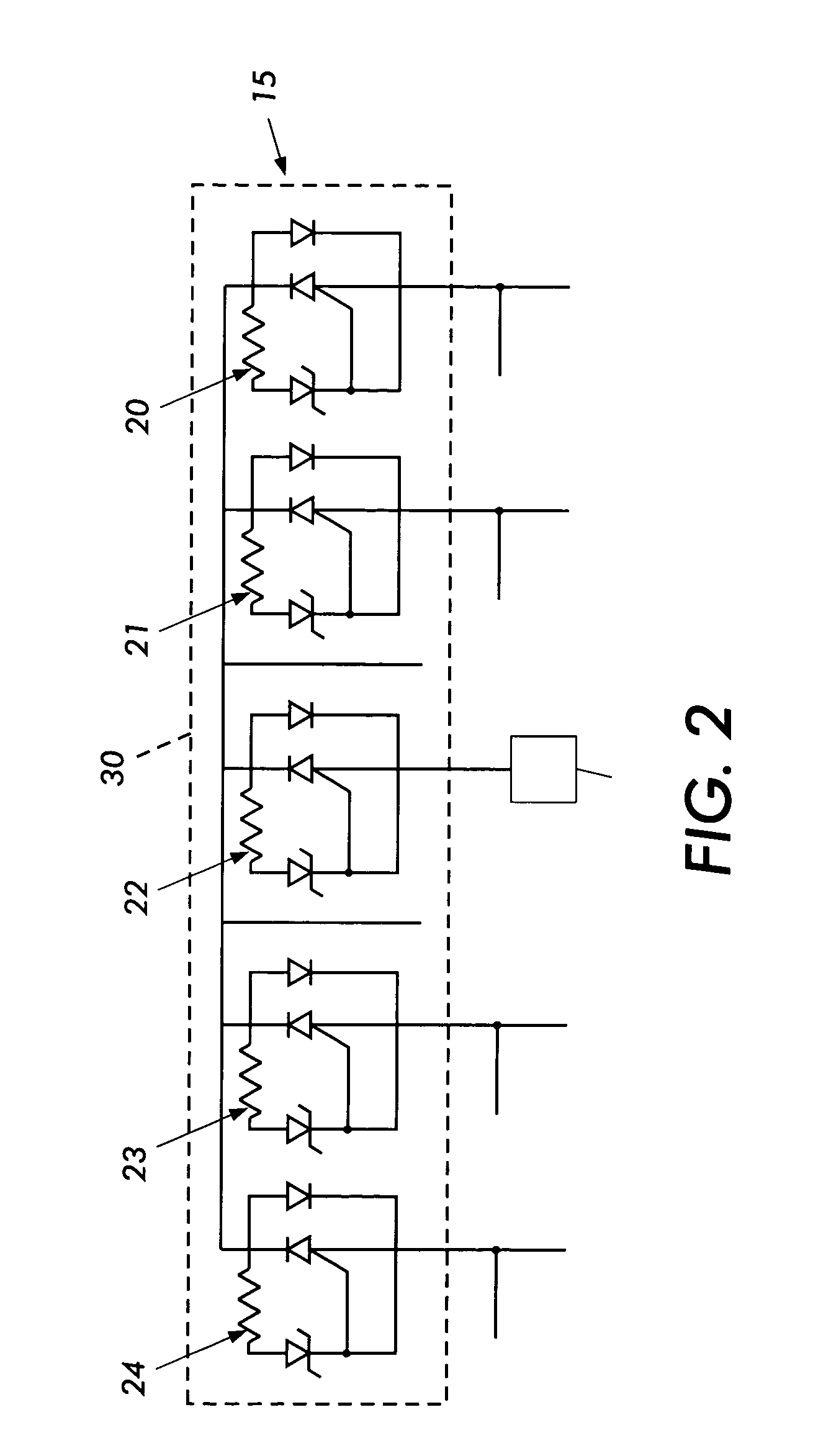
However, such protection, provided by zener diodes and capacitors placed at the input of the medical device, increases the congestion of the medical device circuits, at least one zener diode and one capacitor per input / output connection or interface.
This is contrary to the desire for increased miniaturization of implantable medical devices.
Further, when such protection is provided, interconnect wire length for connecting such protection circuitry and pins of the interfaces to the medical device circuitry that performs desired functions for the medical device tends to be undesirably long.
The excessive wire length may lead to signal loss and undesirable inductive effects.
Additionally, the radio frequency (radio-frequency) energy that is inductively coupled into the wire causes intense heating along the length of the wire, and at the electrodes that are attached to the heart wall.
A further result of this ablation and scarring is that the sensitive node that the electrode is intended to pace with low voltage signals becomes desensitized, so that pacing the patient's heart becomes less reliable, and in some cases fails altogether.
Additionally, the switching of the gradient magnetic fields may also induce unwanted voltages causing problems with the circuitry and potential pacing of the heart.
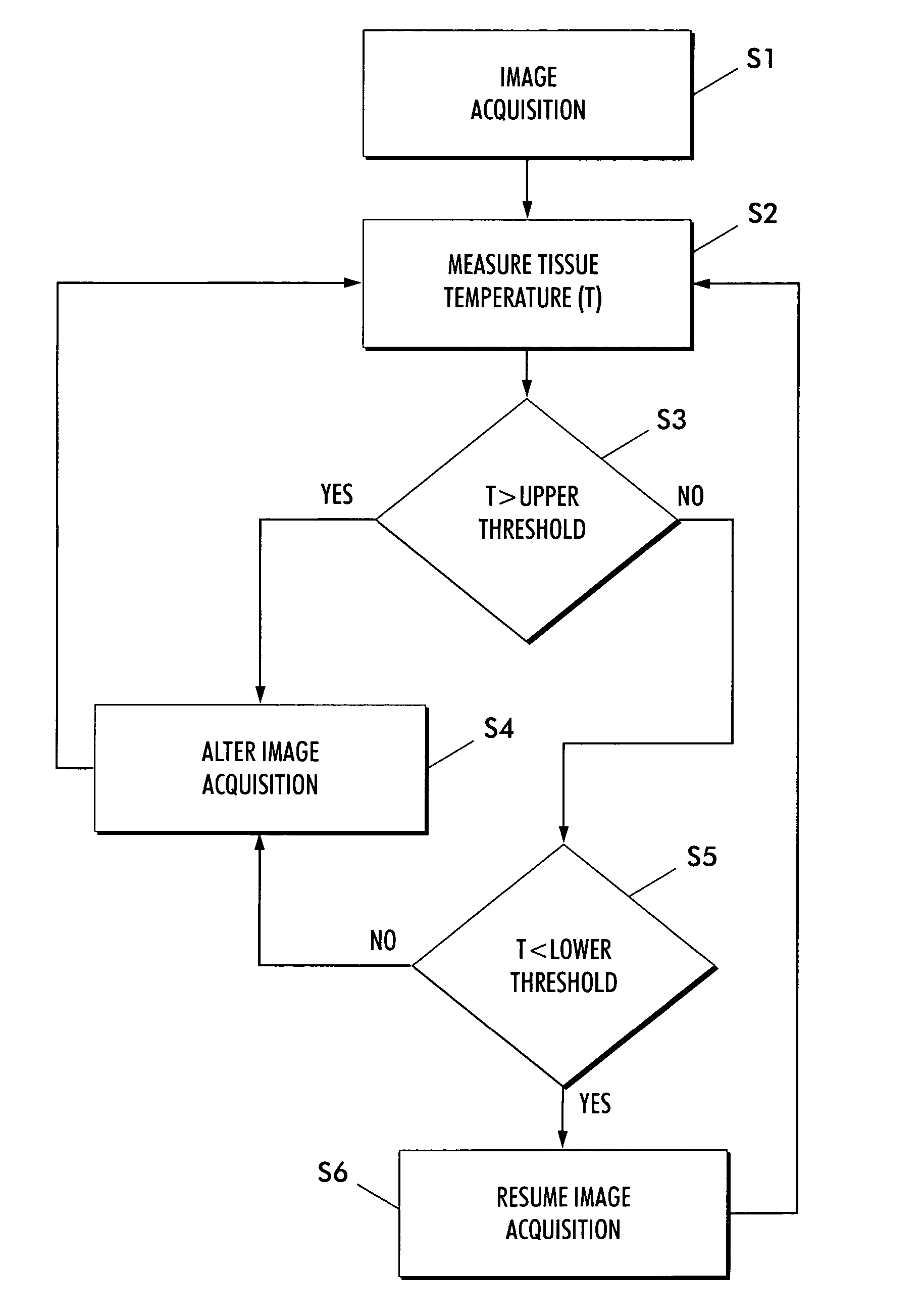
Another problem associated with magnetic-resonance imaging is the temperature change in tissue regions caused by using conventional magnetic-resonance imaging techniques.
The constant changing of alignment of the magnetic moments of the spins in the tissue causes the tissue's temperature to increase, thereby exposing the tissue to possible magnetic-resonance imaging induced thermal damage.
Although, conventional medical assist devices provide some means for protection against electromagnetic interference, these conventional medical assist devices require much circuitry and fail to provide fail-safe protection against radiation produced by magnetic-resonance imaging procedures.
Moreover, the conventional medical assist devices fail to address the possible damage that can be done at the tissue interface due to radio-frequency-induced heating.
Furthermore, the conventional medical assist devices fail to address the unwanted tissue region stimulation that may result from radio-frequency-induced electrical currents.
Lastly, conventional magnetic-resonance imaging processes fail to provide a proper safeguard against potential magnetic-resonance imaging induced thermal damage due to the tissue's exposure to the switching magnetic field gradients and the circularly polarized Radio Frequency Field of the magnetic-resonance imaging process.

Method used


Image

  • Device and method for preventing magnetic resonance imaging induced damage
  • Device and method for preventing magnetic resonance imaging induced damage
  • Device and method for preventing magnetic resonance imaging induced damage

Examples


Embodiment Construction

[0072] As noted above, the present invention is directed to an implantable device, such as a medical assist device, that is immune or hardened to electromagnetic insult or interference.

[0073] For a general understanding of the present invention, reference is made to the drawings. In the drawings, like reference have been used throughout to designate identical elements. In describing the present invention, the following term(s) have been used in the description.

[0074] For the purposes of the description below and the appended claims, the term, medical assist device / system or tissue invasive device / system, refers to any device / system that may enable monitoring of living tissue(s) or living system(s) wherein the monitoring may be, but not limited to an EKG signal, an ECG signal, a glucose level, hormone level, cholesterol level, or magnetic resonance image. The medical assist device / system or tissue invasive device / system may also enable stimulus intervention to provide assistance to l...


PUM


Abstract

An electromagnetic shield has a first patterned or apertured layer having non-conductive materials and conductive material and a second patterned or apertured layer having non-conductive materials and conductive material. The conductive material may be a metal, a carbon composite, or a polymer composite. The non-conductive materials in the first patterned or apertured layer may be randomly located or located in a predetermined segmented pattern such that the non-conductive materials in the first patterned or apertured layer are located in a predetermined segmented pattern with respect to locations of the non-conductive materials in the second patterned or apertured layer.

Description

CROSS REFERENCE TO RELATED PATENT APPLICATIONS[0001] The subject matter of the present application is related to the subject matter of co-pending U.S. patent application Ser. No. 09 / 885,867, filed on Jun. 20, 2001, entitled "Controllable, Wearable magnetic-resonance imaging-Compatible Cardiac Pacemaker With Pulse Carrying Photonic Catheter And VOO Functionality"; co-pending U.S. patent application Ser. No. 09 / 885,868, filed on Jun. 20, 2001, entitled "Controllable, Wearable magnetic-resonance imaging-Compatible Cardiac Pacemaker With Power Carrying Photonic Catheter And VOO Functionality"; co-pending U.S. patent application Ser. No. 10 / 037,513, filed on Jan. 4, 2002, entitled "Optical Pulse Generator For Battery Powered Photonic Pacemakers And Other Light Driven Medical Stimulation Equipment"; co-pending U.S. patent application Ser. No. 10 / 037,720, filed on Jan. 4, 2002, entitled "Opto-Electric Coupling Device For Photonic Pacemakers And Other Opto-Electric Medical Stimulation Equip...

Claims


Application Information

Patent Timeline
IPC IPC(8): G01R33/28G01R33/48G01R33/54
CPCA61N2001/086G01R33/285Y10T428/24322G01R33/4804Y10T428/24331G01R33/288A61N1/086
Inventor CONNELLY, PATRICK R.HELFER, JEFFREY L.
Owner MEDTRONIC INC
Learn More