
Screening kit for detection of grades of cervical cancer and process for the preparation thereof

a screening kit and cervical cancer technology, applied in the direction of material analysis, instruments, measurement devices, etc., can solve the problems of insufficient cytology training for microscopic analysis of pap smears, inability to train cytologists, and inability to meet the needs of cytotechnologists, etc., to achieve great potential

Active Publication Date: 2022-04-26
COUNCIL OF SCI & IND RES
View PDF9 Cites 0 Cited by
  • Summary
  • Abstract
  • Description
  • Claims
  • Application Information

AI Technical Summary

Benefits of technology

The kit provides a reliable, non-invasive, and cost-effective method for cervical cancer screening with high sensitivity and specificity, demonstrated by significant spectral differences and accurate prediction accuracy using chemometric analysis, potentially reducing the reliance on skilled personnel and improving early detection rates.

Problems solved by technology

However, in India and many other low resource countries none of these measures have been implemented so far and cervical cancer continues to take extraordinary toll on the lives of our women.
The major impediment for implementing screening programme by Pap smear is the lack of trained cytologists for microscopic analysis of the Pap smears of the eligible women of the community.
Because skilled cytotechnologists are less available and as it is a tune consuming technique, some other alternative techniques are in high demand.
However, Raman scattering suffers the disadvantage of extremely poor efficiency due to its inherently small cross-section.
Besides, the Raman spectra of biological samples are often superimposed on top of a strong fluorescence background that may be huge and make it difficult to extract the Raman signals.
So in medical diagnosis these disadvantages make it a great challenge for practical applications of conventional Raman spectroscopy.
Label-free SERS produces huge and complex data sets and it necessitates more refined analytical processes to mine substantial information from the spectral data.
A great inconsistency in the intensity and spatial scattering generated due to the variable deposition and orientation of gold nanoparticles reduces the clarity of spectral data obtained.
Collection of tissue sample needs surgical procedures and hence cannot be suggested for screening purpose.

Method used


Image

  • Screening kit for detection of grades of cervical cancer and process for the preparation thereof
  • Screening kit for detection of grades of cervical cancer and process for the preparation thereof
  • Screening kit for detection of grades of cervical cancer and process for the preparation thereof

Examples


examples

[0046]The following examples are given by way of illustration of the present invention and therefore should not be construed to limit the scope of the present invention.

example-1

Preparation of SERS Based Screening Kit:

[0047](i) Synthesis and Optimization of the concentration of Gold Nanoparticle (AuNPs) for label free detection.[0048]Gold nanoparticles (AuNPs, size around 40-50 nm) was prepared by well-established citrate reduction method. The characterization of the synthesized nanoparticles were performed through UV-vis spectroscopy, Dynamic Light Scattering (DLS) and High Resolution Transmission Electron Microscopy (HRTEM). The size was approximately 40-50 nm which serves as an optimal performing SERS substrate.[0049](ii) Optimum concentration of AuNPs which will provide maximum Raman signal intensity; Optimum concentration has been evaluated (AuNPs 8 to 10×1013 particles / ml) which provided the maximum SERS intensity.[0050](iii) A preservative fluid was prepared for preservation and fixation of the sample collected comprising of >50% Ethanol, Methanol, Isopropanol, Formaldehyde, Saline solution, Di-potassium hydrogen phosphate.[0051](iv) A density gradie...

example-2

SERS Fingerprint from Exfoliated Cells

[0054]The exfoliated cell samples were collected in the preservative solution comprising of >50% Ethanol, Methanol, Isopropanol, Formaldehyde, Saline solution, Di-potassium hydrogen phosphate. Density gradient centrifugation was performed using sucrose as gradient solution to enrich the epithelial cells in the cervical smear samples. The pellet obtained was resuspended in the PBS buffer comprising Sodium Chloride, Potassium Chloride, Disodium phosphate, Potassium dihydrogen phosphate. The resuspended solution was dropped in the glass slide coated with Poly L-lysine, APES [(3-Aminopropyl) triethoxy silane. After 10-30 minutes incubation, the excess fluid is discarded and the slide is stored in absolute ethanol till SERS measurement. Single cell has been focused bright field and spectra have been taken from different fields of the cell. A number of spectra have been taken from random locations of the cells and also from nucleus after morphological...


PUM

PropertyMeasurementUnit
sizeaaaaaaaaaa
poweraaaaaaaaaa
integration timeaaaaaaaaaa

Abstract

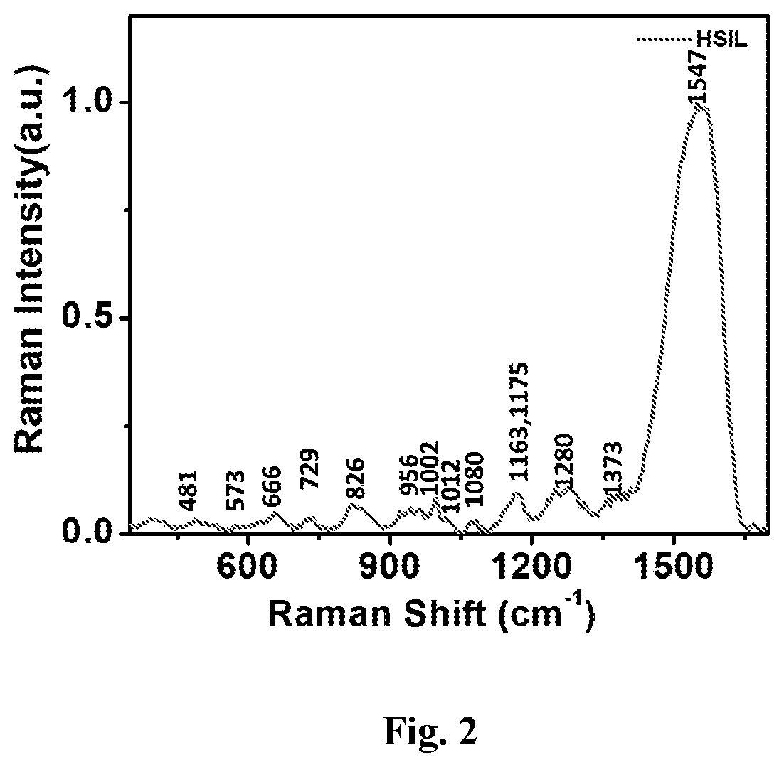
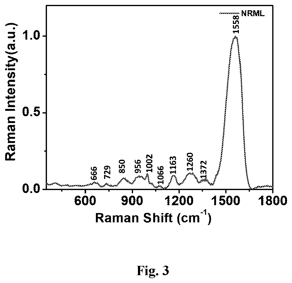
The capabilities of using gold nanoparticle as surface-enhanced Raman scattering (SERS) substrate to obtain cervical smear harvested cells biochemical information for non-invasive cervical precancerous detection were presented in this patent document. A SERS reagent and a platform has been developed and optimized for the generation of a differential spectral fingerprinting for cervical cancer detection. SERS measurements were performed on three group's cervical exfoliated cell samples: one group from patients (n=36) with pathologically confirmed cervical cancer and another group with high-grade squamous intraepithelial lesion (HSIL) (n=41) and the last group from healthy volunteers (control subjects, n=47). Tentative assignments of the Raman bands in the measured SERS spectra suggested interesting cancer specific biomolecular changes, including an increase in the relative amounts of amino acids, nucleic acid, carotenoid contents in the cell samples of cervical cancer patients as compared to that of healthy subjects. The results from this study demonstrated that gold nanoparticle based SERS substrate harvested exfoliated cervical smear cell analysis has tremendous potential for the non-invasive detection of cervical precancerous lesions.

Description

RELATED APPLICATIONS[0001]This application is a national phase application of International Application No. PCT / IN2019 / 050540, filed on Jul. 24, 2019, which claims the benefit of Indian Patent Application No, 201811028087, filed on Jul. 26, 2018. The entire contents of these applications are hereby incorporated by reference.FIELD OF INVENTION[0002]The present invention relates to a screening kit for detection of different grades of cervical cancer. More, particularly the present invention relates to efficient platform developed to identify pre-cancerous lesions of the cervix viz.: (high-grade squamous intraepithelial lesion (HSIL) and cervical squamous cell carcinoma (CSCC) in the cells exfoliated from uterine cervix using a differential Raman spectral pattern.BACKGROUND OF THE INVENTION[0003]Cancer of the uterine cervix is one of the most common cancers among women worldwide and the second most prevalent cancers among women in India. It is considered as a preventable cancer as cerv...

Claims


Application Information

Patent Timeline
Patent Type & Authority Patents(United States)
IPC IPC(8): G01N33/574G01N21/65G01N1/40G01N1/28B82Y40/00
CPCG01N33/57411G01N1/2813G01N1/4077G01N21/658B82Y40/00G01N2001/4083
Inventor MAITI, KAUSTABH KUMARKARUNAKARAN, VARSHASUJATHAN, KUNJURAMAN
Owner COUNCIL OF SCI & IND RES
Learn More