Eureka AIR delivers breakthrough ideas for toughest innovation challenges, trusted by R&D personnel around the world.

African swine fever virus P12 protein nanoparticle and preparation method and application thereof

An African swine fever virus and nanoparticle technology, applied in the field of African swine fever virus P12 protein nanoparticles and their preparation, can solve the problems of low protective efficacy, no protective effect, and inability to resist virulent attacks.

Active Publication Date: 2021-03-30
非零和(北京)投资管理有限公司
View PDF3 Cites 2 Cited by
  • Summary
  • Abstract
  • Description
  • Claims
  • Application Information

AI Technical Summary

Problems solved by technology

[0003] In the 1960s, people first tried to use inactivated ASF vaccines, but most of them had no protective effect
In 2014, Blome et al. used the latest adjuvant combined with ASF inactivated vaccine immunization, but still could not resist the virulent attack
[0005] Through discussion and analysis of existing technologies, the current ASFV subunit vaccines, nucleic acid vaccines and virus live vector vaccines have low protective efficacy

Method used


Image

  • African swine fever virus P12 protein nanoparticle and preparation method and application thereof
  • African swine fever virus P12 protein nanoparticle and preparation method and application thereof
  • African swine fever virus P12 protein nanoparticle and preparation method and application thereof

Examples


Embodiment 1

[0042] Embodiment 1 plasmid construction

[0043] This embodiment provides the amino acid sequence of African swine fever virus P12 protein nanoparticle (GSTP1-MT3-P12), as shown in SEQ ID NO:4. Entrust Qingke Biological Company to synthesize the DNA fragment of GSTP1-MT3-P12 with His tag fused at the C-terminus, its nucleic acid sequence is shown in SEQ ID NO: 5 (wherein, the 13th-987th bits encode the DNA fragment shown in SEQ ID NO: 4 sequence, the 988-1005th encoding His tag), and constructed into the plasmid vector pPICZaA, the map of the constructed plasmid is shown in figure 1 As indicated, the plasmid was named pPICZaA-GSTP1-MT3-P12. The plasmid vector pPICZaA contains the α-factor secretion signal peptide sequence before inserting the fragment. The sequenced correct monoclonal strain was cultivated, and the plasmid pPICZaA-GSTP1-MT3-P12 was extracted for use.

[0044] Amino acid sequence of GSTP1-MT3-P12 (SEQ ID NO: 4):

[0045] DPETCPCPSGGSCTCADSCKCEGCKCTSCKKSCCS...

Embodiment 2

[0046] Example 2 Induced expression of protein nanoparticles

[0047] After the plasmid prepared in Example 1 was linearized, it was electroporated into X-33 yeast competent cells for inducible expression. The specific steps are as follows:

[0048] (1) Plasmid linearization

[0049] The plasmid pPICZaA-GSTP1-MT3-P12 prepared in Example 1 was taken and digested with linearase SacI. The restriction system was as follows:

[0050]

[0051] After mixing, centrifuge at 12000 rpm at room temperature for 1 min to shake the liquid to the bottom of the tube, and bathe in water at 37 °C for 4 h.

[0052] After digestion, take 2 μL, and conduct 1% agarose gel electrophoresis with the original plasmid to identify the linearization result. After identification, the plasmid has been digested completely. Use the plasmid kit to recover the digested plasmid and use it in the subsequent test steps. .

[0053] (2) Electroporation (aseptic operation)

[0054] Add 10μL of the linearized pl...

Embodiment 3

[0058] Example 3 SDS-PAGE identification

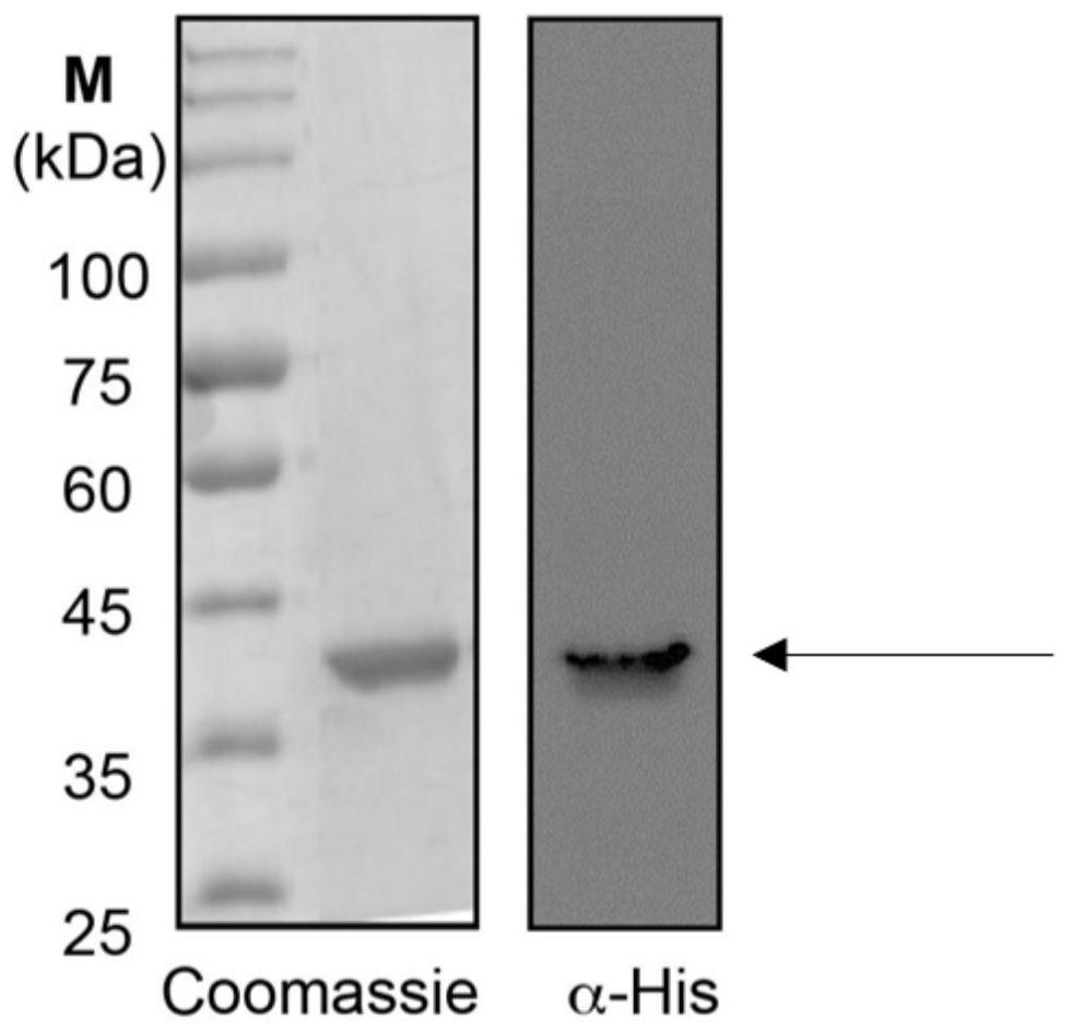
[0059] Take 1 mL of the culture product prepared in Example 2, centrifuge in a centrifuge at the maximum speed for 2 min at room temperature, take the supernatant, mix the supernatant with Loading buffer to make a loading sample, carry out SDS-PAGE identification, and analyze the expression level. SDS-PAGE identification results see figure 2 As shown in (left), it was identified that the target protein was successfully expressed.


PUM

PropertyMeasurementUnit
diameteraaaaaaaaaa
particle diameteraaaaaaaaaa

Abstract

The invention relates to an African swine fever virus P12 protein nanoparticle and a preparation method and application thereof, the protein nanoparticle is formed by self-assembly of a protein monomer, and the protein monomer is a fusion protein sequentially containing metallothionein, glutathione thiotransferase and P12 protein from amino acid to a carboxyl terminal. The GSTP1-MT3 protein and the P12 antigen of the African swine fever virus are subjected to fusion expression, and in a pichia pastoris expression system, the protein nanoparticle can be spontaneously formed through induction offerrous ions. Immunogenicity determination is carried out in the mouse body, and the P12 protein nanoparticle can cause very strong immune response and has great potential to be developed into a newsafe and effective African swine fever virus vaccine.

Description

technical field [0001] The invention relates to the field of biotechnology, in particular to an African swine fever virus P12 protein nanoparticle and a preparation method and application thereof. Background technique [0002] African swine fever (African swine fever, ASF) is an acute, hemorrhagic, highly contagious infectious disease caused by African swine fever virus (ASFV) infecting domestic pigs or wild boars, characterized by short course of disease, high fever and Hemorrhagic lesions, acute infection mortality rate up to 100%, a serious threat to the global pig industry. [0003] In the 1960s, people first tried to use inactivated ASF vaccines, but most of them had no protective effect. In 2014, Blome et al. used the latest adjuvant combined with ASF inactivated vaccine for immunization, but still could not resist the virulent attack. Using subunit vaccines prepared by protective antigens (p72, p54, CD2v, etc.), immunized pigs can only provide partial protection, bu...

Claims


Application Information

Patent Timeline
Patent Type & Authority Applications(China)
IPC IPC(8): C07K19/00C12N15/62C12P21/00A61K47/69A61K39/12A61P31/20
CPCC07K14/005C07K14/825C12N9/13A61K47/6931A61K39/12A61P31/20C12Y208/01C07K2319/00C12N2710/12022C12N2710/12034Y02A50/30
Inventor 林坚朱新杰李紫晨郑梦竹沈显贵
Owner 非零和(北京)投资管理有限公司
Why Eureka
Eureka Blog
Learn More