
PLGA cytoskeleton for articular cartilage repair and preparation method and application of PLGA cytoskeleton for articular cartilage repair

A technology of cell scaffolds and chondrocytes, applied in the field of medicine, can solve problems such as unsatisfactory treatment results, poor bionic effects, and long treatment cycles

Active Publication Date: 2019-08-16
HARBIN MEDICAL UNIVERSITY
View PDF9 Cites 5 Cited by
  • Summary
  • Abstract
  • Description
  • Claims
  • Application Information

AI Technical Summary

Problems solved by technology

[0008] Chondrocyte transplantation technology to repair cartilage damage has made great progress in recent years. Unfortunately, due to the small size of the initial cartilage tissue sample, slow-proliferating chondrocytes must undergo long-term expansion to reach a certain number, and the treatment cycle is long. And unfavorable factors such as excessive absorption of induced matrix, poor cell recognition signal of matrix material, poor bionic effect, and dedifferentiation of cells lead to unsatisfactory treatment results

Method used


Image

  • PLGA cytoskeleton for articular cartilage repair and preparation method and application of PLGA cytoskeleton for articular cartilage repair
  • PLGA cytoskeleton for articular cartilage repair and preparation method and application of PLGA cytoskeleton for articular cartilage repair
  • PLGA cytoskeleton for articular cartilage repair and preparation method and application of PLGA cytoskeleton for articular cartilage repair

Examples


Embodiment 1

[0077] Embodiment 1: Preparation of PLGA porous scaffold

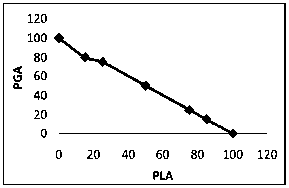
[0078] 1. Preparation of PLGA porous scaffold. The scaffold part of the articular cartilage repair material uses polylactic acid-glycolic acid (PLGA) as the matrix material, which is composed of organic polymer polylactic acid (PLA) and polyglycolic acid (PGA), according to a certain gradient ratio (polylactic acid (PLA): Polyglycolic acid (PGA) = 100:0, 85:15, 75:25, 50:50, 25:75, 15:85, 0:100) polymerization product ( figure 1 ).

[0079] Polylactic acid (PLA), polyglycolic acid (PGA), polylactic acid-glycolic acid (PLGA) monomer chemical structure formula is as follows:

[0080] Polylactic acid [poly(lactic acid), PLA]:

[0081] Polyglycolic acid [poly(glycolic acid), PGA]:

[0082] Polylactic-co-glycolic acid [poly(lactic-co-glycolic acid), PLGA]:

[0083] Prepare the PLGA porous scaffold by freeze-drying technique, the steps are as follows:

[0084] a) PLA / The PGA polymerization product—polylactic ac...

Embodiment 2

[0110] Example 2: Primary culture of adult cells

[0111] 1. Chondrocytes. Adult chondrocytes are derived from articular hyaline cartilage in nonweight-bearing areas.

[0112] a) Cartilage sampling preparation. Items to be prepared include trypsin, type II collagenase, DMEM high-glucose medium, PBS buffer, pipettes, culture dishes or flasks of different capacities, centrifuge tubes, filters, gauze, surgical equipment such as tweezers, scissors, etc. for disinfection after supplies.

[0113] b) Cartilage sampling. After anesthesia, disinfect with povidone iodine and deiodine with 75% alcohol, take about 50-100 mg of articular hyaline cartilage from non-weight-bearing parts, and put it into a petri dish filled with PBS buffer.

[0114] c) Repeated pipetting and washing with cold PBS buffer containing antibiotics 3 times. After the third wash, let stand for 5 minutes, discard the upper liquid and floating tissue, and move the cartilage tissue into a clean sterile vessel.

...

Embodiment 3

[0134] Example 3: Preparation of platelet-rich plasma (PRP) and its release of growth-promoting factors

[0135] 1. Using a 20ml sterile syringe and a vacuum blood collection tube containing ACD-A anticoagulant, draw about 18ml of blood at room temperature (22°C), and put the blood into four 5ml centrifuge tubes, 4ml in each tube. Platelet-rich plasma preparation should be completed within 4 to 6 hours.

[0136] 2. Preparation of platelet-rich plasma. Platelet-rich plasma with effective concentration was prepared by secondary centrifugation, and the activity levels of cytokines PDGF and TGF-β1 in platelet plasma prepared by this method increased significantly (P<0.01).

[0137] a) Place 4ml of each tube symmetrically in a centrifuge, centrifuge for the first time at 200-400g×10min, take 3mm of plasma above and below the buffy coat after centrifugation, and put it in another centrifuge tube,

[0138] b) Perform a second centrifugation at 200-400 g×10 min, remove the supernata...


PUM


Abstract

The invention discloses a PLGA cytoskeleton for articular cartilage repair and a preparation method and application of the PLGA cytoskeleton for articular cartilage repair. The PLGA cytoskeleton comprises a PLGA porous scaffold, cartilage cells, mesenchymal stem cells and adipose-derived stem cells, and the cartilage cells, the mesenchymal stem cells and the adipose-derived stem cells are plantedin the PLGA porous scaffold. The PLGA porous scaffold is a hierarchical composite scaffold formed by PLA (polylactic acid) and PGA (polyglycolic acid) polymerization products in different polymerization ratios and comprises three areas including an outside area, a middle area and an inside area. The cartilage cells are subjected to injection inoculation in pores of the outside area of the PLGA porous scaffold; the cartilage cells and the adipose-derived stem cells are subjected to injection inoculation in pores of the middle area; the cartilage cells and the mesenchymal stem cells are subjected to injection inoculation in pores of the inside area of the PLGA porous scaffold. By combination of a gradient-ratio artificially synthesized high polymer material and natural biological protein, ahierarchical composite structure is achieved. According to experiments, the PLGA cytoskeleton has a great repair effect on cartilage defect.

Description

technical field [0001] The present invention relates to an articular cartilage repair material and its preparation method and application, in particular to a PLGA cell scaffold made by combining a composite scaffold with mechanical strength, tissue compatibility and biodegradability and adult cells , can be used for clinical treatment of cartilage damage caused by various reasons. The invention belongs to the technical field of medicine. Background technique [0002] Cartilage damage is a common and frequently-occurring disease in clinical practice, especially joint damage is very common in people over 50 years old. Diseased joints occur in different parts of the body, and can be single or multiple, with varying degrees of dysfunction. Different degrees of inflammatory reactions and cartilage defects can be found under arthroscopy. Under normal circumstances, doctors will treat symptoms, such as pain relief, anti-inflammation, physical therapy, etc.; if the lesion continu...

Claims


Application Information

Patent Timeline
IPC IPC(8): A61L27/38A61L27/18A61L27/22A61L27/24A61L27/56
CPCA61L27/18A61L27/222A61L27/24A61L27/3817A61L27/3834A61L27/56A61L2430/06C08L67/04C08L77/04C08L89/00
Inventor 王克维王文波孙殿军
Owner HARBIN MEDICAL UNIVERSITY
Learn More