Preparation method of photoelectric chemical biosensor for microRNA detection
A biosensor, photoelectrochemical technology, applied in the direction of material electrochemical variables, scientific instruments, instruments, etc., can solve the problems of high electron-hole recombination rate, cadmium ion toxicity, poor photostability, etc., and achieve high sensitivity and stability good, specific effect
- Summary
- Abstract
- Description
- Claims
- Application Information
AI Technical Summary
Problems solved by technology
Method used
Image
Examples
Embodiment 1
[0034] Synthesis and characterization of cyclometalated iridium complex optoelectronic materials:
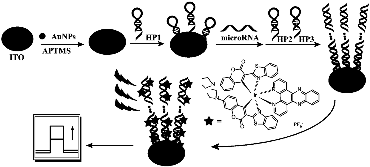
[0035] Weigh IrCl 3 ·3H 2 O solid (0.34g, 1mmol) was dissolved in water (5mL), stirred to fully dissolve, added coumarin-6 (0.77g, 2.2mmol) and ethylene glycol ether (15mL), and the reaction mixture was placed under a nitrogen atmosphere React at 120°C for 24h and cool to room temperature. Filter, wash with water, absolute ethanol and acetone to obtain a chlorine-bridged intermediate. Weigh the obtained chlorine-bridged intermediate (0.14 g, 0.08mmol), dipyrido[3,2-a:2',3'-c]phenazine (0.06g, 0.2mmol), and add to ethylenedi Alcohol ether (10mL). React at 120° C. for 15 h under nitrogen protection, and cool to room temperature. The solvent was removed by evaporation under reduced pressure and purified by column chromatography (200-300 mesh, eluent: dichloromethane / methanol=10 / 1) to obtain a dark red solid (0.061g, 0.046mmol, 58%). 1 H NMR (CDCl 3,500MHz)δ:9.69(d,J=9.0Hz,2H...
Embodiment 2
[0037] Preparation of ITO working electrode:
[0038] The ITO electrode was ultrasonically cleaned with acetone, ethanol, and ultrapure water in sequence, and then fully dried under a nitrogen atmosphere. Immerse the ITO electrode in 1mL ultrapure water, 0.2mLNH 4 OH (30%), 0.2mLH 2 o 2 (30%) in the mixed solution, take it out after soaking for 10min, continue to fully clean with ultrapure water, fully dry in nitrogen atmosphere, soak the above-mentioned treated ITO electrode in the ethanol solution of 3-aminopropyltrimethoxysiloxane Medium (5% (V / V)), 12 hours at room temperature. Fully wash with ethanol, fully dry in a nitrogen atmosphere, and then place it in an oven at 110° C. for 1 hour. Subsequently, it was soaked in the prepared colloidal gold solution and assembled for 12 hours, fully cleaned with ultrapure water, and dried under nitrogen. The capture hairpin HP1 was dissolved in SPSC buffer solution (1M NaCl, 50mM NaCl 2 HPO 4 , 10 mM tris(2-carboxyethyl)phosph...
Embodiment 3
[0040] Preparation of photoelectrochemical biosensors for microRNA detection:
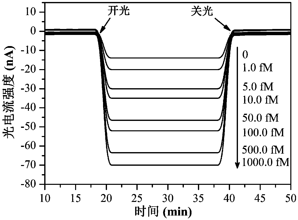
[0041] 20 μL of different concentrations of microRNA-122b was dropped onto the ITO working electrode prepared in Example 2, incubated at 37° C. for 2 h, fully washed with Tris-HCl (0.01 M) buffer, and dried under nitrogen atmosphere. Then 20 μL of the mixture of hairpin HP2 and HP3 (1 μM) was added dropwise, incubated at 37°C for 2 hours, and after being fully washed with Tris-HCl (0.01M) buffer, 10 μL of the cyclometalated iridium complex prepared in Example 1 was added dropwise. Substance solution (solvent: DMF / PBS=1:20, 0.01 μM) was incubated for 40 minutes, and then washed with buffer solution to remove non-specifically adsorbed signal substances for the detection of photoelectric signals.
PUM
 Abstract
Description
Claims
Application Information
 - R&D
- Intellectual Property
- Life Sciences
- Materials
- Tech Scout
- Unparalleled Data Quality
- Higher Quality Content
- 60% Fewer Hallucinations
Browse by: Latest US Patents, China's latest patents, Technical Efficacy Thesaurus, Application Domain, Technology Topic, Popular Technical Reports.
© 2025 PatSnap. All rights reserved.Legal|Privacy policy|Modern Slavery Act Transparency Statement|Sitemap|About US| Contact US: help@patsnap.com


